. 2007 Sep 15;366(1):185-96.
doi: 10.1016/j.virol.2007.04.017. Epub 2007 May 16.

Oral administration of live virus protects susceptible mice from developing Theiler's virus-induced demyelinating disease

Affiliations

Oral administration of live virus protects susceptible mice from developing Theiler's virus-induced demyelinating disease

Bong-Su Kang et al. Virology. .

Abstract

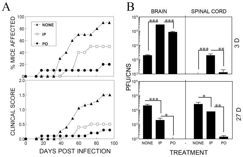
Intracerebral infection of susceptible mouse strains with Theiler's murine encephalomyelitis virus (TMEV) results in an immune-mediated demyelinating disease similar to human multiple sclerosis. TMEV infection is widely spread via fecal-oral routes among wild mouse populations, yet these infected mice rarely develop clinical disease. Oral vaccination has often been used to protect the host against many different infectious agents, although the underlying protective mechanism of prior oral exposure is still unknown. To understand the mechanisms involved in protection from demyelinating disease following previous oral infection, immune parameters and disease progression of mice perorally infected with TMEV were compared with those of mice immunized intraperitoneally following intracerebral infection. Mice infected perorally, but not intraperitoneally, prior to CNS viral infection showed lower chronic viral persistence in the CNS and reduced TMEV-induced demyelinating disease. However, a prolonged period of post-oral infection was necessary for effective protection. Mice orally pre-exposed to the virus displayed markedly elevated levels of antibody response to TMEV in the serum, although T cell responses to TMEV in the periphery were not significantly different between perorally and intraperitoneally immunized mice. In addition, orally vaccinated mice showed higher levels of early CNS-infiltration of B cells producing anti-TMEV antibody as well as virus-specific CD4(+) and CD8(+) T cells in the CNS compared to intraperitoneally immunized mice. Therefore, the generation of a sufficient level of protective immune responses appears to require a prolonged time period to confer protection from TMEV-induced demyelinating disease.


Figures

Figure 1
Figure 1. Requirement of greater than 45 days after oral immunization for effective protection from TMEV-IDD
Female SJL/J mice were orally vaccinated with 1×10⁷ PFU TMEV BeAn at 30 (n=10), 45 (n=10) or 51 (n=6) days prior to intracerebral infection with 1×10⁶ PFU TMEV. All mouse groups were intracerebrally infected with TMEV on the same day at 15 wk of age. Peroral (PO) immunization resulted in significantly lower incidence of TMEV-IDD compared to those non-immunized. Mice immunized at 45 days or earlier (51 days) prior to intracerebral (ic) infection were effectively protected, but mice immunized at 30 days prior to infection were not. Differences in disease incidences between the non-immunized group and the orally immunized group are as follows, based on a paired, two-tailed Student’s t test with Welch correction between 28 and 56 d post infection: at −30 d, p>0.05 (not significant); −45 d, p<0.01 (very significant); and −51 d, p<0.01 (very significant).
Figure 2
Figure 2. Protection from TMEV-IDD by oral, but not intraperitoneal, immunization
Female SJL/J mice (n=10 each group) were perorally (po) or intraperitoneally (ip) administered with 1×10⁷ PFU intact BeAn at 45 days prior to intracerebral infection. Untreated mice were intracerebrally infected as a control group. A. Oral immunization resulted in a significantly lower incidence of TMEV-IDD compared to intraperitoneally immunized mice or the untreated control group. Differences among the experimental groups are significant based on ANOVA Tukey-Kramer multiple comparison test between days 39 and 95. **p<0.01, untreated (NONE) vs intraperitoneally (IP) immunized groups; ***p<0.001, NONE vs perorally (PO) immunized groups; *p<0.05, IP vs PO. B. Viral persistence levels in the CNS (brains and spinal cords) of these groups (n=3–4) were assessed by plaque assay at 3 and 27 d post CNS viral infection. TMEV levels at both 3 and 27 days post-infection were significantly lower in PO immunized mice compared to those of IP immunized or unimmunized mice. *, p<0.05; **, p<0.01; and ***; p<0.001.
Figure 3
Figure 3. Peripheral T cell responses to TMEV antigens in mice vaccinated via different routes
A. Proliferative responses of splenic T cells to TMEV antigens in mice orally or intraperitoneally vaccinated with live TMEV (n=3). Splenocytes from mice at days 15/& −30d, 45/0d, 52/+22d or 115/+70d (post immunization/intracerebral infection, respectively) were cultured for 4 d in the presence of UV-inactivated TMEV or synthetic peptides bearing the predominant CD4+ T cell epitopes (VP1 233–250, VP2 74–86, and VP3 24–37). B. IFNγ-producing splenic CD4+ T cells in response to TMEV capsid epitopes determined by ELISPOT assays. C. IFNγ-producing CD8+ T cells in response to TMEV epitopes (VP111–20, VP3159–166 and VP3173–181) determined by ELISPOT assays. NS, not significant; *, p<0.05; **, p<0.01; and ***, p<0.001.
Figure 4
Figure 4. Levels of T cells specific to TMEV antigens in the CNS of mice vaccinated via different routes
A. Flow cytometric analyses of T cells among the CNS mononuclear cells from vaccinated or untreated mice at 3, 6, and 27 d post intracerebral TMEV infection. The proportion of IFNγ-producing cells in response to the mixture of CD4+ T cell epitope peptides and CD8+ T cell epitope peptides was determined by flow cytometry following intracellular cytokine staining. One representative of three separate experiments with similar results is shown here. B. The assessment of IFNγ-producing T cells infiltrating the CNS of vaccinated mice in response to individual TMEV epitope peptides by ELISPOT assay at 6 and 14 d post CNS TMEV infection. The statistic values represent the significance between vaccinated and non-vaccinated mice and the values inside the parentheses represent the difference between orally vaccinated and peritoneally vaccinated mice. *, p<0.05; **, p<0.01; and ***, p<0.001. When the values of three CD8+ T cell epitopes (VP111–20, VP3159–166 and VP3173–181) were pooled, ***, NONE vs IP; ***, NONE vs PO; **, IP vs PO at 6 d post infection and *, NONE vs IP; ***, NONE vs PO; *, IP vs PO at 14 d post infection.
Figure 5
Figure 5. Antibody levels to TMEV in mice vaccinated orally and peritoneally
A. Mice were bled at 17/−30d, 47/0d, 54/+7d, 69/+22d, 117/+70d or 159/+112d post-immunization followed by intracerebral infection, respectively. Antibody Levels to TMEV in pooled serum (n=5) were analyzed by ELISA. Sera from orally vaccinated mice show markedly higher levels of antibodies to TMEV compared to those from peritoneally vaccinated mice (p<0.001) in the early stages of viral infection (47/0 and 54/+7 d post immunization/infection). B. The isotype determination of anti-TMEV antibody in pooled sera from mice at 47/0 d post immunization/infection. Serum IgG subclasses (IgG1 and IgG2a) were determined by ELISA. C. Antibody titers detected in saliva reactive to UV-inactivated TMEV. Saliva washes were collected from mice at 17/−30d and 54/7d post immunization/infection. Samples were pooled and analyzed by ELISA. Total IgA levels were not different between the groups (data not shown).
Figure 6
Figure 6. TMEV-specific B cells in the CNS and periphery during the course of viral infection
A. Flow cytometric analysis of B cells infiltrating the CNS of mice (47 d prior to viral infection) at 50/3, 53/6 and 74/27 d post immunization/infection, respectively. The percentages of CD19+ cells in the CNS MNC from non-vaccinated, orally and peritoneally vaccinated mice were determined at 3, 6, and 27 d post infection. B. Enumeration of anti-TMEV antibody-producing cells in the CNS and periphery during the course of viral infection. *, p<0.05; **, p<0.01; and ***, p<0.001. A representative of three separate experiments is shown here.
Figure 7
Figure 7. Epitope reactivity and the potential role of antibodies in virus-infected mice
A. Reactivity of anti-TMEV antibodies to linear epitopes in sera from mice (n=4) at 47/0 d and 103/+56 d post immunization/infection. Antibodies to A1C (VP1262–276) and A3A (VP324–37) were relatively higher in orally vaccinated mice at 47/0 d compared to peritoneally vaccinated mice; the level of antibodies to A1C selectively increased after viral infection regardless of immunization. B. Effect of transfusion of antibodies specific to the A1C epitope on the development of demyelinating disease. Naïve SJL/J mice were repeatedly immunized with A1C-KLH and the serum was transfused into SJL/J mice (n=10/group) before (−5, 0 and 5 d) or after (12, 19, 25 d) intracerebral viral infection. Differences between groups that received serum antibodies before and after viral infection (p<0.002) and between the group that received antibodies after viral infection and the group that did not receive any antibodies (P=0.01) were very significant based on a paired, two-tailed Student’s t test between 33 and 61 d post infection. Also, the difference between the group that received antibodies prior to viral infection and the group that did not receive antibodies was significant (P<0.03) based on a paired, two-tailed Student’s t test between 47–75 d post infection. Similar results were observed in a separate additional experiment.

Similar articles

Cited by

References

    1. Begolka WS, Haynes LM, Olson JK, Padilla J, Neville KL, Dal Canto M, Palma J, Kim BS, Miller SD. CD8-deficient SJL mice display enhanced susceptibility to Theiler’s virus infection and increased demyelinating pathology. J Neurovirol. 2001;7:409–20. - PMC - PubMed
    1. Borrow P, Welsh CJ, Nash AA. Study of the mechanisms by which CD4+ T cells contribute to protection in Theiler’s murine encephalomyelitis. Immunology. 1993;80:502–6. - PMC - PubMed
    1. Burton D. Antibodies, viruses and vaccines. Nat Rev Immunol. 2002;2:706–13. - PubMed
    1. Cash E, Bandeira A, Chirinian S, Brahic M. Characterization of B lymphocytes present in the demyelinating lesions induced by Theiler’s virus [published erratum appears in J Immunol 1989 Sep 15;143(6):2081] J Immunol. 1989;143:984–8. - PubMed
    1. Cox RJ, Brokstad KA, Ogra P. Influenza virus: immunity and vaccination strategies. Comparison of the immune response to inactivated and live, attenuated influenza vaccines. Scand J Immunol. 2004;59:1–15. - PubMed

Publication types

MeSH terms