

Conditional Kif3a ablation causes abnormal hedgehog signaling topography, growth plate dysfunction, and excessive bone and cartilage formation during mouse skeletogenesis
- PMID: 17507416
- PMCID: PMC2776720
- DOI: 10.1242/dev.001586
Conditional Kif3a ablation causes abnormal hedgehog signaling topography, growth plate dysfunction, and excessive bone and cartilage formation during mouse skeletogenesis
Abstract
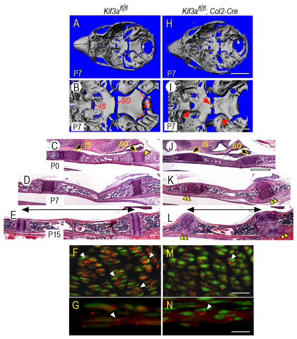
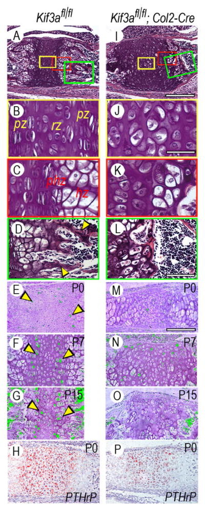
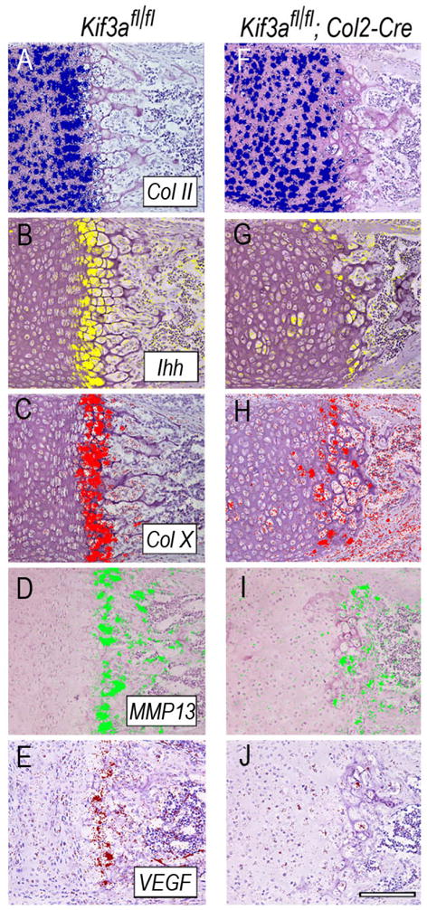
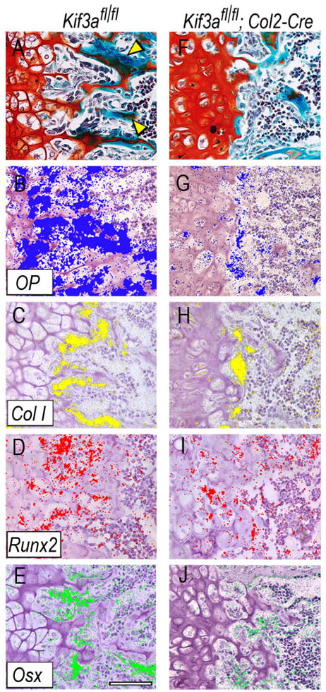
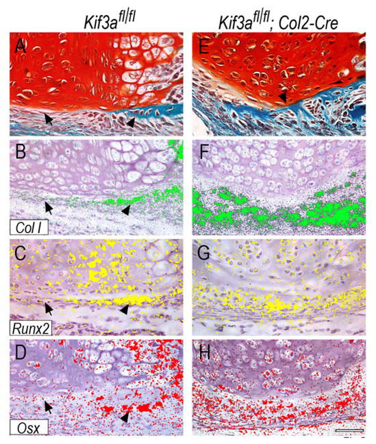
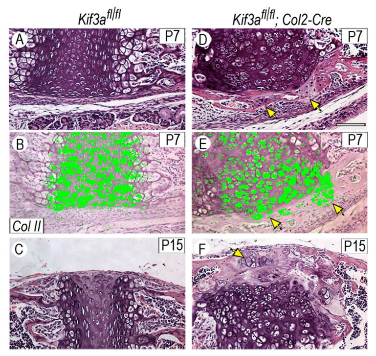
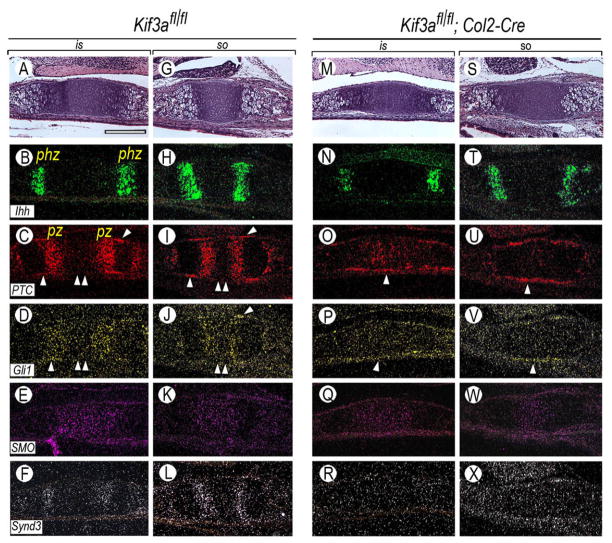
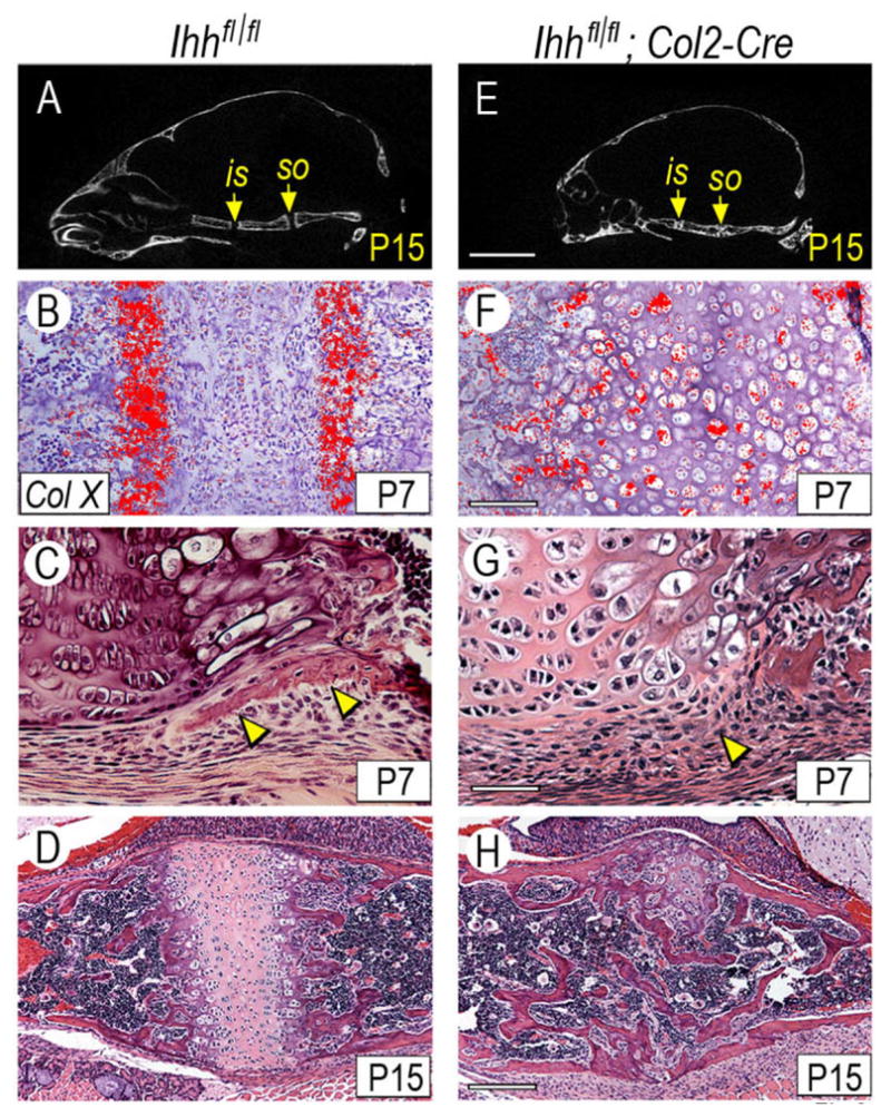
The motor protein Kif3a and primary cilia regulate important developmental processes, but their roles in skeletogenesis remain ill-defined. Here we created mice deficient in Kif3a in cartilage and focused on the cranial base and synchondroses. Kif3a deficiency caused cranial base growth retardation and dysmorphogenesis, which were evident in neonatal animals by anatomical and micro-computed tomography (microCT) inspection. Kif3a deficiency also changed synchondrosis growth plate organization and function, and the severity of these changes increased over time. By postnatal day (P)7, mutant growth plates lacked typical zones of chondrocyte proliferation and hypertrophy, and were instead composed of chondrocytes with an unusual phenotype characterized by strong collagen II (Col2a1) gene expression but barely detectable expression of Indian hedgehog (Ihh), collagen X (Col10a1), Vegf (Vegfa), MMP-13 (Mmp13) and osterix (Sp7). Concurrently, unexpected developmental events occurred in perichondrial tissues, including excessive intramembranous ossification all along the perichondrial border and the formation of ectopic cartilage masses. Looking for possible culprits for these latter processes, we analyzed hedgehog signalling topography and intensity by monitoring the expression of the hedgehog effectors Patched 1 and Gli1, and of the hedgehog-binding cell-surface component syndecan 3. Compared with controls, hedgehog signaling was quite feeble within mutant growth plates as early as P0, but was actually higher and was widespread all along mutant perichondrial tissues. Lastly, we studied postnatal mice deficient in Ihh in cartilage; their cranial base defects only minimally resembled those in Kif3a-deficient mice. In summary, Kif3a and primary cilia make unique contributions to cranial base development and synchondrosis growth plate function. Their deficiency causes abnormal topography of hedgehog signaling, growth plate dysfunction, and un-physiologic responses and processes in perichondrial tissues, including ectopic cartilage formation and excessive intramembranous ossification.
Figures
Similar articles
-
TMJ development and growth require primary cilia function.J Dent Res. 2011 Aug;90(8):988-94. doi: 10.1177/0022034511409407. Epub 2011 May 12. J Dent Res. 2011. PMID: 21566205 Free PMC article.
-
Roles of the primary cilium component Polaris in synchondrosis development.J Dent Res. 2009 Jun;88(6):545-50. doi: 10.1177/0022034509337775. J Dent Res. 2009. PMID: 19587160 Free PMC article.
-
Indian and sonic hedgehogs regulate synchondrosis growth plate and cranial base development and function.Dev Biol. 2006 Nov 1;299(1):272-82. doi: 10.1016/j.ydbio.2006.07.028. Epub 2006 Jul 29. Dev Biol. 2006. PMID: 16935278
-
Key Roles of Gli1 and Ihh Signaling in Craniofacial Development.Stem Cells Dev. 2024 Jun;33(11-12):251-261. doi: 10.1089/scd.2024.0036. Epub 2024 May 3. Stem Cells Dev. 2024. PMID: 38623785 Review.
-
Developmental Regulation of the Growth Plate and Cranial Synchondrosis.J Dent Res. 2016 Oct;95(11):1221-9. doi: 10.1177/0022034516651823. Epub 2016 Jun 1. J Dent Res. 2016. PMID: 27250655 Free PMC article. Review.
Cited by
-
Presphenoidal synchondrosis fusion in DBA/2J mice.Mamm Genome. 2013 Feb;24(1-2):54-62. doi: 10.1007/s00335-012-9437-8. Epub 2012 Nov 21. Mamm Genome. 2013. PMID: 23179633 Free PMC article.
-
The intraflagellar transport protein IFT80 is required for cilia formation and osteogenesis.Bone. 2012 Sep;51(3):407-17. doi: 10.1016/j.bone.2012.06.021. Epub 2012 Jul 4. Bone. 2012. PMID: 22771375 Free PMC article.
-
TMJ development and growth require primary cilia function.J Dent Res. 2011 Aug;90(8):988-94. doi: 10.1177/0022034511409407. Epub 2011 May 12. J Dent Res. 2011. PMID: 21566205 Free PMC article.
-
Polycystin-1 regulates skeletogenesis through stimulation of the osteoblast-specific transcription factor RUNX2-II.J Biol Chem. 2008 May 2;283(18):12624-34. doi: 10.1074/jbc.M710407200. Epub 2008 Mar 5. J Biol Chem. 2008. PMID: 18321855 Free PMC article.
-
Function and regulation of primary cilia and intraflagellar transport proteins in the skeleton.Ann N Y Acad Sci. 2015 Jan;1335(1):78-99. doi: 10.1111/nyas.12463. Epub 2014 Jun 24. Ann N Y Acad Sci. 2015. PMID: 24961486 Free PMC article. Review.
References
-
- Ansley SJ, Badano JL, Blacque OE, Hill J, Hoskins BE, Leitch CC, Kim JC, Ross AJ, Eichers ER, Teslovich TM, et al. Basal body dysfunction is a likely cause of pleiotropic Bardet-Biedl syndrome. Nature. 2003;425:628–633. - PubMed
-
- Bellaiche Y, The I, Perrimon N. Tout-velu is a Drosophila homologue of the putative tumor suppressor EXT-1 and is needed for Hh diffusion. Nature. 1998;394:85–88. - PubMed
-
- Bernfield M, Gotte M, Park PW, Reizes O, Fitzgerald ML, Lincecum J, Zako M. Functions of cell surface heparan sulfate proteoglycans. Annu Rev Biochem. 1999;68:729–777. - PubMed
-
- Bitgood MJ, McMahon AP. Hedgehog and Bmp genes are coexpressed at many diverse sites of cell-cell interaction in the mouse embryo. Dev Biol. 1995;172:126–138. - PubMed
Publication types
MeSH terms
Substances
Grants and funding
LinkOut - more resources
Full Text Sources
Molecular Biology Databases
Research Materials
