. 2007 Aug;81(15):7941-59.
doi: 10.1128/JVI.02848-06. Epub 2007 May 16.

Lipid rafts of primary endothelial cells are essential for Kaposi's sarcoma-associated herpesvirus/human herpesvirus 8-induced phosphatidylinositol 3-kinase and RhoA-GTPases critical for microtubule dynamics and nuclear delivery of viral DNA but dispensable for binding and entry

Affiliations

Lipid rafts of primary endothelial cells are essential for Kaposi's sarcoma-associated herpesvirus/human herpesvirus 8-induced phosphatidylinositol 3-kinase and RhoA-GTPases critical for microtubule dynamics and nuclear delivery of viral DNA but dispensable for binding and entry

Hari Raghu et al. J Virol. 2007 Aug.

Abstract

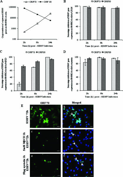
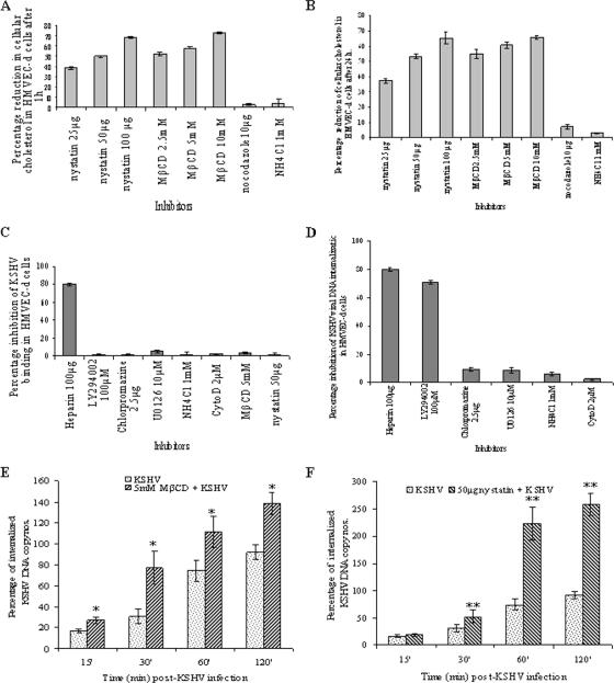
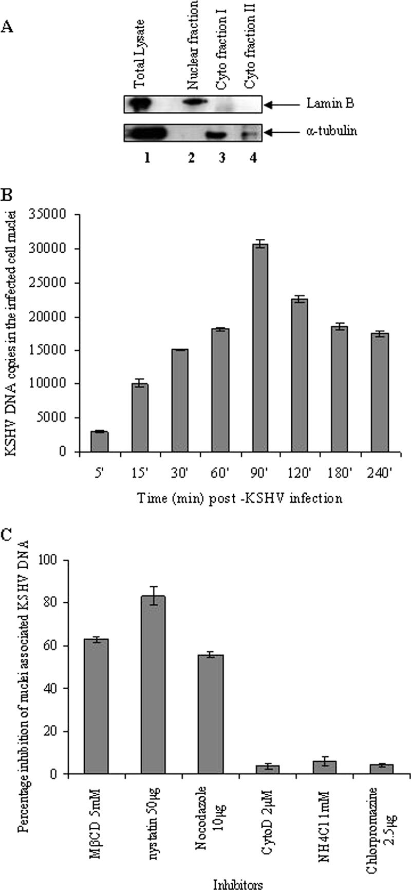
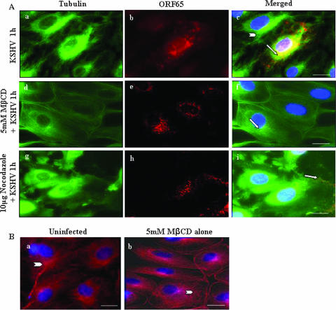
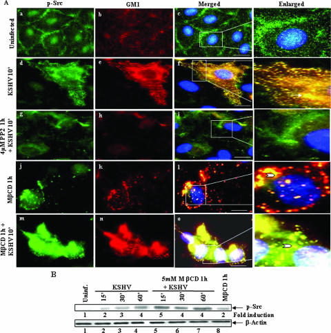
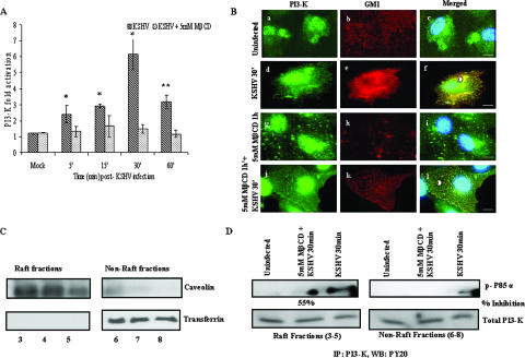
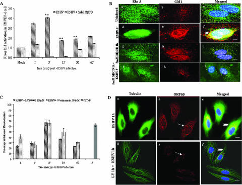
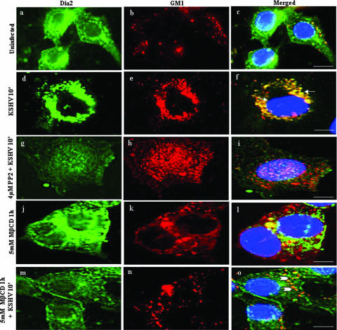
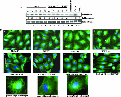
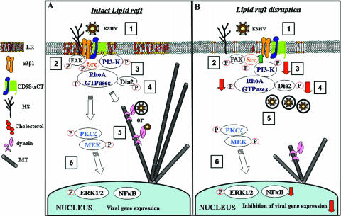
Early during de novo infection of human microvascular dermal endothelial (HMVEC-d) cells, Kaposi's sarcoma-associated herpesvirus (KSHV) (human herpesvirus 8 [HHV-8]) induces the host cell's preexisting FAK, Src, phosphatidylinositol 3-kinase (PI3-K), Rho-GTPases, Diaphanous-2 (Dia-2), Ezrin, protein kinase C-zeta, extracellular signal-regulated kinase 1/2 (ERK1/2), and NF-kappaB signal pathways that are critical for virus entry, nuclear delivery of viral DNA, and initiation of viral gene expression. Since several of these signal molecules are known to be associated with lipid raft (LR) domains, we investigated the role of LR during KSHV infection of HMVEC-d cells. Pretreatment of cells with LR-disrupting agents methyl beta-cyclo dextrin (MbetaCD) or nystatin significantly inhibited the expression of viral latent (ORF73) and lytic (ORF50) genes. LR disruption did not affect KSHV binding but increased viral DNA internalization. In contrast, association of internalized viral capsids with microtubules (MTs) and the quantity of infected nucleus-associated viral DNA were significantly reduced. Disorganized and disrupted MTs and thick rounded plasma membranes were observed in MbetaCD-treated cells. LR disruption did not affect KSHV-induced FAK and ERK1/2 phosphorylation; in contrast, it increased the phosphorylation of Src, significantly reduced the KSHV-induced PI3-K and RhoA-GTPase and NF-kappaB activation, and reduced the colocalizations of PI3-K and RhoA-GTPase with LRs. Biochemical characterization demonstrated the association of activated PI3-K with LR fractions which was inhibited by MbetaCD treatment. RhoA-GTPase activation was inhibited by PI3-K inhibitors, demonstrating that PI3-K is upstream to RhoA-GTPase. In addition, colocalization of Dia-2, a RhoA-GTPase activated molecule involved in MT activation, with LR was reduced. KSHV-RhoA-GTPase mediated acetylation and aggregation of MTs were also reduced. Taken together, these studies suggest that LRs of endothelial cells play critical roles in KSHV infection and gene expression, probably due to their roles in modulating KSHV-induced PI3-K, RhoA-GTPase, and Dia-2 molecules essential for postbinding and entry stages of infection such as modulation of microtubular dynamics, movement of virus in the cytoplasm, and nuclear delivery of viral DNA.


Figures

FIG. 1.
FIG. 1.
Effect of lipid raft-disrupting agents on KSHV gene expression. HMVEC-d cells were infected with KSHV alone (A) or pretreated with 10 μM U0126 for 1 h prior to infection (B) or pretreated with 5 mM MβCD for 1 h and then infected (C) or cells treated with 50 μg of nystatin for 1 h and infected (D). After infection with KSHV (10 DNA copies/cell), total RNA was isolated at 2, 8, and 24 h p.i. and 50 ng of DNase-treated RNA/μl was subjected to real-time RT-PCR with ORF73 and ORF50 gene specific primers and TaqMan probes. Known concentrations of DNase-treated in vitro-transcribed ORF50 and ORF73 transcripts were used in a real-time RT-PCR to construct a standard graph from which the relative copy numbers of viral transcripts were calculated and normalized with GAPDH. Panels B, C, and D show histograms depicting the percent inhibition in RNA copy numbers for KSHV ORF73 and ORF50 genes in the presence of 10 μM U0126, 5 mM MβCD, and 50 μg of nystatin, respectively. Each reaction was done in duplicate, and each point represents the average standard deviation (SD) of three independent experiments. (E) Immunofluorescence observation of KSHV-ORF73 expression in lipid raft-disrupted cells. HMVEC-d cells (90% confluent) grown in eight-well chamber slides were infected with 10 DNA copies of KSHV (subpanels a and b)/cell or pretreated with 5 mM MβCD for 1 h and infected (subpanels c and d) or pretreated with 50 μg of nystatin for 1 h and infected (subpanels e and f). After infection for 2 h, complete medium was added, followed by incubation for 72 h. The cells were fixed in 3.7% paraformaldehyde for 15 min at room temperature, permeabilized, and incubated overnight with rabbit anti-ORF73 antibodies (1:80) in 5% BSA. Cells were washed with PBS, incubated with anti-rabbit Alexa Fluor 488 antibodies for 1 h at room temperature, washed, mounted with antifade agent containing DAPI, and visualized under a fluorescence microscope. Arrows show nuclear staining of ORF73 protein (LANA-1) (subpanels b, d, and f). Scale bars, 10 μm.
FIG. 2.
FIG. 2.
Effect of MβCD and nystatin on KSHV infection. (A and B) Cholesterol depletion/chelation by MβCD and nystatin. HMVEC-d cells were treated with various concentrations of MβCD and nystatin for 1 h, and cholesterol levels were measured at 1 h (A) and 24 h (B) after treatment. The percentage reduction in the cholesterol upon drug treatment was calculated with respect to the total cholesterol in the untreated cells taken as 100%. Each reaction was done in duplicate, and each point represents the mean ± the SD of three independent experiments. (C) Disruption of lipid raft does not affect KSHV binding. HMVEC-d cells were either untreated or pretreated with various nontoxic concentrations of LY294002, NH4Cl, chlorpromazine, U0126, cytochalasin D, MβCD, or nystatin for 1 h. Cells were kept at 4°C for 1 h and incubated with a fixed concentration of [3H]thymidine-labeled virus in RPMI 1640 for 1 h with gentle rotation. As a control, [3H]thymidine-labeled KSHV was preincubated with 100 μg of heparin/ml for 1 h at 37°C before being added to the cells. After incubation, cells were washed, lysed, and precipitated with trichloroacetic acid, and the cell-associated virus radioactivity (in cpm) was counted. The cell-associated virus cpm in the presence of each different treatment was calculated as the percentage inhibition of virus binding. Each reaction was done in triplicate, and each point represents the average ± the SD of three independent experiments. (D) Chlorpromazine and microfilament depolymerizing agents does not affect KSHV DNA internalization in HMVEC-d cells. HMVEC-d cells grown in six-well plates were either untreated or preincubated with various nontoxic concentrations of nocodazole, chlorpromazine, U0126, LY294002, or NH4Cl. Cells were incubated with KSHV for 2 h, washed twice with PBS to remove unbound virus, treated with trypsin-EDTA for 5 min at 37°C to remove the bound noninternalized virus, and washed, and the total DNA was isolated. As a control, KSHV was preincubated with 100 μg of heparin/ml for 1 h at 37°C before being added to the cells. KSHV ORF73 DNA copies were estimated by real-time DNA PCR. The data are represented as the percentage of the inhibition of KSHV DNA internalization obtained when the cells were incubated with virus alone. Each reaction was done in duplicate, and each bar represents the average ± the SD of three experiments. (E and F) Lipid raft-disrupting agents increase the internalization of KSHV DNA in HMVEC-d cells. HMVEC-d cells after treatment with 5 mM MβCD (E) or 50 μg of nystatin (F) for 1 h were washed and infected with 10 MOI of KSHV. At different time points, internalized viral DNA was quantitated as described for Fig. 3B. The total viral DNA detected at 120 min in virus alone is considered as 100%. *, P < 0.01; **, P < 0.001.
FIG. 3.
FIG. 3.
The lipid raft is crucial for the nuclear delivery of KSHV DNA. (A) Kinetics of KSHV DNA delivery into the infected cell nuclei. Uninfected and KSHV-infected HMVEC-d cells were washed, suspended in PBS, treated with 0.25% trypsin-EDTA to remove noninternalized virus, washed, suspended in lysis buffer, and allowed to swell on ice for 5 min. Nuclei and cytoplasmic fractions were prepared (44). (A) Total cell lysate (lane 1), purified nuclear fractions (lane 2), and cytoplasmic fractions I and II (lanes 3 and 4, respectively) were tested with monoclonal antibodies against lamin B and α-tubulin. (B) Kinetics of KSHV DNA delivery into the infected cell nuclei. Nuclear fractions from HMVEC-d cells infected with KSHV at 10 MOI for the indicated time points were isolated, and the total DNA was isolated, normalized to 100 ng/5 μl, and analyzed by real-time PCR with KSHV ORF73 primers. Each reaction was done in duplicate, and each bar represents the mean ± the SD for three experiments. (C) Lipid raft disruption reduces the nuclear delivery of KSHV DNA. HMVEC-d cells preincubated with nontoxic doses of MβCD, nystatin, nocodazole, cytochalasin D, NH4Cl, and chlorpromazine for 1 h were infected with KSHV (10 DNA copies/cell) in the presence of inhibitors. The purification of the nuclear fractions and real-time DNA PCR estimating the numbers of KSHV ORF73 copies was done as described in Materials and Methods. The data are presented as the percentage of inhibition of infected cell nucleus-associated KSHV DNA relative to that of cells infected with virus alone. Each reaction was done in duplicate, and each bar represents the mean ± the SD for three experiments.
FIG. 4.
FIG. 4.
Disruption of the lipid raft affects the trafficking of KSHV capsid. (A) Untreated HMVEC-d cells (Aa to c), cells preincubated with 5 mM MβCD for 1 h (Ad to f), and cells preincubated with 10 μg of nocodazole for 1 h (Ag to i) were infected with 100 MOI of KSHV for 1 h at 37°C, washed, fixed, permeabilized, reacted with rabbit anti-KSHV ORF65 antibodies and mouse anti-tubulin monoclonal antibodies, and detected by goat anti-rabbit IgG-Alexa Fluor 594 and goat anti-mouse Alexa Fluor 488 antibodies, respectively. The nuclei were stained with DAPI (blue). Arrows indicate ORF65 capsid staining, and arrowheads indicate MTs. Magnification, ×80. Scale bar, 20 μm. (B) Untreated HMVEC-d cells (Ba) and cells preincubated with 5 mM MβCD for 1 h (Bb) were stained with mouse anti-tubulin monoclonal antibodies and goat anti-mouse Alexa Fluor 594 antibodies. Scale bar, 10 μm.
FIG. 5.
FIG. 5.
(A) Src colocalizes with lipid rafts in KSHV-infected endothelial cells. HMVEC-d cells grown in eight-well chamber slides were serum starved for 6 to 8 h and infected for 10 min with or without 5 mM MβCD treatments. Cells were washed, labeled for lipid raft marker GM1, fixed, permeabilized, and blocked with 5% BSA for 1 h. These cells were washed, labeled with anti-p-Src antibodies for 1 h at room temperature, washed, and stained for 1 h with Alexa Fluor 488-conjugated secondary antibodies (p-Src) and Alexa Fluor 594 (GM1) at room temperature. Subpanels: a to c, uninfected cells; d to f, cells infected for 10 min; g to i, cells pretreated with PP2 (Src inhibitor) for 1 h and infected with KSHV for 10 min; j to l, cells pretreated with 5 mM MβCD alone for 60 min; m to o, cells pretreated with 5 mM MβCD for 60 min and infected with KSHV for 10 min. Stained cells were viewed with appropriate filters under a fluorescence microscope. The insets in panels c, f, i, l, and o show an enlarged view of colocalization. Arrows indicate colocalization of p-Src with GM1. Arrowheads indicate colocalization of p-Src with GM1 after MβCD treatment. Scale bars, 10 μm. (B) Effect of lipid raft disruption on KSHV-induced Src phosphorylation. Serum-starved HMVEC-d cells were pretreated with 5 mM MβCD for 1 h and infected with KSHV (10 MOI) for the indicated time points. Cells were lysed in radioimmunoprecipitation assay (RIPA) lysis buffer containing protease inhibitors and cellular debris removed by centrifugation at 15,000 × g for 20 min at 4°C. Equal amounts of protein samples were resolved by SDS-7.5% polyacrylamide gel electrophoresis, transferred onto nitrocellulose membrane, and probed with anti-phospho Src (PY418) antibodies and total β-actin antibodies. Immunoreactive band intensities were assessed and are expressed as increased fold phosphorylation of Src over uninfected cells. Each blot is representative of a minimum of three separate experiments.
FIG. 6.
FIG. 6.
Lipid raft disruption reduces KSHV-induced PI3-K activation and colocalization with lipid rafts. (A) HMVEC-d cells cultured in 96-well plates were serum starved for 6 to 8 h, treated with or without 5 mM MβCD for 1 h, and infected with 10 MOI of KSHV for the indicated time points. Total and phospho PI3-K levels were measured in triplicate by using the phospho and total PI3-K p85 antibodies and the FACE PI3-kinase kit. The data are represented as a bar graph showing fold activation of phospho PI3-K in HMVEC-d cells before and after 5 mM MβCD treatment. Each bar represents the mean ± the SD for three experiments. *, P < 0.01; **, P < 0.001. (B) HMVEC-d cells grown in eight-well chamber slides were serum starved for 6 to 8 h, treated with MβCD for 1 h or untreated, and infected for 30 min. Cells were washed, labeled for lipid raft marker GM1, fixed, permeabilized, and blocked with 5% BSA for 1 h. Cells were washed, labeled with anti-p85 antibodies overnight, washed, and stained with Alexa Fluor 488-conjugated secondary antibodies. These cells were washed, stained with DAPI, mounted, and analyzed. Subpanels: a to c, uninfected cells; d to f, cells infected for 30 min; g to i, cells pretreated with 5 mM MβCD alone for 60 min; j to l, cells pretreated with 5 mM MβCD for 60 min and infected for 30 min. Stained cells were viewed with appropriate filters under a fluorescence microscope. Arrowheads indicate colocalization of PI3-K with lipid rafts. Scale bars, 20 μm. (C) Characterization of lipid raft fractions. HMVEC-d cells were treated with 5 mM MβCD for 1 h, washed or left untreated, and then infected with 10 MOI KSHV for 30 min and washed. Cells were lysed, and raft and nonraft fractions were collected as described in Materials and Methods. Fractions were characterized as raft fractions or nonraft fractions by Western blotting with caveolin as a raft marker and transferrin as a nonraft marker. Fractions 3 to 5 were pooled as raft fractions, and fractions 6 to 8 were taken as nonraft fractions. (D) Association of PI3-K with lipid rafts upon KSHV infection. Portions (150 μg) of protein from the pooled raft and nonraft fractions were immunoprecipitated with PI3-K p85α (Z-8) antibody for 2 h at 4°C. The immune complexes were washed four times with ice-cold RIPA buffer containing protease inhibitors, and bound proteins were eluted by boiling in 50 μl of 2× Laemmli buffer for 3 min and subjected to Western blot analysis with anti-phosphotyrosine PY20 antibody (upper panel [p-P85α]) and for total PI3-K (lower panel). The percent inhibition of PI3-K activity in the raft fraction after MβCD treatment was calculated by comparison with PI3-K activity in the untreated infected lysate.
FIG. 7.
FIG. 7.
Lipid raft disruption reduces KSHV-induced RhoA-GTPase activation and colocalization with lipid rafts. (A) HMVEC-d cells were serum starved for 6 to 8 h, treated with 5 mM MβCD for 1 h, and infected with 10 MOI of KSHV for the indicated time points. RhoA-GTPase levels were measured by G-LISA. A histogram depicts the fold change of RhoA activation. Each reaction was done in duplicate, and each bar represents the mean ± the SD for three experiments. **, P < 0.001. (B) HMVEC-d cells grown in eight-well chamber slides were serum starved for 6 to 8 h, treated with MβCD for 1 h or untreated, and infected for 5 min. Cells were washed, labeled for lipid raft marker GM1, fixed, permeabilized, and blocked with 5% BSA for 1 h. Cells were washed, labeled with anti-RhoA antibodies, washed, stained with Alexa Fluor 488-conjugated secondary antibodies, washed, stained with DAPI, mounted, and analyzed. Subpanels: a to c, uninfected cells; d to f, cells infected for 5 min; g to i, cells pretreated with 5 mM MβCD alone for 60 min; j to l, cells pretreated with 5 mM MβCD and infected for 5 min. Stained cells were viewed with appropriate filters under a fluorescence microscope. Arrowheads indicate colocalization of RhoA with GM1. (C) RhoA activation by KSHV in cells pretreated with PI3-K inhibitors. HMVEC-d cells were serum starved for 6 to 8 h, left untreated or pretreated with 100 μM LY294002 or with 500 nM wortmannin, washed, and infected with 10 MOI of KSHV for the indicated time points. RhoA-GTPase levels were measured by G-LISA. A histogram depicts the percent inhibition of RhoA activation compared to untreated infected cells at the respective time points. Each reaction was done in duplicate, and each bar represents the mean ± the SD for three experiments. (D) Cytoplasmic trafficking of KSHV capsid in cells pretreated with PI3-K inhibitor. HMVEC-d cells grown in eight-chamber slides were left untreated or pretreated with 100 μM LY294002 for 1 h and then infected with 100 MOI of KSHV for 1 h at 37°C washed, fixed, permeabilized, reacted with rabbit anti-KSHV ORF65 antibodies and mouse anti-tubulin monoclonal antibodies for 1 h at 4°C, and detected by goat anti-mouse IgG-Alexa Fluor 488 and goat anti-rabbit Alexa Fluor 594 antibodies. The nuclei were stained with DAPI (blue). Arrows indicate ORF65 capsid staining, and arrowheads indicate MTs. The images were obtained on a Nikon 80i epifluorescence microscope, Magnification, ×40. Scale bar, 10 μm.
FIG. 8.
FIG. 8.
Lipid raft disruption reduces KSHV-induced RhoA-GTPase-dependent Dia-2 colocalization with lipid rafts. Serum-starved HMVEC-d cells grown in eight-well chamber slides were infected for 10 min with or without 5 mM MβCD treatments. Cells were washed, labeled for lipid raft marker GM1, fixed, permeabilized, and blocked with 5% BSA for 1 h. Cells were washed, labeled with anti-Dia-2 with Alexa Fluor 488-conjugated secondary antibody for Dia-2 and Alexa Fluor 594 for GM1 for 1 h at room temperature. Subpanels: a to c, uninfected; d to f, cells infected for 10 min; g to i, cells pretreated with PP2 (Src inhibitor) for 1 h and infected; j to l, cells pretreated with 5 mM MβCD alone for 1 h; m to o, cells pretreated with 5 mM MβCD for 1 h and infected for 10 min. Arrows indicate the localization of Dia-2 with GM1. Arrowheads indicate the disruption of Dia-2 and GM1 after treatment with 5 mM MβCD. Scale bars, 10 μm.
FIG. 9.
FIG. 9.
Effect of lipid raft disruption on KSHV-induced MT acetylation. (A) Lipid raft disruption decreases hyperacetylation of tubulin in endothelial cells. Serum-starved HMVEC-d cells were either mock infected or infected with 10 MOI of KSHV for the indicated times and then lysed with RIPA lysis buffer. A total of 10 μg of lysate was resolved by SDS-10% polyacrylamide gel electrophoresis and immunoblotted with monoclonal antibody to acetylated tubulin (upper panel) or total tubulin (lower panel). Immunoreactive bands were developed by standard enhanced chemiluminescence reactions. The middle panel shows the fold phosphorylation of acetylated tubulin calculated with mock-infected HMVEC-d cells taken as 1. (B) Lipid raft disruption reduces the hyperacetylation of tubulin in endothelial cells. Serum-starved HMVEC-d cells were either mock treated (a) or infected with 10 MOI of KSHV (b to d). At different time points, the cells were washed, fixed in 4% formaldehyde, washed, and permeabilized in 0.5% Triton X-100 at room temperature. The cells were incubated for 30 min with 1% BSA and then with mouse anti-acetylated tubulin monoclonal antibody (diluted 1:500 in 1% BSA) at room temperature for 1 h. Cells were washed, incubated with anti-mouse Alexa Fluor 488 for 1 h, washed, mounted with slow fade agent containing DAPI, and visualized under a fluorescence microscope. Narrow arrowheads indicate bundles of acetylated tubulin upon infection. Scale bar, 20 μm. The insets at the bottom represents enlarged views of panels c, e, and g, respectively.
FIG. 10.
FIG. 10.
Model depicting the overlapping dynamic phases of KSHV-induced preexisting signal pathways and viral infection in HMVEC-d cells with intact lipid raft (A) and in lipid raft-disrupted cells (B). In phase 1, KSHV binds to adherent target cell HS molecules via its envelope glycoproteins gpK8.1A and gB, followed by interaction with α3β1 integrin via gB, interaction to CD98-xCT molecules, and possibly to other yet-to-be-identified molecules. Interactions with cell surface receptors trigger the cascades of host cell preexisting signal pathways (phases 2 to 4), which overlaps with virus entry by endocytosis. Activation of FAK and Src leads to the activation of PI3-K and Rho-GTPases, and RhoA activates Dia-2. Activated Src, PI3-K, Rho-GTPases, and Dia-2 colocalize with lipid raft domains. These signal cascades lead to the formation of endocytic vesicles, their movement in the cytoplasm, MT acetylation, and modulation of MT dynamics. In phase 5, the endosome moves in the cytoplasm, and the capsid is released, probably facilitated by the induced signaling pathways such as PKC-ζ. The endocytic vesicles with virus or released capsid or tegument complexes bind to dynein motor components, transported along the RhoA GTPase modulated MT to reach the nuclear vicinity, and deliver the viral DNA into the nucleus for viral gene expression (phase 6). When lipid rafts are disrupted (B), FAK and ERK activation is not altered. Increased Src phosphorylation occurs, which facilitates more viral internalization. Decreased activation of PI3-K and decreased association with lipid raft occurs, leading to reduced RhoA-GTPase and Dia-2 activation, reduced association with lipid raft domains, and reduced MT stabilization. The reduction in the activation of lipid raft-associated signal molecules such as PI3-K, RhoA-GTPase, and Dia-2 molecules essential for postbinding and entry stages of infection such as modulation of microtubular dynamics, movement of virus in the cytoplasm, and nuclear delivery of viral DNA, together with reduced NF-κB activation, probably account for the inhibition in the viral gene expression after lipid raft disruption. The present study thus demonstrates that lipid rafts of endothelial cells play critical roles in the postentry stage of KSHV infection.

Similar articles

Cited by

References

    1. Ahn, S., J. Kim, C. L. Lucaveche, M. C. Reedy, L. M. Luttrell, R. J. Lefkowitz, and Y. Daaka. 2002. Src-dependent tyrosine phosphorylation regulates dynamin self-assembly and ligand-induced endocytosis of the epidermal growth factor receptor. J. Biol. Chem. 277:26642-26651. - PubMed
    1. Akula, S. M., P. P. Naranatt, N. S. Walia, F. Z. Wang, B. Fegley, and B. Chandran. 2003. Kaposi's sarcoma-associated herpesvirus (human herpesvirus 8) infection of human fibroblast cells occurs through endocytosis. J. Virol. 77:7978-7990. - PMC - PubMed
    1. Akula, S. M., N. P. Pramod, F. Z. Wang, and B. Chandran. 2001. Human herpesvirus 8 envelope-associated glycoprotein B interacts with heparan sulfate-like moieties. Virology 284:235-249. - PubMed
    1. Akula, S. M., N. P. Pramod, F. Z. Wang, and B. Chandran. 2002. Integrin α3β1 (CD 49c/29) is a cellular receptor for Kaposi's sarcoma-associated herpesvirus (KSHV/HHV-8) entry into the target cells. Cell 108:407-419. - PubMed
    1. Akula, S. M., F. Z. Wang, J. Vieira, and B. Chandran. 2001. Human herpesvirus 8 interaction with target cells involves heparan sulfate. Virology 282:245-255. - PubMed

Publication types

MeSH terms

LinkOut - more resources