. 2007 Aug;81(15):7833-43.
doi: 10.1128/JVI.00580-07. Epub 2007 May 16.

Surface-exposed adeno-associated virus Vp1-NLS capsid fusion protein rescues infectivity of noninfectious wild-type Vp2/Vp3 and Vp3-only capsids but not that of fivefold pore mutant virions

Affiliations

Surface-exposed adeno-associated virus Vp1-NLS capsid fusion protein rescues infectivity of noninfectious wild-type Vp2/Vp3 and Vp3-only capsids but not that of fivefold pore mutant virions

Joshua C Grieger et al. J Virol. 2007 Aug.

Abstract

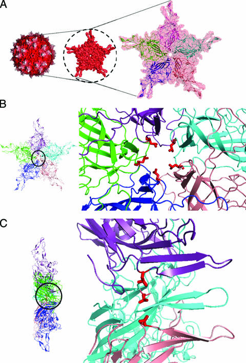
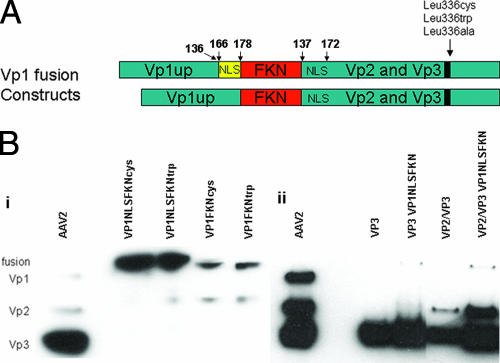
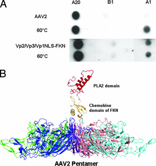
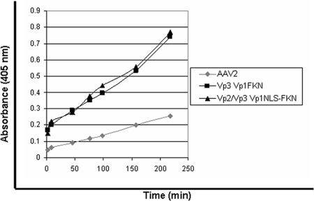
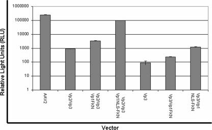
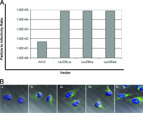
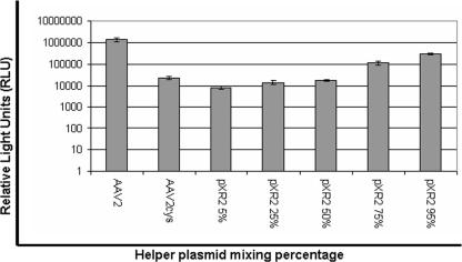
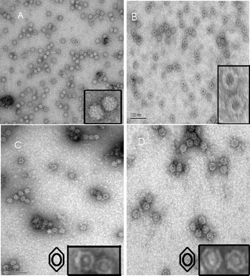
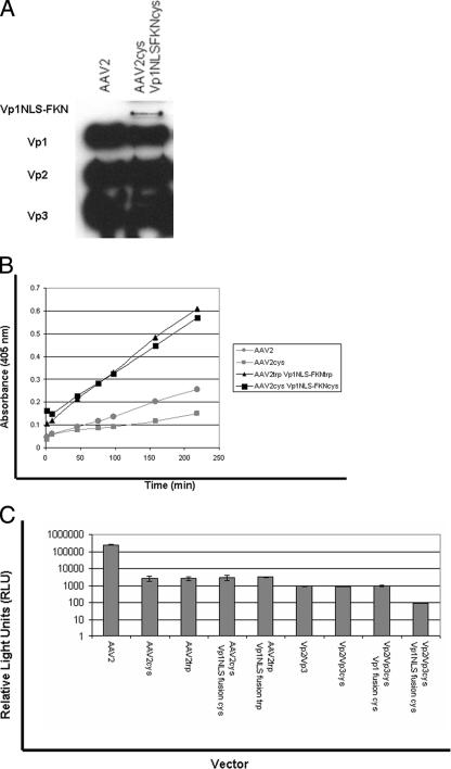
Over the past 2 decades, significant effort has been dedicated to the development of adeno-associated virus (AAV) as a vector for human gene therapy. However, understanding of the virus with respect to the functional domains of the capsid remains incomplete. In this study, the goal was to further examine the role of the unique Vp1 N terminus, the N terminus plus the recently identified nuclear localization signal (NLS) (J. C. Grieger, S. Snowdy, and R. J. Samulski, J. Virol 80:5199-5210, 2006), and the virion pore at the fivefold axis in infection. We generated two Vp1 fusion proteins (Vp1 and Vp1NLS) linked to the 8-kDa chemokine domain of rat fractalkine (FKN) for the purpose of surface exposure upon assembly of the virion, as previously described (K. H. Warrington, Jr., O. S. Gorbatyuk, J. K. Harrison, S. R. Opie, S. Zolotukhin, and N. Muzyczka, J. Virol 78:6595-6609, 2004). The unique Vp1 N termini were found to be exposed on the surfaces of these capsids and maintained their phospholipase A2 (PLA2) activity, as determined by native dot blot Western and PLA2 assays, respectively. Incorporation of the fusions into AAV type 2 capsids lacking a wild-type Vp1, i.e., Vp2/Vp3 and Vp3 capsid only, increased infectivity by 3- to 5-fold (Vp1FKN) and 10- to 100-fold (Vp1NLSFKN), respectively. However, the surface-exposed fusions did not restore infectivity to AAV virions containing mutations at a conserved leucine (Leu336Ala, Leu336Cys, or Leu336Trp) located at the base of the fivefold pore. EM analyses suggest that Leu336 may play a role in global structural changes to the virion directly impacting downstream conformational changes essential for infectivity and not only have local effects within the pore, as previously suggested.


Figures

FIG. 1.
FIG. 1.
AAV2 crystal structure. (A) Surface topology of the whole capsid, viewed down the icosahedral fivefold symmetry axis, that contains a pore (left) bounded by five symmetry-related Vp subunits shown in salmon, blue, green, purple, and cyan ribbons in the close-up view on the right. The depth-cued view (left) shows the tops of the three protrusions surrounding the icosahedral threefold axes in white and the rest of the capsid in red. (B) Top and (C) side views of the icosahedral fivefold-symmetry-related Vps (in the same colors as in panel A) with a close-up of the pore on the right. Leu336, located at the inside base of the pore, is highlighted in red in panels B and C.
FIG. 2.
FIG. 2.
(A) Schematic representation of the Vp1FKN and Vp1NLSFKN fusion proteins generated for this study. The numbers above the constructs represent amino acid sequence. (B) Western blots of (i) cell lysate that had been previously transfected with the Vp1 fusion proteins and (ii) purified AAV2 virions with and without the Vp1NLSFKN fusion protein. The MAb B1 was used as the primary antibody in the Western blot analysis of the capsid proteins.
FIG. 3.
FIG. 3.
(A) Native dot blot Western analysis of purified AAV2 and Vp2/Vp3/Vp1NLSFKN. A20 (MAb to intact AAV2 virions), B1 (MAb to individual capsid proteins), and A1 (MAb to the unique N terminus of VP1) were used to assess the surface exposure of wt VP1 for AAV2 when heated to 60°C and Vp1NLSFKN when incorporated into Vp2/Vp3-only particles at room temperature. (B) Schematic representation of the surface-exposed domains of the Vp1 fusion proteins. This is not a predicted structure but was compiled to provide a visualization of the possible juxtaposition of the FKN and PLA2 protein regions above the AAV2 fivefold pore region. It was generated from the crystal structure of the AAV2 Vp3 protein (Protein Data Bank [PDB] identifier 1LP3), the structure of the chemokine domain of FKN (PDB identifier 1F2L), and a homologous model of the AAV2 PLA2 domain generated by sequence comparison to the PLA2 domain structure of bee venom using the SWISS MODEL algorithm (; http://www.expasy.org). The FKN and PLA2 domain structures were visually “docked” above the fivefold pore in the AAV2 Vp3 pentamer in the PyMol program (W. L. DeLano, The PyMol molecular graphics system, DeLano Scientific, San Carlos, CA, 2002).
FIG. 4.
FIG. 4.
PLA2 assay. The PLA2 assay was carried out at room temperature, using AAV2 as the negative control, to assess the surface exposure and functionality of the Vp1 fusion proteins according to the manufacturer's protocol.
FIG. 5.
FIG. 5.
Luciferase transduction assay for comparison of wt AAV2 virions and virions containing VP1 fusion proteins in substitution for wt VP1. HeLa cells were infected at 1 × 10⁴ vg/cell with each vector (n = 4). Luciferase activity was then measured 24 h postinfection in accordance with the manufacturer's instructions (Promega).
FIG. 6.
FIG. 6.
(A) Infectious-center assay data to assess the particle-to-infectivity ratio between wt AAV2 virions and the Leu336 mutant virions. (B) Confocal-microscopy images of (i) A20 negative control and (ii) AAV2-ala-, (iii) AAV2-cys-, (iv) AAV2-trp-, and (v) wt AAV2-infected HeLa cells. MAB A20 was used to assess the locations of the virions 4 h postinfection.
FIG. 7.
FIG. 7.
Luciferase transduction assay for comparison of wt AAV2 virions and chimeric virions composed of various concentrations (95%, 75%, 50%, 25%, and 5%) of the Leu336-cys capsid protein (n = 4).
FIG. 8.
FIG. 8.
Negative-stain EM images of (A) wt AAV2, (B) AAV2-ala, (C) AAV2-cys, and (D) AAV2-trp that identified the unique staining pattern of the Leu336 mutant virions. The insets are magnified close-ups of the AAV2 virions. Symbols in panels C and D represent the staining pattern/profile of the fivefold mutant virlons.
FIG. 9.
FIG. 9.
(A) Western blot of purified Leu336 mutant containing the Vp1NLSFKN fusion protein that was previously transfected 1:20 to the helper plasmid. (B) PLA2 assay. The PLA2 assay was carried out at room temperature, using AAV2 and AAV2-cys as negative controls to assess the surface exposure and functionality of the Vp1 fusion proteins when incorporated into the Leu336 mutant virions according to the manufacturer's protocol. (C) Luciferase transduction assay for comparison of wt AAV2 and Leu336 mutant virions containing the VP1 fusion proteins and Leu336 mutant virions containing VP1 fusion proteins in substitution for wt VP1 (n = 4).

References

    1. Agbandje, M., S. Kajigaya, R. McKenna, N. S. Young, and M. G. Rossmann. 1994. The structure of human parvovirus B19 at 8 Å resolution. Virology 203:106-115. - PubMed
    1. Agbandje, M., R. McKenna, M. G. Rossmann, M. L. Strassheim, and C. R. Parrish. 1993. Structure determination of feline panleukopenia virus empty particles. Proteins 16:155-171. - PubMed
    1. Agbandje-McKenna, M., A. L. Llamas-Saiz, F. Wang, P. Tattersall, and M. G. Rossmann. 1998. Functional implications of the structure of the murine parvovirus, minute virus of mice. Structure 6:1369-1381. - PubMed
    1. Asokan, A., J. B. Hamra, L. Govindasamy, M. Agbandje-McKenna, and R. J. Samulski. 2006. Adeno-associated virus type 2 contains an integrin α5β1 binding domain essential for viral cell entry. J. Virol. 80:8961-8969. - PMC - PubMed
    1. Atchison, R. W., B. C. Casto, and W. M. Hammon. 1965. Adenovirus-associated defective virus particles. Science 149:754-756. - PubMed

Publication types

MeSH terms