. 2007 Jul;81(14):7683-94.
doi: 10.1128/JVI.02839-06. Epub 2007 May 16.

Roles of phosphatidylinositol 3-kinase and NF-kappaB in human cytomegalovirus-mediated monocyte diapedesis and adhesion: strategy for viral persistence

Affiliations

Roles of phosphatidylinositol 3-kinase and NF-kappaB in human cytomegalovirus-mediated monocyte diapedesis and adhesion: strategy for viral persistence

M Shane Smith et al. J Virol. 2007 Jul.

Abstract

Infected peripheral blood monocytes are proposed to play a key role in the hematogenous dissemination of human cytomegalovirus (HCMV) to tissues, a critical step in the establishment of HCMV persistence and the development of HCMV-associated diseases. We recently provided evidence for a unique strategy involved in viral dissemination: HCMV infection of primary human monocytes promotes their transendothelial migration and differentiation into proinflammatory macrophages permissive for the replication of the original input virus. To decipher the mechanism of hematogenous spread, we focused on the viral dysregulation of early cellular processes involved in transendothelial migration. Here, we present evidence that both phosphatidylinositol 3-kinase [PI(3)K] and NF-kappaB activities were crucial for the HCMV induction of monocyte motility and firm adhesion to endothelial cells. We found that the beta(1) integrins, the beta(2) integrins, intracellular adhesion molecule 1 (ICAM-1), and ICAM-3 were upregulated following HCMV infection and that they played a key role in the firm adhesion of infected monocytes to the endothelium. The viral regulation of adhesion molecule expression is complex, with PI(3)K and NF-kappaB affecting the expression of each adhesion molecule at different stages of the expression cascade. Our data demonstrate key roles for PI(3)K and NF-kappaB signaling in the HCMV-induced cellular changes in monocytes and identify the biological rationale for the activation of these pathways in infected monocytes, which together suggest a mechanism for how HCMV promotes viral spread to and persistence within host organs.


Figures
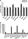
FIG. 1.
FIG. 1.
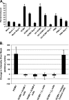
HCMV promotes transendothelial migration and motility of monocytes in a PI(3)K-dependent and NF-κB-dependent manner. (A) CellTracker green CMFDA-labeled monocytes were pretreated with LY (50 μM), Bay11 (5 μM), or DMSO for 45 min and then HCMV infected (MOI of 15), mock infected, or PMA treated (10 ng/ml) for 3 h. Labeled monocytes (2.5 × 10⁴) were added to cell culture inserts containing confluent monolayers of HMECs. HCMV-induced monocyte diapedesis is dependent on both PI(3)K and NF-κB activity. The ratios of cells undergoing diapedesis to those cells that were stationary at the monolayer surface were determined by fluorescence microscopy after 24 h in culture. The results are plotted as means ± SD for 15 random fields of view. The results are representative of three independent experiments with separate blood donors. (B) Monocytes were plated on colloidal gold-coated coverslips for 1 hour. Cells were subsequently pretreated with LY (50 μM), Bay11 (5 μM), or DMSO for 45 min and then HCMV infected (MOI of 15), UV-HCMV treated (equivalent MOI of 15), mock infected, or PMA treated (10 ng/ml). Cells were incubated on coverslips for 6 h and fixed with 3% paraformaldehyde. Individual cell track images were captured. The average area (square arbitrary units) of colloidal gold cleared by 20 monocytes was determined for each experimental arm. Results are plotted as means ± SEM for 20 cells per experimental arm. The results are representative of three independent experiments with separate human blood donors. In both panels, lanes marked with an asterisk denote significance (P < 0.01); depending on the experiment, the HCMV-infected, UV-HCMV-treated, and PMA-treated samples are significantly different from the mock-treated samples and those samples treated with LY, Bay11, or cytochalasin D (Cyt D).
FIG. 2.
FIG. 2.
HCMV promotes firm adhesion of monocytes to endothelial cells. (A) HCMV infection of monocytes promotes firm adhesion to endothelial cells in a PI(3)K-dependent and NF-κB-dependent manner. CellTracker green CMFDA-labeled monocytes were pretreated with LY (50 μM), Bay11 (5 μM), or DMSO for 45 min and then HCMV infected (MOI of 15), mock infected, or PMA treated (10 ng/ml). Monocytes were cultured nonadherently for 6 h. Labeled monocytes (2.5 × 10⁵ per well) were then added to cell culture inserts containing confluent monolayers of HMECs in 24-well plates and incubated for 30 min. Cells were then washed four times, and fluorescence intensities were determined with a fluorescence plate reader. The percentage of adherent cells represents the ratio of the adjusted fluorescence intensities of an experimental arm to the total input cell-adjusted fluorescence intensities. The experiment was performed in triplicate. The results are plotted as means ± SEM. The results are representative of three independent experiments with separate human blood donors. Lanes marked with an asterisk denote significance (P < 0.01); the HCMV-infected, UV-HCMV-treated, and PMA-treated samples are significantly different from the mock-treated samples and those samples treated with LY or Bay11. (B) CD29, CD18, ICAM-1, and ICAM-3 are required for HCMV-induced firm adhesion of monocytes to endothelial cells. CellTracker green CMFDA-labeled monocytes were treated and cultured as described above. Prior to addition of monocytes to endothelial cells, monocytes were incubated with neutralizing Abs against CD29, CD18, ICAM-1, or ICAM-3 or with an IgG1 isotype control Ab for 1 h at room temperature. Labeled monocytes (2.5 × 10⁵ per well) were then added to cell culture inserts containing confluent monolayers of HMECs in 24-well plates and incubated for 30 min. Cells were then washed four times and fluorescence intensities determined with a fluorescence plate reader in triplicate. To determine the changes in intensity over mock-infected monocytes (arbitrary units), the adjusted fluorescence intensities of mock-infected monocytes were subtracted from the adjusted fluorescence intensities of each experimental arm. The results are plotted as means ± SEM. The results are representative of three independent experiments with separate human blood donors. The HCMV-infected group and the isotype control-treated HCMV-infected group are significantly different from the CD29, CD18, ICAM-1, and ICAM-3 neutralizing MAb-treated groups (P < 0.01). This significance is denoted in the appropriate lanes via an asterisk.
FIG. 3.
FIG. 3.
Regulation of CD29, CD18, ICAM-1, and ICAM-3 mRNA expression in HCMV-infected monocytes. Monocytes were pretreated with LY (50 μM), Bay11 (5 μM), or DMSO for 45 min and then HCMV infected (MOI of 15), mock infected, or PMA treated (10 ng/ml). Cells were then cultured nonadherently for 6 h, and total RNA was harvested from each sample. RT-PCR was performed for CD29, CD18, ICAM-1, and ICAM-3. The results are representative of three independent experiments with separate human blood donors.
FIG. 4.
FIG. 4.
Regulation of CD29, CD18, CD11a, CD11b, CD11c, ICAM-1, and ICAM-3 protein expression in HCMV-infected monocytes. Monocytes were pretreated with LY (50 μM), Bay11 (5 μM), or DMSO for 45 min and then HCMV infected (MOI of 15), mock infected, or PMA treated (10 ng/ml). Cells were then cultured nonadherently for 6 h, and protein was harvested and solubilized. Samples for the analysis of CD29, CD18, CD11a, CD11b, and CD11c were separated by continuous native PAGE with equal protein loading. Samples for the analysis of ICAM-1 and ICAM-3 were separated by sodium dodecyl sulfate-PAGE with equal protein loading based on actin band intensity. Blots were probed with the different Abs stated in the figure.
FIG. 5.
FIG. 5.
HCMV infection induces cell surface expression of CD29, CD18, ICAM-1, and ICAM-3. Monocytes were plated on fibronectin-coated coverslips for 2 h and subsequently HCMV infected (MOI of 15), mock infected, or PMA treated (10 ng/ml) and cultured for 24 h. Monocytes were fixed at 24 hpi and incubated with MAbs against CD29, CD18, ICAM-1, or ICAM-3. Cells were then incubated with the appropriate FITC-conjugated secondary Ab to label the adhesion receptors, permeabilized, and stained with Alexa Fluor 546 phalloidin (depicted in blue) to stain actin and TO-PRO-3 (depicted in red) to stain the nucleus, according to the manufacturer's protocol. Cells were examined and photographed with a confocal microscope (×3,000 magnification; 1,000× optical, 3× digital zoom). Results are representative of three independent experiments with different human blood donors.
FIG. 6.
FIG. 6.
Confirmation of HCMV-induced cell surface translocation of CD29, CD18, ICAM-1, and ICAM-3. Monocytes were pretreated with LY (50 μM), Bay11 (5 μM), or DMSO for 45 min and then HCMV infected (MOI of 15) or mock infected. Cells were then cultured nonadherently for 24 h. Cells were fixed and incubated with MAbs against CD29, CD18, ICAM-1, or ICAM-3 and examined by flow cytometry for CD29 expression (A), CD18 expression (B), ICAM-1 expression (C), or ICAM-3 expression (D). Results are representative of three independent experiments with separate blood donors.
FIG. 7.
FIG. 7.
HCMV infection of monocytes promotes the translocation of CD29, CD18, and ICAM-3 from the cytosol to the cell membrane. Monocytes were treated as stated in the legend for Fig. 5, with the following exceptions: (i) cells were pretreated with LY (50 μM), Bay11 (5 μM), or DMSO for 45 min prior to infection (MOI of 15) or mock treatment; (ii) prior to incubation of monocytes with Abs against the adhesion receptors, monocytes were permeabilized with Triton X-100; and (iii) TO-PRO-3 was the only other stain utilized. Cells were examined and photographed with a confocal microscope (×3,000 magnification; 1,000× optical, 3× digital zoom). Results are representative of three independent experiments with separate blood donors.

References

    1. Albrecht-Buehler, G. 1977. The phagokinetic tracks of 3T3 cells. Cell 11:395-404. - PubMed
    1. Baillie, J., D. A. Sahlender, and J. H. Sinclair. 2003. Human cytomegalovirus infection inhibits tumor necrosis factor alpha (TNF-α) signaling by targeting the 55-kilodalton TNF-α receptor. J. Virol. 77:7007-7016. - PMC - PubMed
    1. Barja-Fidalgo, C., A. L. Coelho, R. Saldanha-Gama, E. Helal-Neto, A. Mariano-Oliveira, and M. S. Freitas. 2005. Disintegrins: integrin selective ligands which activate integrin-coupled signaling and modulate leukocyte functions. Braz. J. Med. Biol. Res. 38:1513-1520. - PubMed
    1. Bentz, G. L., M. Jarquin-Pardo, G. Chan, M. S. Smith, C. Sinzger, and A. D. Yurochko. 2006. Human cytomegalovirus (HCMV) infection of endothelial cells promotes naïve monocyte extravasation and transfer of productive virus to enhance hematogenous dissemination of HCMV. J. Virol. 80:11539-11555. - PMC - PubMed
    1. Boehme, K. W., M. Guerrero, and T. Compton. 2006. Human cytomegalovirus envelope glycoproteins B and H are necessary for TLR2 activation in permissive cells. J. Immunol. 177:7094-7102. - PubMed

Publication types