. 2007 Jul;81(14):7476-90.
doi: 10.1128/JVI.00308-07. Epub 2007 May 16.

Productive human immunodeficiency virus type 1 assembly takes place at the plasma membrane

Affiliations

Productive human immunodeficiency virus type 1 assembly takes place at the plasma membrane

Andrés Finzi et al. J Virol. 2007 Jul.

Abstract

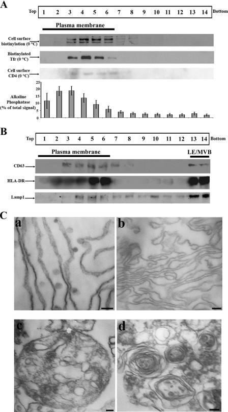
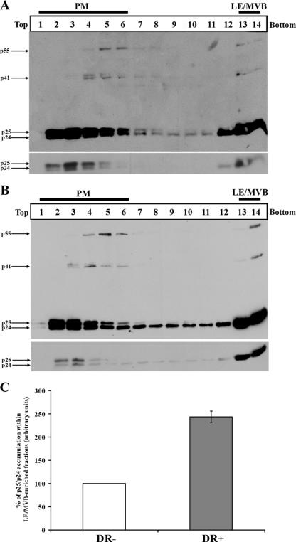
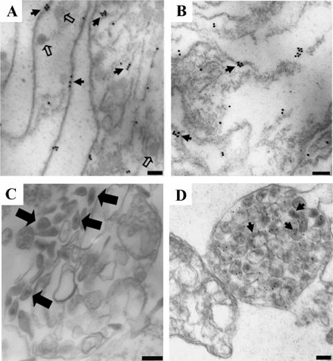
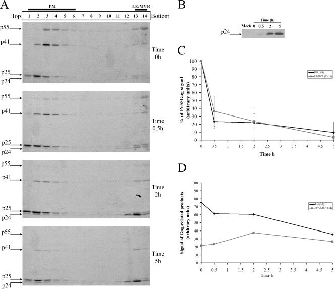
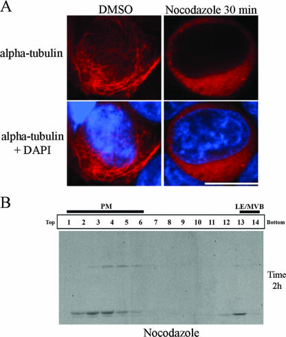
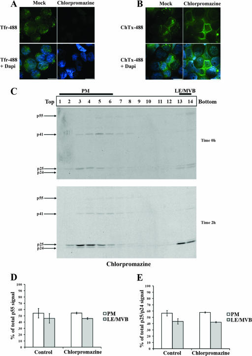
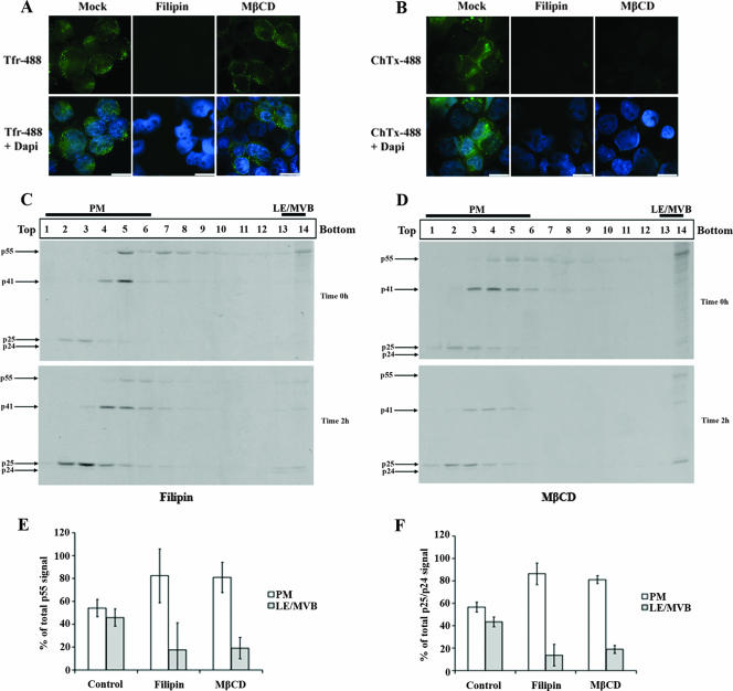
Gag proteins are necessary and sufficient to direct human immunodeficiency virus type 1 (HIV-1) particle assembly and budding. Recent evidence suggests that Gag targeting to late endosomal/multivesicular body (LE/MVB) compartments occurs prior to viral particle budding at the plasma membrane (PM). However, the route that Gag follows before reaching its steady-state destinations still remains a subject of debate. Using a subcellular fractionation method that separates PM from LE/MVB combined with pulse-chase labeling, we analyzed Gag trafficking in HIV-1-producing HEK 293T cells. Our results reveal that the majority of newly synthesized Gag is primarily targeted to the PM. While PM-targeted Gag was efficiently released, a significant fraction of the remaining cell surface-associated Gag was found to be subsequently internalized to LE/MVB, where it accumulated, thus accounting for the majority of LE/MVB-associated Gag. Importantly, this accumulation of Gag in LE/MVB was found to be cholesterol dependent since it was sensitive to the sterol-binding drugs filipin and methyl-beta-cyclodextrin. These results point towards the PM as being the primary site of productive HIV-1 assembly in cells that also support Gag accumulation in intracellular compartments.


Figures

FIG. 1.
FIG. 1.
PM separation from LE/MVB after iodixanol gradient-based subcellular fractionation. HEK 293T cells were transfected with the HxBc2 provirus and mechanically homogenized 48 h later. PNS production and subcellular fractionation were performed as described in Materials and Methods. (A) Plasma membrane location in the gradient. Cells were cell surface biotinylated or incubated with biotinylated Tfr at 0°C. Alternatively cells were cotransfected with a CD4 expressor and cell surface CD4 was labeled with an anti-CD4 Ab at 0°C; detection of the CD4-bound Ab was performed by Western blotting as indicated in Materials and Methods. Biotinylated cell surface proteins and biotinylated Tfr were analyzed in each fraction by Western blotting as described in Materials and Methods. Finally, the presence of alkaline phosphatase was evaluated by incubating each fraction with its specific substrate as described in Materials and Methods. Data shown for alkaline phosphatase represent the average ± standard deviation of more than 10 independent experiments. (B) LE/MVB marker migration was analyzed by Western blotting using specific anti-CD63 and Lamp1 Abs or by immunoprecipitation/Western blotting using HLA-DR Abs. Data shown in panels A and B are representative of at least two independent experiments. (C) Fractions 2 and 3, 6 and 7, and 13 and 14 were pooled, fixed, ultracentrifuged, and processed for conventional EM. PM structures were highly enriched in fractions 2 and 3 (a) and 6 and 7 (b). Fractions 13 and 14 were highly enriched in multivesicular bodies (c) and multilamellar structures (d). Scale bars represent 200 nm in panel a and 100 nm in panels b to d. Data shown are representative of at least two independent experiments.
FIG. 2.
FIG. 2.
HIV-1 Gag steady-state distribution after subcellular fractionation. HEK 293T cells were cotransfected with the HxBc2 provirus together with either HLA-DR or empty vector and mechanically homogenized 48 h later. PNS was processed as described in Materials and Methods. Fourteen fractions were collected from the top of the gradient, and Gag and its cleavage products were analyzed by Western blotting using a monoclonal anti-p24 Ab. (A) Steady-state Gag distribution after subcellular fractionation. (B) Steady-state distribution of Gag upon HLA-DR expression. The lower panels represent lower exposure of the mature (p25/p24) Gag products. Data shown are representative of at least two independent experiments. (C) Quantification of the relative amounts of mature Gag products (p25/p24) detected in PM- or LE/MVB-enriched fractions in the presence or absence of HLA-DR. Signals obtained for HLA-DR-negative samples were arbitrarily set to 100. Data shown are means ± standard deviations of two independent experiments.
FIG. 3.
FIG. 3.
Ultrastructural analysis of HIV-1 Gag association with different subcellular compartments. HEK 293T cells were transfected with the HxBc2 provirus together with an HLA-DR expressor and mechanically homogenized 48 h later. PNS production and subcellular fractionation were performed as described in Materials and Methods. Fourteen fractions were collected from the top of the gradient; fractions 2 and 3, 6 and 7, and 13 and 14 were pooled, fixed, ultracentrifuged, and observed by conventional transmission EM (C) or processed for immunogold staining with a rabbit polyclonal anti-p24 Ab followed by incubation with a goat anti-rabbit serum coupled to 18-nm (A and B) or 12-nm (D) gold beads. Filamentous structures staining positive for p24 were exclusively observed in fractions 2 and 3 (A) and 6 and 7 (B). Fractions 13 and 14 were enriched in HIV-1 particle-containing LE/MVB (C) as indicated by a positive p24 staining (D). Caveola-like structures are indicated by small open arrows. Small solid arrows indicate CA staining, whereas large solid arrows indicate mature virus. Scale bars represent 100 nm. Data shown are representative of at least two independent experiments.
FIG. 4.
FIG. 4.
Subcellular trafficking of HIV-1 Gag. (A) HEK 293T cells were transfected with the HxBc2 provirus. Two days after transfection, cells were metabolically labeled with [35S]Met-Cys for 10 min and chased for various times prior to cell lysis and subcellular fractionation by Optiprep gradient centrifugation as described in Materials and Methods. Fourteen fractions were collected from the top of the gradient, and Gag-related products in each fraction were detected by immunoprecipitation using a monoclonal anti-p24 Ab. (B) Viral release during the pulse-chase analysis. Virion lysates were immunoprecipitated as for panel A. Data shown are representative of five independent experiments. (C) Quantification of the relative amounts of Pr55gag in PM- and LE/MVB-enriched fractions (fractions 1 to 6 and 13 and 14, respectively) over time. Pr55gag signal obtained at time zero h in PM- or LE/MVB-enriched fractions was arbitrarily set to 100. (D) Quantification of the relative amounts of Gag-related products in PM- or LE/MVB-enriched fractions over time. Quantifications were performed using ImageQuant software 5.0.
FIG. 5.
FIG. 5.
Microtubule depolymerization does not affect HIV-1 Gag targeting to PM-enriched fractions. HEK 293T cells were transfected with the HxBc2 provirus. Two days after transfection, cells were incubated with nocodazole (10 μg/ml) for 30 min before being processed for immunofluorescence with an anti-α-tubulin (A). In parallel, cells were pretreated with nocodazole (10 μg/ml) for 30 min before being metabolically labeled with [35S]Met-Cys for 10 min and chased for 2 h in presence of the drug. Cell lysis and subcellular fractionation were performed as described in Materials and Methods. Fourteen fractions were collected from the top of the gradient, and Gag-related products in each fraction were detected by immunoprecipitation using a monoclonal anti-p24 Ab (B). DMSO, dimethyl sulfoxide; DAPI, 4′,6′-diamidino-2-phenylindole. The data shown are representative of two independent experiments.
FIG. 6.
FIG. 6.
Chlorpromazine, an inhibitor of clathrin-dependent endocytosis, does not affect HIV-1 Gag trafficking in HEK 293T cells. HEK 293T cells were transfected with the HxBc2 provirus. Two days after transfection, cells were pretreated with chlorpromazine (10 μg/ml) for 30 min before incubation for another 30 min at 0°C with (A) Tfr- or (B) ChTxβ-Alexa-488 conjugates. Tfr or ChTxβ internalization was allowed for 10 min at 37°C. Chlorpromazine was maintained throughout the experiment before samples were analyzed by immunofluorescence microscopy (bar, 10 μm) as described in Materials and Methods. Data shown are representative of two independent experiments. (C) In parallel, cells were metabolically labeled with [35S]Met-Cys for 10 min and chased for 2 h in the presence of chlorpromazine prior to cell lysis and subcellular fractionation by Optiprep gradient centrifugation. Gag-related products in Optiprep fractions were detected by immunoprecipitation using a monoclonal anti-p24 Ab for each collected fraction. (D) Quantification of the relative amount of Pr55gag detected in PM- and LE/MVB-associated fractions (1 to 6 and 13 and 14, respectively) at time zero h of chase. (E) Quantification of the relative amounts of mature Gag products (p25/p24) detected in PM or LE/MVB-associated fractions at time 2 h of chase. Gag-related signals were quantified using a PhosphorImager equipped with ImageQuant software 5.0. The data shown are means ± standard deviations of the results of five or two independent experiments: for control or chlorpromazine-treated cells, respectively.
FIG. 7.
FIG. 7.
Effect of cholesterol-binding drugs on HIV-1 Gag trafficking in HEK 293T cells. HEK 293T cells were transfected with the HxBc2 provirus. Two days after transfection, cells were pretreated with filipin (4 μg/ml) or MβCD (8 mM) for 30 min before incubation for another 30 min at 0°C with (A) Tfr- or (B) ChTxβ-Alexa-488 conjugates. Tfr or ChTxβ internalization was allowed for 10 min at 37°C. Endocytosis inhibitors were maintained throughout the experiment. Samples were subsequently analyzed by immunofluorescence microscopy (bar, 10 μm) as described in Materials and Methods. Data shown are representative of two independent experiments. In parallel, cells were metabolically labeled with [35S]Met-Cys for 10 min and chased for 2 h in the presence of (C) filipin or (D) MβCD prior to cell lysis and subcellular fractionation by Optiprep gradient centrifugation. Gag-related products in Optiprep fractions were detected by immunoprecipitation using a monoclonal anti-p24 Ab for each collected fraction. (E) Quantification of the relative amounts of Pr55gag detected in PM- and LE/MVB-associated fractions (1 to 6 and 13 and 14, respectively) at zero h of chase. (F) Quantification of the relative amounts of mature Gag products (p25/p24) detected in PM- or MVB-associated fractions at 2 h of chase. Gag-related signals were quantified as described in the legend to Fig. 6. Data shown are means ± standard deviations of the results of five control, three filipin, and two MβCD independent experiments.

References

    1. Abedinpour, P., and B. Jergil. 2003. Isolation of a caveolae-enriched fraction from rat lung by affinity partitioning and sucrose gradient centrifugation. Anal. Biochem. 313:1-8. - PubMed
    1. Aniento, F., N. Emans, G. Griffiths, and J. Gruenberg. 1993. Cytoplasmic dynein-dependent vesicular transport from early to late endosomes. J. Cell Biol. 123:1373-1387. - PMC - PubMed
    1. Beriault, V., J. F. Clement, K. Levesque, C. Lebel, X. Yong, B. Chabot, E. A. Cohen, A. W. Cochrane, W. F. Rigby, and A. J. Mouland. 2004. A late role for the association of hnRNP A2 with the HIV-1 hnRNP A2 response elements in genomic RNA, Gag, and Vpr localization. J. Biol. Chem. 279:44141-44153. - PubMed
    1. Bieniasz, P. D. 2006. Late budding domains and host proteins in enveloped virus release. Virology 344:55-63. - PubMed
    1. Bomsel, M., R. Parton, S. A. Kuznetsov, T. A. Schroer, and J. Gruenberg. 1990. Microtubule- and motor-dependent fusion in vitro between apical and basolateral endocytic vesicles from MDCK cells. Cell 62:719-731. - PubMed

Publication types

Substances

LinkOut - more resources