. 2007 Jul;81(14):7766-75.
doi: 10.1128/JVI.00633-07. Epub 2007 May 16.

DNA immunization using highly conserved murine cytomegalovirus genes encoding homologs of human cytomegalovirus UL54 (DNA polymerase) and UL105 (helicase) elicits strong CD8 T-cell responses and is protective against systemic challenge

Affiliations

DNA immunization using highly conserved murine cytomegalovirus genes encoding homologs of human cytomegalovirus UL54 (DNA polymerase) and UL105 (helicase) elicits strong CD8 T-cell responses and is protective against systemic challenge

Christopher S Morello et al. J Virol. 2007 Jul.

Abstract

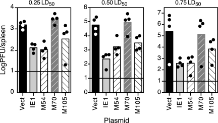
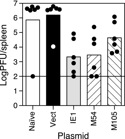
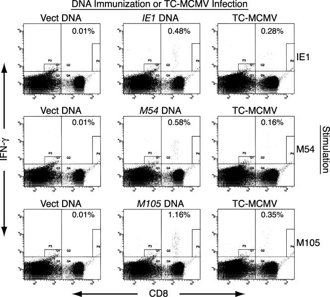
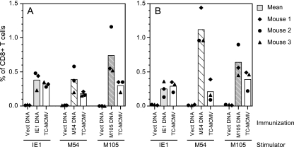
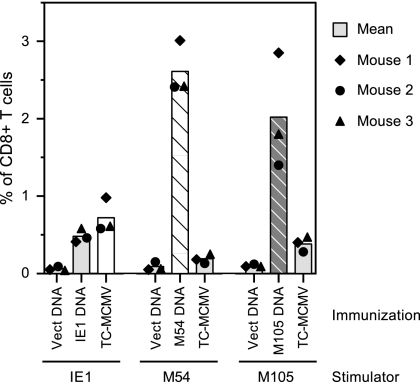
Human cytomegalovirus (HCMV) establishes a lifelong infection with the potential for reinfection or viral transmission even in the presence of strong and diverse CD8 T-lymphocyte responses. This suggests that the CMVs skew the host T-cell response in order to favor viral persistence. In this study, we hypothesized that the essential, nonstructural proteins that are highly conserved among the CMVs may represent a novel class of T-cell targets for vaccine-mediated protection due to their requirements for expression and sequence stability, but that the observed subdominance of these antigens in the CMV-infected host results from the virus limiting the T-cell responses to otherwise-protective specificities. We found that DNA immunization of mice with the murine CMV (MCMV) homologs of HCMV DNA polymerase (M54) or helicase (M105) was protective against virus replication in the spleen following systemic challenge, with the protection level elicited by the M54 DNA being comparable to that of DNA expressing the immunodominant IE1 (pp89). Intracellular gamma interferon staining of CD8 T cells from mice immunized with either the M54 or M105 DNAs showed strong primary responses that recalled rapidly after viral challenge. M54- and M105-specific CD8 T cells were detected after the primary MCMV infection, but their levels were not consistently above the background level. The conserved, essential proteins of the CMVs thus represent a novel class of CD8 T-cell targets that may contribute to a successful HCMV vaccine strategy.


Figures

FIG. 1.
FIG. 1.
In vitro and in vivo expression of the MCMV homologs of HCMV DNA polymerase (M54), primase (M70), and helicase (M105). (A) The MCMV ORFs, cloned into pcDNA3.1/V5-His-TOPO, were expressed using the T7 promoter by coupled in vitro transcription/translation reactions (TNT T7 Quick) with [35S]methionine. A portion of each reaction mixture was subjected to reducing SDS-PAGE on a 7% polyacrylamide gel, and the labeled proteins were detected by autoradiography. The numbers and lines on the left indicate the positions and relative molecular masses (in kDa) of the proteins in the marker (not shown). Vect indicates the TNT reaction performed with the DNA vector alone. The predicted molecular masses for the encoded proteins are M54, 128.8 kDa; M70 (untagged), 109.6 kDa; and M105, 111.4 kDa. (B) After the M70 clone was mutated to yield the V5- and six-His-tagged M70*, plasmids were transiently transfected into COS-7 cells and the whole cells were lysed and solubilized in reducing SDS-PAGE sample buffer 48 h posttransfection. The lysate proteins were resolved by SDS-PAGE as described above and electroblotted to a nitrocellulose membrane that was subsequently probed with a mouse anti-V5 monoclonal antibody. The numbers on the left are as described for panel A, and Vect indicates the lysate from cells transfected with empty plasmid vector. The predicted molecular mass for the M70* protein is 114.7 kDa.
FIG. 2.
FIG. 2.
DNA immunization with M54 (DNA polymerase) or M105 (helicase) is consistently protective against challenge virus replication in the spleen. Four BALB/c mice per group were i.d. immunized in the shaved flank three times in 2 weeks with either empty pc3Δneo vector DNA (Vect) or DNA encoding IE1, M54, M70, or M105, as described in Materials and Methods. Two weeks after the last immunization, the mice were i.p. challenged with one of the three doses of SG-MCMV shown. On day 6 postchallenge, the spleens were removed and homogenized for determination of their MCMV titers. The bars and circles represent the means of the log10 values of the virus titers for each group and the log10 values of the individual virus titers for each of the spleens, respectively. The horizontal lines indicate the limits of detection for the plaque assay.
FIG. 3.
FIG. 3.
Protection against virus replication in the spleen elicited by M54 and M105 in an independent experiment. Six BALB/c mice per group were either left untreated (Naïve) or i.d. immunized in the tail with 50 μg of empty pc3Δneo vector DNA (Vect) or DNA encoding IE1, M54, or M105. Three weeks after the last immunization, the mice were i.p. challenged with 0.5 × LD50 of SG-MCMV, and on day 6 postchallenge, the spleens were harvested and titered as described in the Fig. 2 legend. The bars, circles, and horizontal line are as described in the Fig. 2 legend.
FIG. 4.
FIG. 4.
Flow cytometric analyses of the CD8 T-cell responses in BALB/c mice immunized with DNA expressing IE1, M54, or M105 by using transiently transfected stimulator cells. The mice were i.d. immunized four times in 3 weeks with either empty plasmid vector (Vect DNA) or plasmid DNA expressing IE1, M54, or M105. To measure the CD8 T-cell responses elicited by MCMV infection, another group of mice was i.p. infected with 1.2 × 10⁵ PFU of TC-MCMV. Two weeks after the last DNA immunization or MCMV infection, the mice were sacrificed for ICS assay. As described in Materials and Methods, the splenocytes were incubated in the presence of brefeldin A with BALB SV40 (H-2d) stimulator cells that were transfected 48 h earlier with plasmid DNA expressing either IE1, M54, or M105, as shown (Stimulation). After stimulation, the splenocytes were surface stained with a phycoerythrin-Cy5-conjugated anti-mouse-CD8a, fixed, stained intracellularly with fluorescein isothiocyanate-conjugated anti-mouse-IFN-γ antibodies, and analyzed by flow cytometry. The scatter plot for one mouse per immunization/infection and stimulation group is shown, arbitrarily chosen as mouse 2. The percentages shown are the percentage of CD8+ T cells that were IFN-γ positive after stimulation, with the cell number for CD8 and IFN-γ double-positive cells calculated as the cell number in quadrant Q2 minus the background staining in gate P4.
FIG. 5.
FIG. 5.
CD8 T-cell responses in BALB/c mice immunized with DNA expressing IE1, M54, or M105. (A) Mice were i.d. immunized in the tail four times in 3 weeks or i.p. infected with 1.2 × 10⁵ PFU of TC-MCMV as described in the Fig. 4 legend, and 2 weeks after the last DNA immunization or MCMV infection, three mice per group were sacrificed for ICS assay. The complete data for the experiment are shown in Fig. 4, with bars representing the mean percentages of CD8+ T cells that were IFN-γ positive for each vaccine group and symbols representing the individual value for each mouse in the group. Note that splenocytes from the same three TC-MCMV-infected mice were tested with those from each of the three stimulator groups. (B) Mice were i.d. immunized three times in 2 weeks, a fourth immunization was given 3 weeks later, and the ICS assay was performed 3 weeks after the last DNA immunization or 4 weeks after infection with TC-MCMV as described for panel A. Vect DNA, DNA vector alone.
FIG. 6.
FIG. 6.
Levels of CD8 T-cell responses in DNA-immunized or TC-MCMV-infected mice on day 5 after challenge with SG-MCMV. Three BALB/c mice were immunized three times in 2 weeks with either empty vector or plasmid DNA expressing either IE1, M54, or M105. Another group of mice was i.p. infected with TC-MCMV as described in the Fig. 4 legend. Seven weeks after the last DNA immunization or 6 weeks after MCMV infection, the mice were i.p. challenged with 1.2 × 10⁵ PFU of SG-MCMV, and an ICS assay was performed on the splenocytes of these mice on day 5 postchallenge, as described in Materials and Methods. The bars and symbols are as described in the Fig. 5 legend.

References

    1. Arase, H., E. S. Mocarski, A. E. Campbell, A. B. Hill, and L. L. Lanier. 2002. Direct recognition of cytomegalovirus by activating and inhibitory NK cell receptors. Science 296:1323-1326. - PubMed
    1. Brune, W., H. Hengel, and U. H. Koszinowski. 1999. A mouse model for cytomegalovirus infection, p. 19.17.11-19.17.13. In J. E. Coligan, A. M. Kruisbeek, D. H. Margulies, E. M. Shevach, and W. Strober (ed.), Current protocols in immunology, vol. 4. John Wiley & Sons, Inc., New York, NY. - PubMed
    1. Cranmer, L. D., C. L. Clark, C. S. Morello, H. E. Farrell, W. D. Rawlinson, and D. H. Spector. 1996. Identification, analysis, and evolutionary relationships of the putative murine cytomegalovirus homologs of the human cytomegalovirus UL82 (pp71) and UL83 (pp65) matrix phosphoproteins. J. Virol. 70:7929-7939. - PMC - PubMed
    1. Dunn, W., C. Chou, H. Li, R. Hai, D. Patterson, V. Stolc, H. Zhu, and F. Liu. 2003. Functional profiling of a human cytomegalovirus genome. Proc. Natl. Acad. Sci. USA 100:14223-14228. - PMC - PubMed
    1. Elliott, R., C. Clark, D. Jaquish, and D. H. Spector. 1991. Transcription analysis and sequence of the putative murine cytomegalovirus DNA polymerase gene. Virology 185:169-186. - PubMed

Publication types