. 2007 Jul;81(14):7400-9.
doi: 10.1128/JVI.02720-06. Epub 2007 May 16.

Thermolabilizing pseudoreversions in reovirus outer-capsid protein micro 1 rescue the entry defect conferred by a thermostabilizing mutation

Affiliations

Thermolabilizing pseudoreversions in reovirus outer-capsid protein micro 1 rescue the entry defect conferred by a thermostabilizing mutation

Melina A Agosto et al. J Virol. 2007 Jul.

Abstract

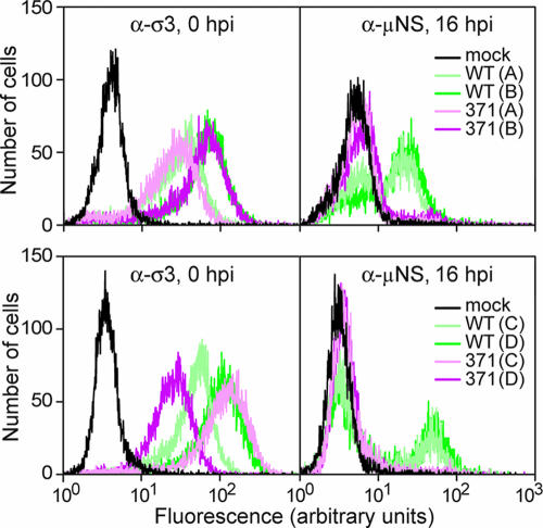
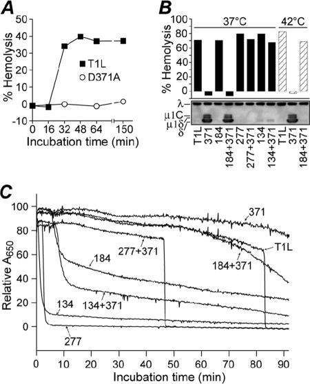
Heat-resistant mutants selected from infectious subvirion particles of mammalian reoviruses have determinative mutations in the major outer-capsid protein micro 1. Here we report the isolation and characterization of intragenic pseudoreversions of one such thermostabilizing mutation. From a plaque that had survived heat selection, a number of viruses with one shared mutation but different second-site mutations were isolated. The effect of the shared mutation alone or in combination with second-site mutations was examined using recoating genetics. The shared mutation, D371A, was found to confer (i) substantial thermostability, (ii) an infectivity defect that followed attachment but preceded viral protein synthesis, and (iii) resistance to micro 1 rearrangement in vitro, with an associated failure to lyse red blood cells. Three different second-site mutations were individually tested in combination with D371A and found to wholly or partially revert these phenotypes. Furthermore, when tested alone in recoated particles, each of these three second-site mutations conferred demonstrable thermolability. This and other evidence suggest that pseudoreversion of micro 1-based thermostabilization can occur by a general mechanism of micro 1-based thermolabilization, not requiring a specific compensatory mutation. The thermostabilizing mutation D371A as well as 9 of the 10 identified second-site mutations are located near contact regions between micro 1 trimers in the reovirus outer capsid. The availability of both thermostabilizing and thermolabilizing mutations in micro 1 should aid in defining the conformational rearrangements and mechanisms involved in membrane penetration during cell entry by this structurally complex nonenveloped animal virus.


Figures

FIG. 1.
FIG. 1.
Diagram of virus clone and virus stock origins and portions of M2 DNA-sequencing electropherograms of the initial, mixed stock of HR mutant IL54-5 and of the previously characterized HR mutant clone IL54-5-a (24). The IL54-5 stock contains a C+T mixture at nucleotide position 580, encoding a mixture of A184 and A184V. Both IL54-5 and IL54-5-a have a C at nucleotide position 1141, encoding D371A.
FIG. 2.
FIG. 2.
Changes in infectivity of reovirus clones after heat treatment of ISVPs at 52°C for 30 min. P2 cell lysate stocks in two separate batches, clones 1 to 15 (A) and 16 to 33 (B), were digested with chymotrypsin to convert virions in the lysates to ISVPs. Infectivity change is expressed as log10(infectious titer), measured by plaque assay, relative to an aliquot of each sample held at room temperature. Results from two independent experiments with each clone are shown as superimposed bars. Stocks of T1L WT and the previously characterized HR mutant clone IL54-5-a were treated and analyzed in parallel with the other clones.
FIG. 3.
FIG. 3.
Effect of double mutations on thermostability. RCs were made with μ1 protein containing double mutations, or each mutation in isolation, identified from clones IL54-5-a (mutations D371A and A184V) (A); IL54-5-8 (mutations D371A and P277T) (B); IL54-5-16 (mutations D371A and S134L) (C); or ID46-2 (mutations Y431C and T325A) (E) or a combination of mutations from IL62-3 and IL54-5-16 (mutations K459E and S134L) (D). After digestion with chymotrypsin to yield pRCs, virus was subjected to treatment at the indicated temperature for 15 min. Infectivity change is expressed as log10(infectious titer), measured by plaque assay, relative to an aliquot of each sample held on ice. Each point represents the mean of two determinations, except points in parentheses, for which one of the replicates resulted in a titer below the limit of detection.
FIG. 4.
FIG. 4.
Effect of double mutations on infectivity. RCs were made with μ1 proteins containing double mutations, or each mutation in isolation, identified from clones IL54-5-a (thermostabilizing mutation D371A and thermolabilizing mutation A184V), IL54-5-8 (thermostabilizing mutation D371A and thermolabilizing mutation P277T), and IL54-5-16 (thermostabilizing mutation D371A and thermolabilizing mutation S134L). Infectivity was measured by plaque assay, and relative infectivity is expressed as the log10 of the particle/PFU ratio of the RC preparation after subtracting log10 of the particle/PFU ratio of the cores used for recoating. RCs made without σ1 (A) and with σ1 (B) were assayed. A184V-RCs with σ1 are omitted because, for unknown reasons, they could not be generated in several attempts. Error bars indicate the means ± the standard deviations of the results from three or more RC preparations, and overlapping bars represent the results from two RC preparations.
FIG. 5.
FIG. 5.
Effect of thermostabilizing mutation D371A on viral protein synthesis. L929 cells were adsorbed with WT- or D371A-RCs for 90 min in the cold and then washed. An aliquot was fixed and labeled with σ3 antibody to measure attachment (left panels), and the rest were shifted to 37°C to allow infection to proceed. At 16 h p.i., cells were fixed, permeabilized, and labeled with anti-μNS serum (right panels). Samples were labeled with a fluorescently conjugated secondary antibody and analyzed by flow cytometry. Results from four different preparations (A, B, C, and D) of WT- and D371A-RCs, in two experiments (top and bottom), are shown.
FIG. 6.
FIG. 6.
Effect of double mutations on hemolysis activity. Chymotrypsin digests of RCs (pRCs) made with μ1 protein containing double mutations, or each mutation in isolation, were mixed with bovine red blood cells and hemolysis buffer and incubated at 37°C, unless indicated otherwise. (A) Representative time course of hemolysis reactions with 4 × 10¹² WT- and D371A-pRCs/ml at 37°C. Hemolysis was measured by calculating the A405 after pelleting unlysed cells. (B) Thirty-minute end points of hemolysis reactions with 3.5 × 10¹² pRCs/ml at 37°C (filled bars) or 42°C (striped bars). Hemolysis (top) was measured by calculating the A405 after pelleting unlysed cells; means of results from two experiments are shown. Conformational change of μ1 in the hemolysis reactions (bottom) was assayed by trypsin digestion on ice and Western blotting with virion-specific serum. Viral proteins λ and μ1 (present as cleavage products μ1C, μ1δ, and δ) are indicated. A representative result is shown. (C) Time course of hemolysis reactions with 2 × 10¹² pRCs/ml at 37°C. Hemolysis was measured by calculating the A650 of whole reactions at 10-second intervals. In this assay, the abrupt reduction in A650 in association with hemolysis reflects the reduced light scattering by lysed cells. A representative experiment is shown.
FIG. 7.
FIG. 7.
(A) The positions of mutations in the primary sequence of T1L μ1 are shown in a line diagram, along with the positions of the δ/φ and μ1N/δ cleavages, which occur during ISVP and ISVP* formation, respectively (28, 29). (B) Locations of μ1, λ2, and σ1 in an electron cryomicroscopy reconstruction of the ISVP (16, 24, 27, 32). Two μ1 trimers related by quasi-twofold symmetry are indicated by yellow outlines. (C through E) Locations of double mutations in the μ1 trimer crystal structure (22) and model of the trimer-trimer interface (39). The two trimers shown are related by quasi-twofold symmetry, analogous to the trimers outlined in panel B. The position of thermostabilizing mutation D371A is shown in red, and that of orthologous thermostabilizing mutation K459E is shown in purple. Second-site mutations are shown in yellow. Views from the side (C), top (D), and bottom (E) are shown.

References

    1. Agosto, M. A., T. Ivanovic, and M. L. Nibert. 2006. Mammalian reovirus, a nonfusogenic nonenveloped virus, forms size-selective pores in a model membrane. Proc. Natl. Acad. Sci. USA 103:16496-16501. - PMC - PubMed
    1. Baer, G. S., and T. S. Dermody. 1997. Mutations in reovirus outer-capsid protein σ3 selected during persistent infections of L cells confer resistance to protease inhibitor E64. J. Virol. 71:4921-4928. - PMC - PubMed
    1. Bodkin, D., M. L. Nibert, and B. N. Fields. 1989. Proteolytic digestion of reovirus in the intestinal lumens of neonatal mice. J. Virol. 63:4676-4681. - PMC - PubMed
    1. Borsa, J., D. G. Long, T. P. Copps, M. D. Sargent, and J. D. Chapman. 1974. Reovirus transcriptase activation in vitro: further studies on the facilitation phenomenon. Intervirology 3:15-35. - PubMed
    1. Broering, T. J., A. M. McCutcheon, V. E. Centonze, and M. L. Nibert. 2000. Reovirus nonstructural protein μNS binds to core particles but does not inhibit their transcription and capping activities. J. Virol. 74:5516-5524. - PMC - PubMed

Publication types

Substances

LinkOut - more resources