. 2007 Jul;14(7):886-93.
doi: 10.1128/CVI.00407-06. Epub 2007 May 16.

Recombinant Mycobacterium bovis bacillus Calmette-Guerin elicits human immunodeficiency virus type 1 envelope-specific T lymphocytes at mucosal sites

Affiliations

Recombinant Mycobacterium bovis bacillus Calmette-Guerin elicits human immunodeficiency virus type 1 envelope-specific T lymphocytes at mucosal sites

Jae-Sung Yu et al. Clin Vaccine Immunol. 2007 Jul.

Abstract

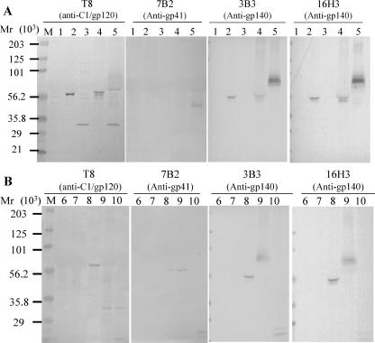
A successful vaccine vector for human immunodeficiency virus type 1 (HIV-1) should induce anti-HIV-1 T-cell immune responses at mucosal sites. We have constructed recombinant Mycobacterium bovis bacillus Calmette-Guérin (rBCG) expressing an HIV-1 group M consensus envelope (Env) either as a surface, intracellular, or secreted protein as an immunogen. rBCG containing HIV-1 env plasmids engineered for secretion induced optimal Env-specific T-cell gamma interferon enzyme-linked immunospot responses in murine spleen, female reproductive tract, and lungs. While rBCG-induced T-cell responses to HIV-1 envelope in spleen were lower than those induced by adenovirus prime/recombinant vaccinia virus (rAd-rVV) boost, rBCG induced comparable responses to rAd-rVV immunization in the female reproductive tract and lungs. T-cell responses induced by rBCG were primarily CD4(+), although rBCG alone did not induce anti-HIV-1 antibody. However, rBCG could prime for a protein boost by HIV-1 envelope protein. Thus, rBCG can serve as a vector for induction of anti-HIV-1 consensus Env cellular responses at mucosal sites.


Figures

FIG. 1.
FIG. 1.
Expression of HIV-1 group M consensus gp120 and gp140 envelope proteins in BCG strains. (A) Expression of intact CON6 gp120 in rBCG-P transformed by the surface expression plasmid pJH152-gp120 (lane 2), the intracellular expression plasmid pJH153-gp120 (lane 3), and the secreted expression plasmid pJH154-gp120 (lane 4) in rBCG-P. The expression of CON6 gp140CF was also determined in rBCG-P transformed by the surface expression plasmid pJH152-gp140CF (lane 5). (B) The secreted expression of CON6 gp120 in pJH154-gp120 (lane 8), the surface expression of gp140CF in pJH152-gp140CF (lane 9), and the intracellular expression of gp140 in pJH153-gp140CF (lane 10) were demonstrated in rBCG-D. Both the intact and partially cleaved gp140 products were shown using gp120 MAb T8 (anti-C1 gp120 region), gp41-specific MAb 7B2, anti-gp140 MAb 3B3, and anti-gp140 MAb 16H3. Lane M is molecular weight standards in thousands (Bio-Rad, CA). Negative controls were untransformed BCG-D (lane 6) and rBCG strains transformed by empty plasmid pJH222 (lanes 1 and 7).
FIG. 2.
FIG. 2.
Antigen-specific T-cell responses induced by different modes of insert expression by rBCG. HIV-1 envelope-specific (A) and BCG vector-specific (B) T-cell responses were assessed by using IFN-γ ELISPOT assays. Mice were immunized i.p. with various doses of rBCG strains or untransformed BCG strains such as BCG-P or BCG-D. The mean (± standard error of the mean) SFC per 106 cells derived from five mice per group are shown on the y axis. Mouse groups immunized with the indicated doses and designs of rBCG or control constructs are shown on the x axis. The mouse group indicated as CON6 was immunized with CON6 protein only. The mouse group indicated as rAd-rVV was primed with rAd-CON6 and boosted with rVV-CON6 as a positive control.
FIG. 3.
FIG. 3.
HIV-1 envelope-specific and vector-specific T-cell responses in spleen, lungs, and FRT of mice immunized with rBCG-P/120secPas. HIV-1-specific (A to C) and BCG vector-specific (D to F) T-cell responses in spleen (A and D), lungs (B and E), and FRT (C and F) were assessed by using IFN-γ ELISPOT assays. “CON6” indicates mice immunized with only CON6 protein. “rAd-rVV” indicates mice that were primed with rAd5 expressing CON6 Env and boosted with rVV expressing CON6 Env, used as positive controls. The mean (± standard error of the mean) SFC per 106 cells are shown on the y axis. The indicated immunization groups and doses are shown on the x axis. Assays were performed and analyzed on pooled lungs and FRT samples as described in Materials and Methods. Statistical significance was assessed by comparing rBCG-P/120secPas with control groups such as BCG-P or CON6 (*, P < 0.0001; #, P < 0.001).
FIG. 4.
FIG. 4.
M. tuberculosis WCL-specific IFN-γ production is dependent on CD4+ splenocytes. In order to access the T-cell population responsible for IFN-γ production following immunization with 120secPas and BCG-P, via the i.p. route, we removed the respective CD4 or CD8 population using magnetically labeled antibodies from the prepared splenocytes. CD4+ T-cell responses were predominant when mice were immunized with rBCG. The mean (± standard error of the mean) SFC per 106 cells derived from 15 mice per group are shown on the y axis.
FIG. 5.
FIG. 5.
HIV envelope-specific isotype induction by rBCG-P/120secPas. IgG subclasses including IgG1 (empty columns), IgG2a (gray columns), IgG2b (hatched columns), and IgG3 (solid columns) of HIV-1 envelope-specific serum antibodies induced by immunization with various immunization regimens (shown on the x axis) were determined by ELISA. The geometric mean log endpoint antibody titers (± standard errors of the means; n = 15) were plotted on the y axis.
FIG. 6.
FIG. 6.
Induction of antibody response with rBCG-P expressing HIV-1 gp120 in BALB/c mice with combinations of immunogen formulations. To evaluate the immunogenicity in mice of the rBCG-P expressing CON6 Env proteins, mice were immunized once with either killed or live rBCG-P/120secPas with or without IFA. The geometric mean endpoint titers (± standard errors of the means) for each group of mice (n = 5 per group) are shown. Abbreviations: 120secPas, rBCG-P expressing HIV-1 CON6 gp120 as the secreted protein; CON6, recombinant CON6 gp140CF protein.

Similar articles

Cited by

References

    1. Brandt, L., J. F. Cunha, A. W. Olsen, B. Chilima, P. Hirsch, R. Appelberg, and P. Andersen. 2002. Failure of the Mycobacterium bovis BCG vaccine: some species of environmental mycobacteria block multiplication of BCG and induction of protective immunity to tuberculosis. Infect. Immun. 70672-678. - PMC - PubMed
    1. Burlein, J. E., C. K. Stover, S. Offutt, and M. S. Hanson. 1994. Expression of foreign genes in mycobacteria. American Society for Microbiology, Washington, DC.
    1. Cayabyab, M. J., A. H. Hovav, T. Hsu, G. R. Krivulka, M. A. Lifton, D. A. Gorgone, G. J. Fennelly, B. F. Haynes, W. R. Jacobs, Jr., and N. L. Letvin. 2006. Generation of CD8+ T-cell responses by a recombinant nonpathogenic Mycobacterium smegmatis vaccine vector expressing human immunodeficiency virus type 1 Env. J. Virol. 801645-1652. - PMC - PubMed
    1. de Lisle, G. W., B. J. Wards, B. M. Buddle, and D. M. Collins. 2005. The efficacy of live tuberculosis vaccines after presensitization with Mycobacterium avium. Tuberculosis (Edinburgh) 8573-79. - PubMed
    1. Finlay, B. B., and P. Cossart. 1997. Exploitation of mammalian host cell functions by bacterial pathogens. Science 276718-725. - PubMed

Publication types

MeSH terms

Substances