Comparative Study
. 2007 May 16;27(20):5291-300.
doi: 10.1523/JNEUROSCI.1069-07.2007.

Effects of treadmill exercise on dopaminergic transmission in the 1-methyl-4-phenyl-1,2,3,6-tetrahydropyridine-lesioned mouse model of basal ganglia injury

Affiliations
Comparative Study

Effects of treadmill exercise on dopaminergic transmission in the 1-methyl-4-phenyl-1,2,3,6-tetrahydropyridine-lesioned mouse model of basal ganglia injury

Giselle M Petzinger et al. J Neurosci. .

Abstract

Studies have suggested that there are beneficial effects of exercise in patients with Parkinson's disease, but the underlying molecular mechanisms responsible for these effects are poorly understood. Studies in rodent models provide a means to examine the effects of exercise on dopaminergic neurotransmission. Using intensive treadmill exercise, we determined changes in striatal dopamine in the 1-methyl-4-phenyl-1,2,3,6-tetrahydropyridine (MPTP)-lesioned mouse. C57BL/6J mice were divided into four groups: (1) saline, (2) saline plus exercise, (3) MPTP, and (4) MPTP plus exercise. Exercise was started 5 d after MPTP lesioning and continued for 28 d. Treadmill running improved motor velocity in both exercise groups. All exercised animals also showed increased latency to fall (improved balance) using the accelerating rotarod compared with nonexercised mice. Using HPLC, we found no difference in striatal dopamine tissue levels between MPTP plus exercise compared with MPTP mice. There was an increase detected in saline plus exercise mice. Analyses using fast-scan cyclic voltammetry showed increased stimulus-evoked release and a decrease in decay of dopamine in the dorsal striatum of MPTP plus exercise mice only. Immunohistochemical staining analysis of striatal tyrosine hydroxylase and dopamine transporter proteins showed decreased expression in MPTP plus exercise mice compared with MPTP mice. There were no differences in mRNA transcript expression in midbrain dopaminergic neurons between these two groups. However, there was diminished transcript expression in saline plus exercise compared with saline mice. Our findings suggest that the benefits of treadmill exercise on motor performance may be accompanied by changes in dopaminergic neurotransmission that are different in the injured (MPTP-lesioned) compared with the noninjured (saline) nigrostriatal system.


Figures

Figure 1.
Figure 1.
Analysis of motor behavior on motorized treadmill. Both saline and MPTP-lesioned mice were run on the motorized treadmill for 28 d (5 d per week). The running velocity of the mice in each group (n = 12) were determined three times per week and compared. The graph demonstrates that (1) MPTP-lesioned mice had a running velocity less than the saline group, where the asterisk indicates significant difference (p < 0.05), and (2) both saline- and MPTP-lesioned groups improved in running velocity, and by the final 2 weeks, the difference between the two groups was not significant (post hoc analysis; p < 0.05; t test). Error bars indicate SEM.
Figure 2.
Figure 2.
Analysis of behavior on rotarod mice from all groups were tested once a week for latency to fall from an accelerating rotarod (mean and SEM in seconds per week). There was a significant effect of exercise (treadmill running) on the mean latency to fall (in seconds) from the accelerating rotarod, where the asterisk represents significant differnce between the exercise and no exercise groups (F (3,44) = 9.587; p < 0.0001). Both MPTP plus exercise and saline plus exercise mice performed better on the rotarod compared with the nonexercised groups. There was no significant effect of MPTP on mean latency to fall (F (3,44) = 0.851; p = 0.504) and no significant interaction between exercise and MPTP on mean latency to fall (F (3,44) = 0.965; p = 0.435).
Figure 3.
Figure 3.
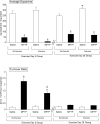
HPLC analysis of striatal dopamine levels and dopamine turnover. This figure shows data from Table 1 for striatal dopamine levels and turnover ratio at both 5 d of exercise (corresponding to 10 d postlesioning), and also at 28 d of exercise (corresponding to 42 d postlesioning). At day 5 of exercise, there was 82–98% depletion of dopamine in all MPTP-treated animals (exercise and no exercise) compared with their respective saline groups (asterisk represents significance at p < 0.0001). There were no significant differences in striatal dopamine levels between exercise and no exercise animals in either the MPTP- or saline-treated animals. Turnover ratio was significantly elevated in all MPTP-compared with saline-treated animals (asterisk represents significance at p < 0.0001). There was a significant decrease in turnover ratio in the MPTP plus exercise compared with the MPTP plus no exercise animals (hash mark represents significance at p < 0.02). At day 28 of exercise, there was >68–78% depletion of striatal dopamine levels in all MPTP-treated animals (exercise and no exercise) compared with their respective saline groups (asterisk represents significance at p < 0.0001). There was a significant increase in striatal dopamine levels in saline plus exercise compared with saline plus no exercise groups only (cross represents significance at p = 0.015). Exercise had no effect on turnover ratio. Error bars indicate SEM.
Figure 4.
Figure 4.
Analysis of dopamine release using fast-scan cyclic voltammetry. The amount of dopamine release was determined at five regions within the striatum (at bregma level ∼1.00 to bregma 0.80) including (1) mid-striatum, (2) dorsomedial, (3) dorsal, (4) dorsolateral, and (5) ventrolateral in representative mice from all four groups (see brain slice insert). A, Comparison of peak dopamine released by a single intrastriatal stimulus (200 A, 0.1 ms). Top, Data comparing saline and saline plus exercise mice. There was no significant difference in dopamine release in any region. Middle, Data comparing saline and MPTP groups. There was a significant decrease in dopamine release in MPTP mice and the asterisk represents significant difference compared with saline (p < 0.001). Bottom, Data comparing MPTP and MPTP plus exercise mice. There was a significant increase in dopamine release in the MPTP plus exercise mice at dorsal sites 3 and 4 compared with MPTP mice, and the asterisk represents significant difference (p < 0.05). B, Comparison of dopamine signal (peak and decay) evoked by intrastriatal stimulation. Top, A plot of average time to peak and decay in the dopamine signal at electrode position 4 for MPTP plus no exercise (n = 7, filled circles) and MPTP plus exercise (n = 11, open circles). The intrastriatal stimulus was delivered at the time indicated by the filled triangle (between 0 and 0.8 s). Data points are normalized to the peak-evoked dopamine signal. The middle panel illustrates the decay phase of the graph (shaded area), which was fit with a single exponential function. Best fit for the average data from MPTP plus no exercise mice (solid line) and MPTP plus exercise mice are shown (dotted line). The bottom panel illustrates the averages of the decay constant (k) obtained by the exponential fit of the decay phase for each recording and are shown for each group (mean ± SEM). MPTP plus exercise mice had a significantly lower decay constant compared with MPTP plus no exercise mice (p < 0.05; t test). A similar trend was seen in the saline plus exercise compared with the saline plus no exercise animals (p = 0.13; t test). Exc, Exercise.
Figure 5.
Figure 5.
Analysis of striatal TH and DAT protein. A, B, The relative expression of striatal TH (A) and DAT (B) was determined using immunohistochemistry and computer-assisted image analysis after 28 d of exercise. Sections at the level of the midstriatum (≥6–8 sections from 4 different mice within each group) were immunostained with an antibody against TH or DAT. Sections were scanned and the relative optical density mean plus SEM for each group was determined. MPTP causes a significant decline in the level of expression of striatal TH and DAT protein compared with saline-treated mice, and the asterisk represents significance at p < 0.001. There is a significant interaction between exercise and MPTP, with a significant decrease in the MPTP plus exercise compared with the MPTP plus no exercise mice, and the hash mark represents significance at p < 0.05.
Figure 6.
Figure 6.
Analysis of TH and DAT mRNA in midbrain nigrostriatal neurons. The relative expression of TH and DAT mRNA in midbrain dopaminergic neurons were determined using in situ hybridization histochemistry followed by grain counting of emulsion-dipped sections. Representative sections showing neurons for the determination of TH mRNA expression are shown in panels A (saline), B (saline plus exercise), C (MPTP), and D (MPTP plus exercise). The respective analysis of the data for either TH or DAT is shown in the graphs below each set of images. At least 120 neurons with grains were counted for each treatment group. MPTP lesioning caused a significant decrease in both TH and DAT transcript compared with the saline-treated groups, and the asterisk represents significance at p < 0.001. Statistical differences were seen between the saline and saline plus exercise groups for both the TH and DAT transcript, and the cross represents significance at p < 0.001. There were no significant differences in transcript expression between the MPTP and MPTP plus exercise groups. Representative sections showing neurons for determination of DAT mRNA expression are shown in E (saline), F (saline plus exercise), G (MPTP), and H (MPTP plus exercise). Error bars indicate SEM.
Figure 7.
Figure 7.
Determination of SNpc dopaminergic cell number. Unbiased stereological counting of TH-ir neurons was performed in mice (n = 3 per group) from all four groups at the end of the 28 d running regimen. MPTP lesioning resulted in an ∼50% decline in SNpc dopaminergic neurons. There was no statistically significant difference between the saline and the saline plus exercise mice as well as no difference between the MPTP and MPTP plus exercise groups, indicating that there was no effect of exercise on the number of SNpc dopaminergic neurons. The asterisk represents significant difference from the saline group (p < 0.0001).

Similar articles

Cited by

References

    1. Akopian G, Walsh JP. Reliable long-lasting depression interacts with variable short-term facilitation to determine corticostriatal paired-pulse plasticity. J Physiol (Lond) 2007;580:225–240. - PMC - PubMed
    1. Bezard E, Dovero S, Imbert C, Boraud T, Gross CE. Spontaneous long-term compensatory dopaminergic sprouting in MPTP-treated mice. Synapse. 2000;38:363–368. - PubMed
    1. Bezard E, Dovero S, Belin D, Duconger S, Jackson-Lewis V, Przedborski S, Piazza PV, Gross CE, Jaber M. Enriched environment confers resistance to 1-methyl-4-phenyl-1,2,3,6-tetrahydropyridine and cocaine: involvement of dopamine transporter and trophic factors. J Neurosci. 2003;23:10999–11007. - PMC - PubMed
    1. Brown LL, Sharp FR. Metabolic mapping of rat striatum: somatotopic organization of sensorimotor activity. Brain Res. 1995;686:207–222. - PubMed
    1. Burke RE, Cadet JL, Kent JD, Karanas AL, Jackson-Lewis V. An assessment of the validity of densitometric measures of striatal tyrosine hydroxylase-positive fibers: relationship to apomorphine-induced rotations in 6-hydroxydopamine lesioned rats. J Neurosci Methods. 1990;35:63–73. - PubMed

Publication types

LinkOut - more resources