Comparative Study
. 2007 May 16;27(20):5313-25.
doi: 10.1523/JNEUROSCI.3934-06.2007.

Wnt-7a induces presynaptic colocalization of alpha 7-nicotinic acetylcholine receptors and adenomatous polyposis coli in hippocampal neurons

Affiliations
Comparative Study

Wnt-7a induces presynaptic colocalization of alpha 7-nicotinic acetylcholine receptors and adenomatous polyposis coli in hippocampal neurons

Ginny G Farías et al. J Neurosci. .

Abstract

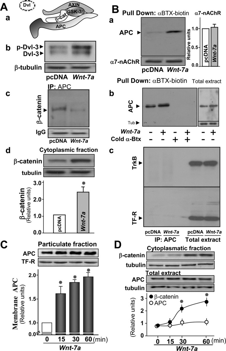
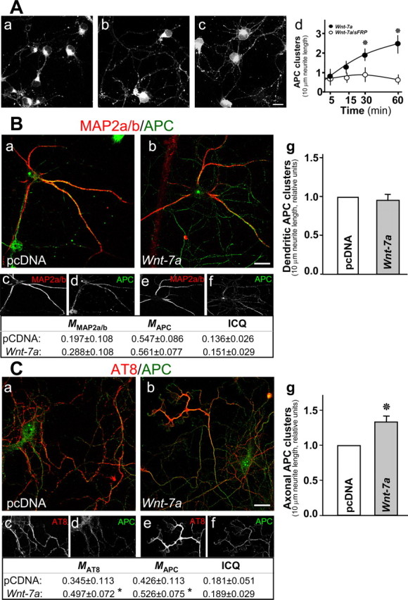
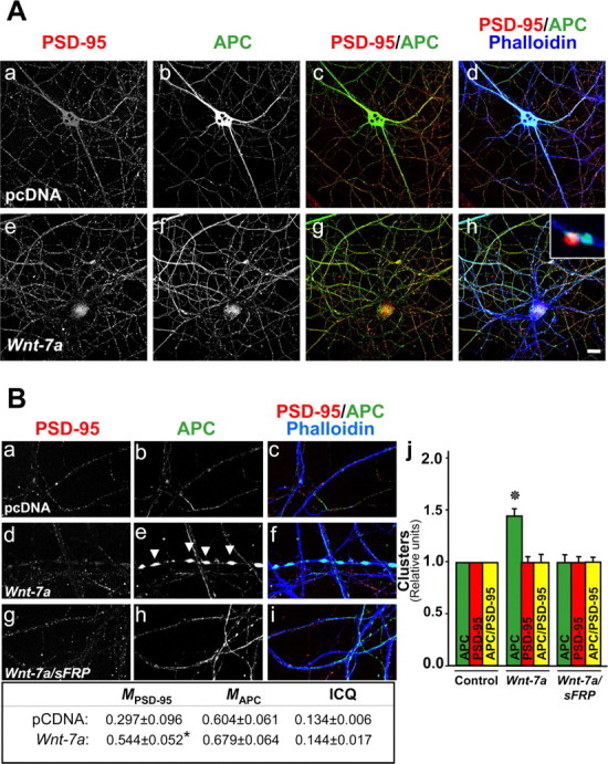
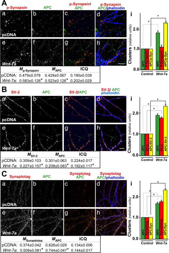
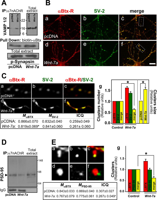
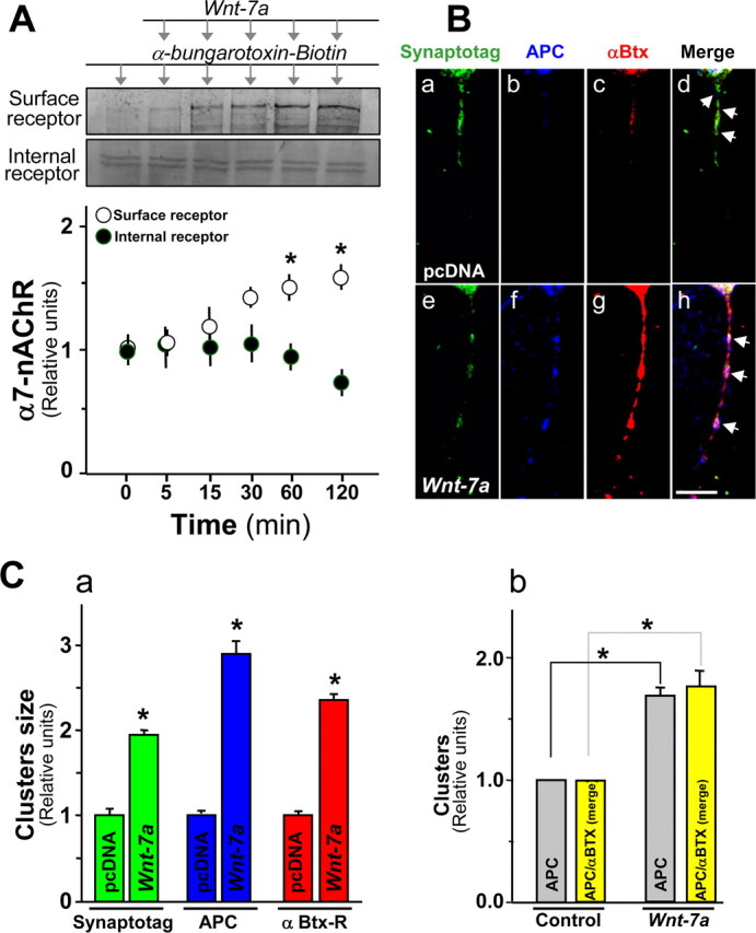
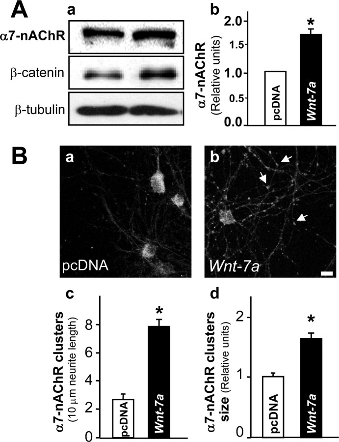
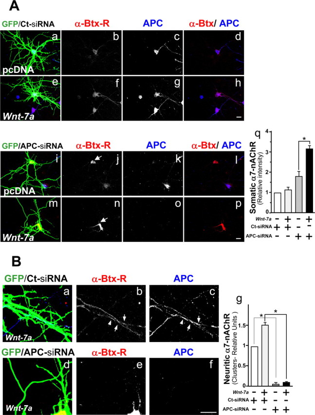
Nicotinic acetylcholine receptors (nAChRs) contribute significantly to hippocampal function. Alpha7-nAChRs are present in presynaptic sites in hippocampal neurons and may influence transmitter release, but the factors that determine their presynaptic localization are unknown. We report here that Wnt-7a, a ligand active in the canonical Wnt signaling pathway, induces dissociation of the adenomatous polyposis coli (APC) protein from the beta-catenin cytoplasmic complex and the interaction of APC with alpha7-nAChRs in hippocampal neurons. Interestingly, Wnt-7a induces the relocalization of APC to membranes, clustering of APC in neurites, and coclustering of APC with different, presynaptic protein markers. Wnt-7a also increases the number and size of coclusters of alpha7-nAChRs and APC in presynaptic terminals. These short-term changes in alpha7-nAChRs occur in the few minutes after ligand exposure and involve translocation to the plasma membrane without affecting total receptor levels. Longer-term exposure to Wnt-7a increases nAChR alpha7 subunit levels in an APC-independent manner and increases clusters of alpha7-nAChRs in neurites via an APC-dependent process. Together, these results demonstrate that stimulation through the canonical Wnt pathway regulates the presynaptic localization of APC and alpha7-nAChRs with APC serving as an intermediary in the alpha7-nAChR relocalization process. Modulation by Wnt signaling may be essential for alpha7-nAChR expression and function in synapses.


Figures

Figure 1.
Figure 1.
APC dissociates from the β-catenin cytoplasmic complex and associates with α7-nAChRs and membrane compartments in hippocampal neurons in the presence of Wnt-7a. Aa, Schematic representation of the β-catenin destruction complex. Ab, Western blot analysis after Wnt-7a treatment of hippocampal neurons for 1 h reveals increased phosphorylation of DVL-3 normalized for sample loading with tubulin (representative of n = 3; pcDNA represents the control condition using conditioned medium without secreted Wnt-7a from transfected cells). Ac, Ad, Immunoprecipitation (IP) of APC and Western detection of β-catenin indicates that the β-catenin/APC complex dissociates after Wnt-7a treatment under conditions in which total β-catenin is not altered (Ac; representative of n = 3), whereas Wnt-7a induces β-catenin stabilization in the cytoplasmic fraction marked by the presence of and normalized for sample loading with tubulin (Ad; n = 4; bar graph shows quantitation). Ba, Bb, APC interaction as determined by Western analysis with α7-nAChRs and assessed after pull down of α7-nAChRs using biotinylated-α-Btx/streptavidin-agarose is increased after exposure to Wnt-7a relative to pcDNA controls and normalized for total α7-nAChRs recovered, which is unchanged by ligand exposure (Ba; n = 4; bar graph shows α7-nAChR quantitation) and is prevented in the presence of excess, fluid-phase Btx under conditions in which total APC levels are not altered (Bb; n = 3). Bc, Immunoprecipitation of APC and detection by Western blot of associated TrkB receptor or TF-R reveals no nonspecific receptor interaction with APC under conditions in which total receptor numbers are high and unchanged by Wnt-7a exposure. C, Subcellular fractionation and Western blot of APC normalized to transferrin receptor levels indicates increases in APC localization to the particulate fraction in a time-dependent form in the presence of Wnt-7a (n = 4). D, Western blot analysis of β-catenin levels in the cytoplasmic fraction and of APC in total extracts indicates that β-catenin levels increase and become stabilized at 30 min and that total APC levels are not affected over the same time course of Wnt-7a treatment (n = 4; normalized to β-tubulin levels). Data are the mean ± SEM of three to six independent experiments, expressed as fold increase over control cells. In all figures, *p < 0.01 by nonpaired Student's t test.
Figure 2.
Figure 2.
Wnt-7a induces clustering of APC in neurites. A, Immunofluorescence for APC indicates that APC clusters as puncta in neurites and that these APC aggregates increase after Wnt-7a treatment for 0 min (Aa), 15 min (Ab), and 60 min (Ac). Scale bar, 10 μm. Ad, Quantification of APC puncta per 100 μm of neurites demonstrates time dependence for increased APC Wnt-7a-induced puncta and sensitivity of puncta formation to cotreatment with sFRP. Data are the mean ± SEM of six independent experiments performed in duplicate. B, Double immunofluorescence for APC and MAP2a/b shows that, under control conditions (pcDNA), APC is localized in the soma and dendrites (Ba, Bc, Bd), whereas Wnt-7a treatment induces neuritic localization of APC but not to MAP2a/b-positive dendrites (Bb, Be, Bf). Table, Colocalization analysis (M) and ICQ values. Bg, The number of dendritic APC clusters is not affected by Wnt-7a treatment relative to control conditions. Scale bar, 10 μm. C, Double immunofluorescence for APC and p-Tau recognized by AT8 antibody shows that Wnt-7a induces APC relocalization to p-Tau-positive axons (Cb, Ce, Cf) relative to control conditions (pcDNA; Ca, Cc, Cd; 3 independent experiments performed in triplicate). Table, colocalization analysis (M) and ICQ values. Cg, Wnt-7a treatment increases the number of APC clusters in axons relative to control conditions. Scale bar, 10 μm.
Figure 3.
Figure 3.
APC is not clustered in postsynaptic sites in hippocampal neurons exposed to Wnt-7a. A, Fluorescence labeling of processes by phalloidin (Ad, Ah) and immunofluorescence labeling for APC (Ab, Af) or PSD-95 (Aa, Ae) are shown in cells subjected to treatments with control (pcDNA; Aa–Ad) or Wnt-7a (Ae–Ah) media. Induction of APC in neurites is observed in the presence of Wnt-7a relative to control conditions (Ab, Ad, Af, Ah), but its localization at synaptic sites is in apposition to PSD-95-labeled postsynaptic sites, indicating a presynaptic localization (Ah, inset). Scale bar, 10 μm. B, Three scan zoom images show PSD-95 staining (Ba, Bd, Bg), APC staining (Bb, Be, Bh), and merged image costaining with phalloidin (Bc, Bf, Bi) illustrating a modest coclustering of APC with PSD-95 under control conditions (Ba–Bc, Bj) but no Wnt-7a-induced APC coclustering with PSD-95 (Bd–Bf, Bj), indicating that Wnt-7a-induced APC clusters are not localized in specialized, postsynaptic sites. Bh, Bj, Cotreatment with sFRP prevented Wnt-7a-induced clustering of APC. Arrows indicate APC clusters formed in the presence of Wnt-7a. Data are the results of four independent experiments performed in duplicate. B, Table, Colocalization analysis (M) and ICQ indicate an increase in the colocalization of PSD-95 with respect to APC but not vice versa. Bj, APC or PSD-95 clusters or coclusters were quantified.
Figure 4.
Figure 4.
Wnt-7a induces clustering of APC in presynaptic sites in hippocampal neurons. A, Immunofluorescence labeling of hippocampal neurons for p-synapsin (Aa, Ae) or APC (Ab, Af) shows induction in coclustering in the presence of Wnt-7a (Ag, Ah) relative to control (pcDNA) treatment (Ac, Ad) and as indicated in quantification (Ai). B, Double labeling of hippocampal neurons for SV-2 (Ba, Be) and APC (Bb, Bf) shows that Wnt-7a induces coclustering (Bg, Bh) relative to control conditions (Bc, Bd) and as quantified (Bi). C, Double labeling for synaptotagmin (Synaptotag; Ca, Ce) and APC (Cb, Cf) shows induction in coclustering in hippocampal neurons exposed to Wnt-7a (Cg, Ch) relative to controls (Cc, Cd) and as indicated by quantification (Ci). Scale bars, 10 μm. Tables, Colocalization analysis (M) and ICQ values.
Figure 5.
Figure 5.
Wnt-7a increases presynaptic but not postsynaptic relocalization of α7-nAChRs in hippocampal neurons. A, Immunoprecipitation (IP) of α7-nAChRs and Western blot for VAMP-1/2, a synaptic vesicle protein, shows that Wnt-7a exposure induces the association of α7-nAChRs with VAMP-1/2 under conditions in which the total levels of VAMP-1/2 are not affected (n = 3). Pull down of α7-nAChRs and Western blot of p-synapsin shows that Wnt-7a treatment induces the association of α7-nAChRs with p-synapsin, another presynaptic protein (n = 3). B, Fluorescent rhodamine-labeled α-Btx (α-Btx-R; Ba, Bd) or SV-2 immunofluorescence (Bb, Be) staining in hippocampal neurons treated with control media (pcDNA; Ba–Bc) or Wnt-7a (Bd–Bf) indicates that Wnt-7a increases the interaction between α7-nAChRs and SV-2 (merged images; Bc, Bf). Scale bar, 10 μm. Ca–Cf, Magnified views of the boxed regions in Bc and Bf. Cg, Wnt-7a induced an increase in the size of α7-nAChR and SV-2 coclusters but not the number of clusters. D, Immunoprecipitation of α7-nAChRs and Western blot for PSD-95 shows α7-nAChR association with PSD-95 in control media (pcDNA)-treated samples and that Wnt-7a treatment decreases this association (representative of n = 3). E, Fluorescent rhodamine-labeled α-Btx (Ea, Ed) or PSD-95 immunofluorescence (Eb, Ee) staining in hippocampal neurons treated with pcDNA (Ea–Ec) or Wnt-7a (Ed–Ef) indicates that Wnt-7a increases the size of α7-nAChR clusters but not coclustering with PSD-95. Eg, Quantification of α-Btx staining (α7-nAChRs) or PSD-95 clusters and of α7-nAChRs contained in PSD-95 clusters, demonstrating a decrease in coclustering after treatment with Wnt-7a ligand. C, E, Tables, Colocalization analysis (M) and ICQ values.
Figure 6.
Figure 6.
APC and α7-nAChRs are coclustered in presynaptic regions in hippocampal neurons exposed to Wnt-7a. A, Effects on surface α7-nAChR levels assessed using biotinylated-α-Btx pull down and Western analysis in hippocampal neurons subjected to Wnt-7a treatments for different times show increases under conditions in which the fraction of α7-nAChRs in the intracellular pool also assessed by immunoblot decreases (n = 4). B, Rhodamine-labeled α-Btx fluorescence (Bc, Bg) or immunofluorescence for APC (Bb, Bf) or synaptotagmin (Synaptotag; Ba, Be) in hippocampal neurons treated with control medium (pcDNA; Ba–Bd) or Wnt-7a (Be–Bh) for 1 h indicates that Wnt-7a induces clustering of synaptotagmin (Be), APC (Bf), and α7-nAChR (Bg). Bh, Arrows, Wnt-7a elevates coclustering of APC and α7-nAChR in presynaptic regions. Scale bar, 5 μm. C, Quantification of size for synaptotagmin (green bars), APC (blue bars), or α7-nAChR (identified by α-Btx-R staining; red bars) clusters contained in neurites of hippocampal neurons (Ca) and quantification of cluster numbers for APC and for APC contained in α-Btx clusters (Cb). Data are the mean ± SEM of five independent experiments. One hundred clusters per treatment per each independent experiment were evaluated using LSM 5 Image Browser.
Figure 7.
Figure 7.
Wnt-7a increases nAChR α7 subunit levels in hippocampal neurons. A, Western blot analysis of hippocampal neurons treated with Wnt-7a (right) or control (pcDNA; left) medium for 24 h was performed to assess effects on nAChR α7 subunit, β-catenin, or β-tubulin (normalization control) levels (Aa) and showed an increase in α7 subunit levels (Ab; n = 5). B, Hippocampal neurons exposed for 24 h either to control (pcDNA; Ba) or Wnt-7a (Bb) treatments were analyzed using immunofluorescence to determine the distribution of total expressed α7-nAChRs. Wnt-7a treatment for 24 h induced preferential localization of α7-nAChR clusters in neurites (arrows). Scale bar, 10 μm. Bc, Quantification of α7-nAChR clusters per 10 μm of neurite length shows that Wnt-7a induces cluster formation in neurites. Data are the mean ± SEM of four independent experiments. Bd, Quantification shows that Wnt-7a induces an increase in the size of clusters containing α7-nAChR. Data are the mean ± SEM of four independent experiments. One hundred clusters per treatment per each independent experiment were evaluated using LSM 5 Image Browser.
Figure 8.
Figure 8.
Wnt-7a treatment increases nAChR α7 subunit levels in an APC-independent manner and alters the neuritic localization of α7-nAChRs in an APC-dependent manner. A, Mouse hippocampal neurons cotransfected at 10 d in vitro with green fluorescent protein and control siRNA (GFP/Ct-siRNA; Aa–Ah) or with GFP and anti-APC siRNA (GFP/APC-siRNA; Ai–Ap) were treated in the presence of control media (pcDNA; Aa–Ad, Ai–Al) or Wnt-7a (Ae–Ah, Am–Ap) for 24 h and were stained with rhodamine-labeled α-Btx (α-Btx-R; Ab, Af, Aj, An) or anti-APC (Ac, Ag, Ak, Ao). Arrows show regions of α-Btx-R stain. GFP images were overexposed to visualize neurites. Scale bar, 10 μm. Aq, Somatic levels of α7-nAChR staining. B, Zoom images of Wnt-7a-treated (24 h) GFP/Ct-siRNA (Ba–Bc) or GFP/APC-siRNA (Bd–Bf) mouse hippocampal neurons stained with α-Btx-R (Bb, Be) or anti-APC (Bc, Bf) as well as a quantitation of neuritic α7-nAChR clusters (Bg). Arrows show clusters of α7-nAChR and APC in control transfected neurons. Aq, Neurons transfected with APC siRNA show an increase in the soma of α-Btx-R stain in the presence of control media relative to levels in neurons transfected with control siRNA, and Wnt-7a treatment induces an additional increase in somal α-Btx-R staining in neurons transfected with APC siRNA but not in neurons transfected with control siRNA. Bg, In neurons transfected with inactive siRNA and exposed to Wnt-7a, an increase in the neuritic localization of α7-nAChR is observed relative to similarly transfected neurons in control media, but neuritic α7-nAChRs are very low and insensitive to Wnt treatment in APC-deficient cells. Data are the mean ± SEM of three independent experiments performed in triplicate, expressed as fold increase over control cells.

Similar articles

Cited by

References

    1. Aberle H, Bauer A, Stappert J, Kispert A, Kemler R. β-Catenin is a target for the ubiquitin-proteasome pathway. EMBO J. 1997;16:3797–3804. - PMC - PubMed
    1. Ahmad-Annuar A, Ciani L, Simeonidis I, Herreros J, Fredj NB, Rosso SB, Hall A, Brickley S, Salinas PC. Signaling across the synapse: a role for Wnt and Dishevelled in presynaptic assembly and neurotransmitter release. J Cell Biol. 2006;174:127–139. - PMC - PubMed
    1. Alkondon M, Albuquerque EX. Nicotinic acetylcholine receptors α7 and α4β2 subtypes differentially control GABAergic input to CA1 neurons in rat hippocampus. J Neurophysiol. 2001;86:3043–3055. - PubMed
    1. Alvarez AR, Godoy JA, Mullendorff K, Olivares GH, Bronfman M, Inestrosa NC. Wnt-3a overcomes β-amyloid toxicity in rat hippocampal neurons. Exp Cell Res. 2004;297:186–196. - PubMed
    1. Banerjee C, Nyengaard JR, Wevers A, de Vos RA, Jansen Steur EN, Lindstrom J, Pilz K, Nowacki S, Bloch W, Schroder H. Cellular expression of α7 nicotinic acetylcholine receptor protein in the temporal cortex in Alzheimer's and Parkinson's disease—a stereological approach. Neurobiol Dis. 2000;7:666–672. - PubMed

Publication types

MeSH terms

LinkOut - more resources