Comparative Study
. 2007 May 16;27(20):5349-62.
doi: 10.1523/JNEUROSCI.4107-06.2007.

Protein kinase C delta negatively regulates tyrosine hydroxylase activity and dopamine synthesis by enhancing protein phosphatase-2A activity in dopaminergic neurons

Affiliations
Comparative Study

Protein kinase C delta negatively regulates tyrosine hydroxylase activity and dopamine synthesis by enhancing protein phosphatase-2A activity in dopaminergic neurons

Danhui Zhang et al. J Neurosci. .

Abstract

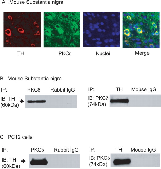
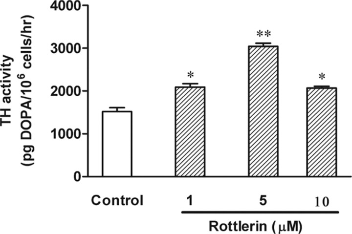
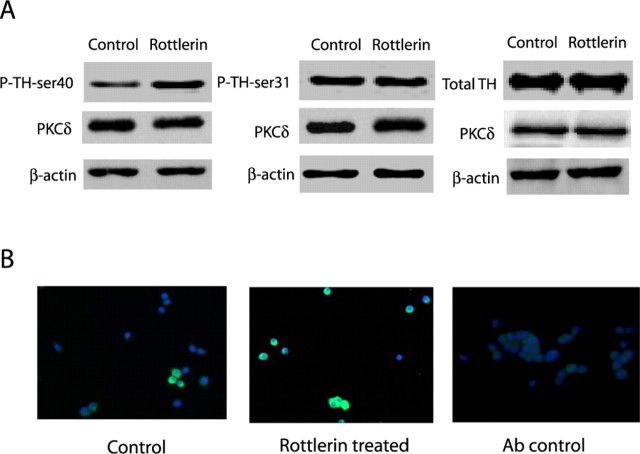
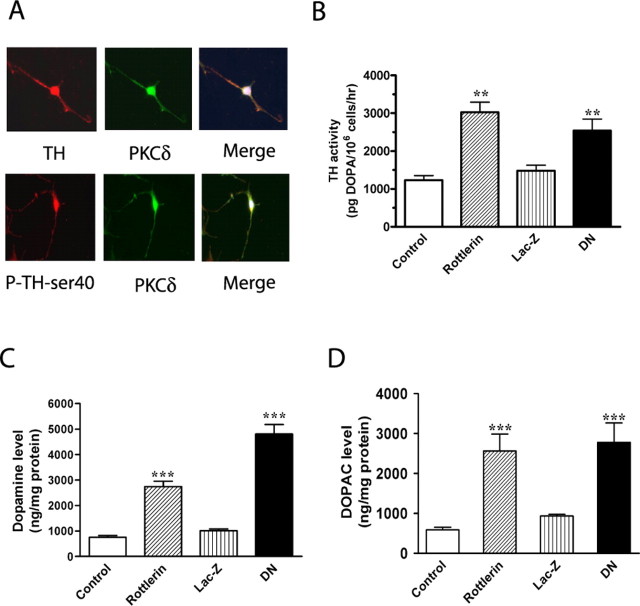
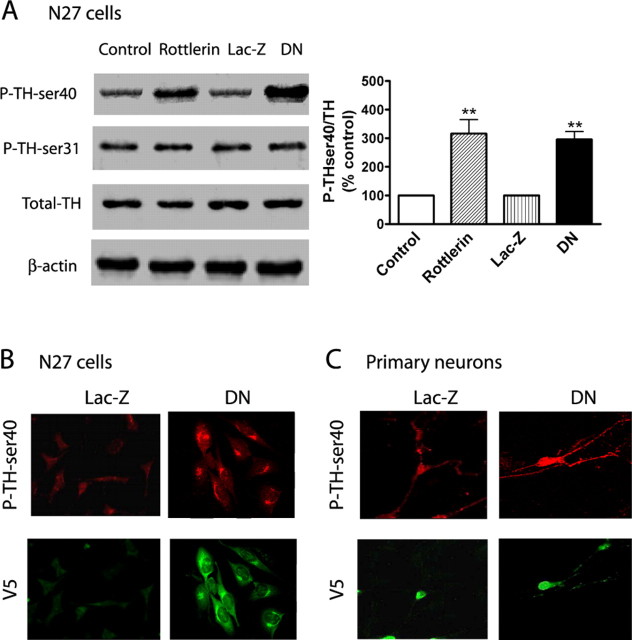
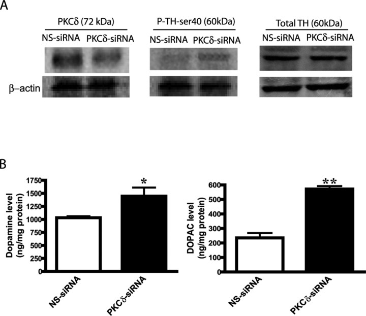
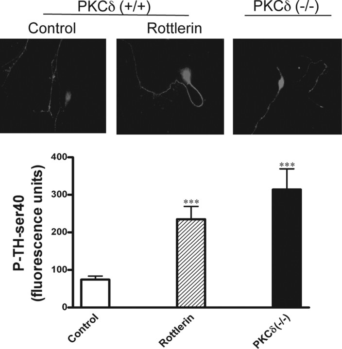
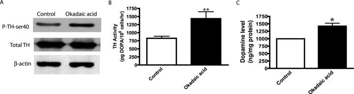
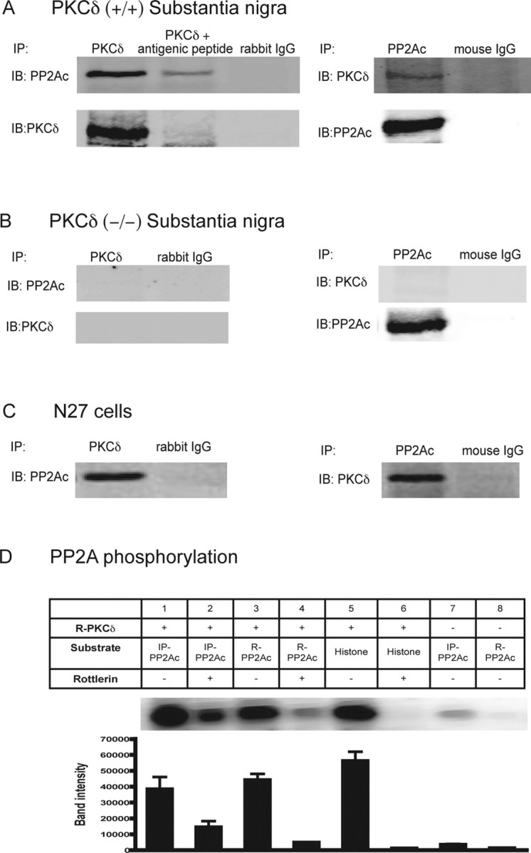
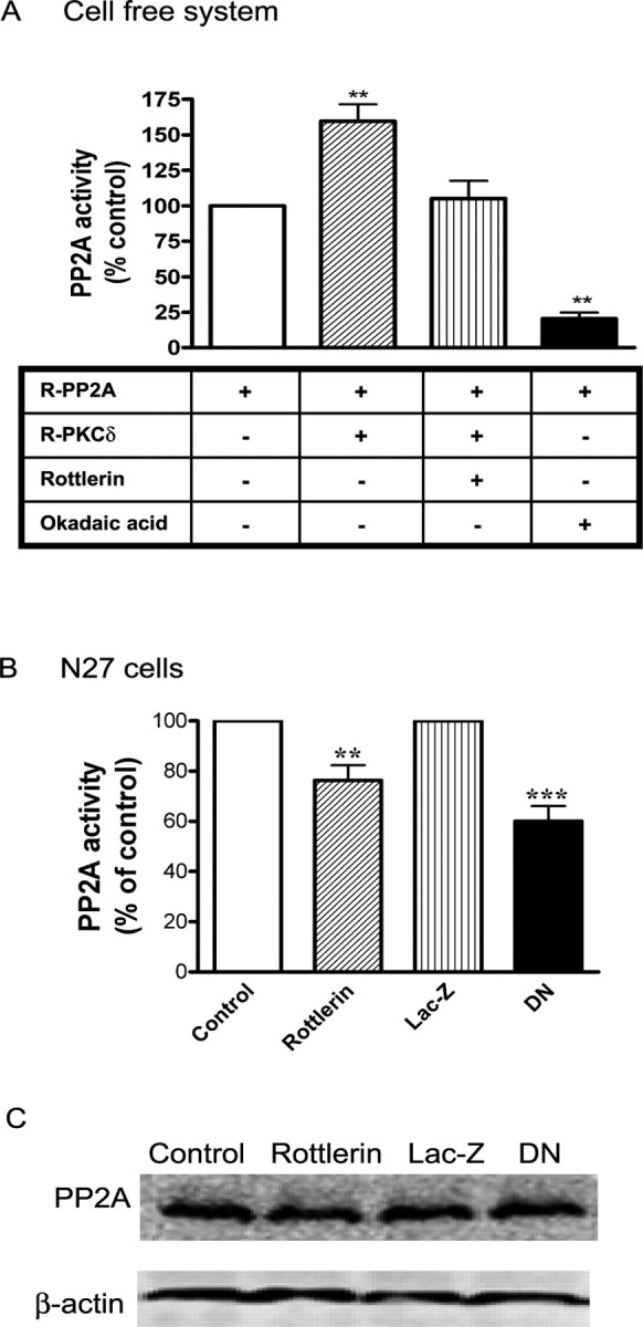
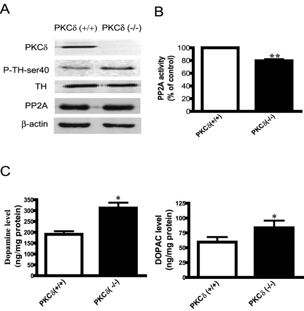
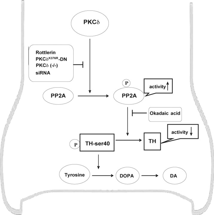
Tyrosine hydroxylase (TH), the rate-limiting enzyme in dopamine synthesis, can be regulated by phosphorylation at multiple serine residues, including serine-40. In the present study, we report a novel interaction between a key member of the novel PKC family, protein kinase Cdelta (PKCdelta), and TH, in which the kinase modulates dopamine synthesis by negatively regulating TH activity via protein phosphatase 2A (PP2A). We observed that PKCdelta is highly expressed in nigral dopaminergic neurons and colocalizes with TH. Interestingly, suppression of PKCdelta activity with the kinase inhibitor rottlerin, PKCdelta-small interfering RNA, or with PKCdelta dominant-negative mutant effectively increased a number of key biochemical events in the dopamine pathway, including TH-ser40 phosphorylation, TH enzymatic activity, and dopamine synthesis in neuronal cell culture models. Additionally, we found that PKCdelta not only physically associates with the PP2A catalytic subunit (PP2Ac) but also phosphorylates the phosphatase to increase its activity. Notably, inhibition of PKCdelta reduced the dephosphorylation activity of PP2A and thereby increased TH-ser40 phosphorylation, TH activity, and dopamine synthesis. To further validate our findings, we used the PKCdelta knock-out (PKCdelta-/-) mouse model. Consistent with other results, we found greater TH-ser40 phosphorylation and reduced PP2A activity in the substantia nigra of PKCdelta-/- mice than in wild-type mice. Importantly, this was accompanied by an increased dopamine level in the striatum of PKCdelta-/- mice. Collectively, these results suggest that PKCdelta phosphorylates PP2Ac to enhance its activity and thereby reduces TH-ser40 phosphorylation and TH activity and ultimately dopamine synthesis.


Figures

Figure 1.
Figure 1.
PKCδ associates with TH in mouse brain and PC12 cells. A, Immunohistochemical analysis. Mouse brain was cut to 20 μm thickness at the level of the substantia nigra and stained with PKCδ polyclonal antibody (Ab) (1:500 dilution) and TH monoclonal Ab (1:500 dilution), followed by incubation with either Alexa 488-conjugated (green; 1:1000) or Cy3-conjugated (red; 1:1000) secondary antibody. Hoechst 33342 (10 μg/ml) was used to stain the nucleus. Red, TH; green, PKCδ; blue, nucleus. B, PKCδ (74 kDa) coimmunoprecipitated with TH in mouse substantia nigra. PKCδ was immunoprecipitated (IP) from mouse substantia nigra lysates by using PKCδ polyclonal Ab (1:100) and immunoblotted (IB) with anti-TH (1:100). In reverse immunoprecipitation studies, TH was immunoprecipitated with mouse monoclonal anti-TH antibody (1:100) and immunoblotted with PKCδ. TH (60 kDa) coimmunoprecipitated with PKCδ in mouse substantia nigra. Similarly, PKCδ (74 kDa) coimmunoprecipitated with TH in mouse substantia nigra. C, PKCδ also coimmunoprecipitated with TH in PC12 cells, and PKCδ was immunoprecipitated (IP) from PC12 cell lysates by using PKCδ polyclonal Ab (1:100) and immunoblotted (IB) with anti-TH (1:100). In reverse immunoprecipitation studies, TH was immunoprecipitated with mouse monoclonal anti-TH antibody (1:100) and immunoblotted with PKCδ. TH (60 kDa) coimmunoprecipitated with PKCδ in PC12 cell lysates. Similarly, PKCδ (74 kDa) coimmunoprecipitated with TH in PC12 cell lysates. Rabbit IgG and mouse IgG were used as negative controls.
Figure 2.
Figure 2.
PKCδ inhibition enhances TH activity. PC12 cells were incubated with the DOPA decarboxylase inhibitor NSD-1015 (2 mm) for 1 h before treatment with the PKCδ inhibitor rottlerin (1–10 μm). DMSO (0.01%) was used as vehicle control. After 3 h of rottlerin treatment, cells were lysed and extracts were used for determining l-DOPA levels by HPLC. Rottlerin treatment increased l-DOPA levels, indicating enhanced TH activity. The data represent mean ± SEM of six to eight individual measurements. Asterisks (*p < 0.05; **p < 0.01) indicate significant differences between rottlerin-treated cells and control cells.
Figure 3.
Figure 3.
PKCδ inhibition enhances TH-ser40 phosphorylation. A, Western blot analysis. PC12 cells were exposed to rottlerin (5 μm) or 0.01% DMS0 (vehicle control) for 3 h. Cell extracts were prepared and separated by SDS-PAGE and transferred to nitrocellulose membrane. Phospho-specific antibodies directed against P-TH-ser40 and P-TH-ser31, and antibodies directed against TH and PKCδ were used for immunoblotting. To confirm equal protein loading in each lane, the membranes were reprobed with β-actin antibody. The immunoblots were visualized using the GE Healthcare ECL detection agents. Densitometric analysis of 60 kDa TH and P-TH-ser40 and P-TH-ser31 bands represents the mean ± SEM from three separate experiments (**p < 0.01). B, Immunocytochemistry. PC12 cells were grown on poly-l-lysine-coated coverslips and then exposed to either 5 μm rottlerin or 0.01% DMS0 for 3 h. After treatment, the cells were fixed and immunostained for P-TH-ser40 followed by staining with Alexa 488-conjugated antibody (green), as described in Materials and Methods. For a negative control, cells were also immunostained with Alexa 488 without primary antibody staining. Immunostained cells were mounted and viewed under a Nikon TE2000 fluorescence microscope, and images were captured with a SPOT digital camera. For in situ quantitative analysis of P-TH-ser40 levels, fluorescence immunoreactivity in cells was measured from six different areas in each group using MetaMorph image analysis software, and data were analyzed with Prism software. Experiments were repeated three times, and representative images are presented.
Figure 4.
Figure 4.
PKCδ inhibition regulates TH activity and DA synthesis in N27 dopaminergic neuronal cells. A, In situ colocalization of TH and PKCδ. Differentiated N27 cells were grown on poly-l-lysine-coated coverslips and processed for double immunostaining with anti-PKCδ monoclonal and anti-TH polyclonal antibodies or anti-PKCδ monoclonal and anti-P-TH-ser40 polyclonal antibodies, as described in Materials and Methods. B, TH activity in PKCδ inhibitor rottlerin-treated cells and PKCδ-DN cells. Differentiated N27 cells stably expressing LacZ or PKCδ-DN were pretreated with 2 mm NSD-1015 for 1 h and lysed, and extracts were used for determining l-DOPA levels by HPLC. Also, cell extracts from N27 cells incubated with 2 mm NSD-1015 for 1 h before treatment with 0.01% DMSO (vehicle control) or rottlerin (5 μm for 3 h) were also used for measuring DOPA levels. C, D, DA synthesis. Cell extracts from N27 cells stably expressing LacZ and PKCδ-DN, or treated with rottlerin (5 μm for 3 h), were used for determining DA and DOPAC levels by HPLC. The data represent a mean ± SEM of six to eight individual measurements. Asterisks (**p < 0.01; ***p < 0.001) indicate significant differences between rottlerin-treated cells and vehicle control cells, or between LacZ- and PKCδ-DN-expressing cells.
Figure 5.
Figure 5.
Effect of PKCδ-DN and rottlerin on TH-ser40 phosphorylation. A, Western blot. Cell extracts from rottlerin-treated, PKCδ-DN, and LacZ-expressing N27 cells were subjected to Western blot analysis, as described in Materials and Methods. P-TH-ser40, P-TH-ser31, and TH levels were detected using appropriate phospho-specific and TH antibodies. Densitometric analysis of 60 kDa P-TH-ser40, P-TH-ser31, and TH bands are shown next to the Western blot image. The data represent the mean ± SEM from three separate experiments (**p < 0.01). B, TH-ser40 phosphorylation in stably expressing PKCδ-DN and LacZ N27 cells. C, Primary mesencephalic cultures transiently expressing PKCδ-DN and LacZ constructs. Briefly, N27 cells and primary mesencephalic neurons expressing PKCδ-DN and LacZ (these constructs express V5 fusion protein) were cultured and processed for double immunostaining of P-TH-ser40 and V5 using specific antibodies. Stained cells were mounted on slides and viewed under a Nikon TE2000 fluorescence microscope, and images were captured with a SPOT digital camera, as described in Materials and Methods. P-TH-ser40 levels were quantitatively analyzed from six different areas in each group using MetaMorph image analysis. Experiments were repeated three times, and representative images are shown. Asterisks (**p < 0.01) indicate significant differences between LacZ- and PKCδ-DN-expressing cells.
Figure 6.
Figure 6.
Effect of RNAi-mediated knockdown of PKCδ on P-TH-ser40 levels and DA synthesis. A, Western blot of TH-ser40 phosphorylation. Briefly, N27 cells were transfected with PKCδ-siRNA and NS-siRNA, and then extracts from siRNA-transfected N27 cells were subjected to Western blot analyses of PKCδ, P-TH-ser40, and total TH. Densitometric analysis of the 74 kDa native PKCδ band and 60 kDa P-TH-ser40 and TH bands were normalized to β-actin levels and are plotted below their respective Western blots. B, DA synthesis. Cell extracts from PKCδ-siRNA and NS-siRNA-transfected N27 cells were used for determining DA and DOPAC levels by HPLC. The data represent a mean ± SEM of six to eight individual measurements. Asterisks (*p < 0.05 and **p < 0.01) indicate significant differences between PKCδ-siRNA- and NS-siRNA-transfected N27 cells.
Figure 7.
Figure 7.
P-TH-ser40 levels in primary mesencephalic cultures from PKCδ knock-out (−/−) animals. Primary mesencephalic neurons were cultured on laminin-coated coverslips from PKCδ (−/−) and PKCδ (+/+) E16–E18 pups. Primary cultures from PKCδ (+/+) animals were also incubated with 5 μm rottlerin for 3 h. Cells were fixed and processed for P-TH-ser40 immunocytochemistry. Cy3-conjugated secondary antibody was used for visualization of P-TH-ser40 and viewed under a Nikon TE2000 fluorescence microscope. P-TH-ser40 levels were quantitatively analyzed from six different areas in each group using MetaMorph image analysis. Asterisks (***p < 0.001) indicate significant differences from controls. Experiments were repeated three times, and representative images are shown.
Figure 8.
Figure 8.
Effect of PP2A inhibition on TH-ser40 phosphorylation, TH activity, and dopamine levels. A, Western blot analysis of P-TH-ser40. N27 cells were treated with 0.01% DMSO (vehicle control) or 2 μm okadaic acid for 2 h, and then cell lysates were subjected to immunoblot for total TH, P-TH-ser40, and β-actin. B, Effect of okadaic acid on TH activity. C, Dopamine content. N27 cells were treated with 2 μm okadaic acid for 2 h. To determine TH activity, N27 cells were incubated with a DOPA decarboxylase inhibitor (2 mm NSD-1015 for 1 h) before okadaic acid treatment. After treatment, cells were lysed and neurochemicals were extracted for determining l-DOPA and dopamine levels by HPLC. The data represent the mean ± SEM from three separate experiments. Asterisks (*p < 0.05; **p < 0.01) indicate significant differences between untreated and okadaic acid-treated N27 cells.
Figure 9.
Figure 9.
PKCδ associates with PP2Ac and modulates PP2A activity. Immunoprecipitation assays: A, PKCδ (+/+) mouse substantia nigra; B, PKCδ (−/−) mouse substantia nigra; and C, N27 cells. Anti-PKCδ polyclonal antibody was used for immunoprecipitation (IP) and immunoblotted (IB) with mouse monoclonal PP2Ac antibody. In reverse immunoprecipitation studies, anti-PP2Ac mouse monoclonal antibody was used for immunoprecipitation followed by immunoblotting with rabbit polyclonal PKCδ antibody. Anti-PKCδ polyclonal antibody was coincubated with antigenic blocking peptide in the immunoprecipitation studies as a negative control. Rabbit IgG and mouse IgG were also used as negative controls. D, In vitro kinase assay, PKCδ phosphorylates PP2Ac. The top panel is a representative autoradiography showing [32P] phosphorylation of the immunoprecipitated PP2Ac (IP-PP2Ac) and recombinant PP2Ac (R-PP2Ac) by recombinant PKCδ (R-PKCδ). PP2Ac immunoprecipitates from N27 cell homogenates represent endogenous PP2Ac. Recombinant PKCδ protein was purchased from Sigma. Immunoprecipitation and phosphorylation assays were performed as described in Materials and Methods. The quantitative data in the bottom panel represent the mean ± SEM from three assays. Histone H1 was used as positive control substrate, and for negative controls, no substrate was added.
Figure 10.
Figure 10.
Effect of PKCδ inhibition on PP2A activity. A, Cell-free system. Recombinant PP2A enzyme (R-PP2A) was incubated with recombinant PKCδ (R- PKCδ) in the presence or absence of 5 μm rottlerin for 1 h. PP2A enzyme activity was measured by using a serine/threonine phosphatase assay kit from Promega. Okadaic acid (2 μm) was used as a positive control to inhibit PP2A activity. B, N27 cells. N27 cells were treated with 0.01% DMSO (vehicle control) or 3 μm rottlerin for 3 h, or expressed LacZ or PKCδ-DN. The data represent a mean ± SEM of four to six individual measurements. Asterisks (**p < 0.01; ***p < 0.001) indicate significant differences between either rottlerin-treated and vehicle control cells or LacZ- and PKCδ-DN-expressing N27 cells. C, Effect of PKCδ inhibition on PP2A protein level. Cell lysates from control, rottlerin-treated N27 cells, or LacZ and PKCδ-DN-expressing N27 cells were used to determine PP2A protein level by Western blot analysis.
Figure 11.
Figure 11.
Increased TH-ser40 phosphorylation, dopamine levels, and PP2A activity in PKCδ (−/−) knock-out animals. A, Western blot analysis of P-TH-ser40. Substantia nigral lysates from PKCδ (−/−) knock-out and PKCδ (+/+) naive animals were subjected to immunoblotting of PKCδ, P-TH-ser40, and total TH. B, PP2A activity. PP2A activity was measured in substantia nigra homogenates obtained from PKCδ (+/+) and PKCδ (−/−) animals. The data represent a mean ± SEM from four to six animals. Asterisks (**p < 0.01) indicate significant differences between PKCδ (+/+) and PKCδ (−/−) animals. C, Neurotransmitter levels. DA and DOPAC levels were determined in the striatal extracts of PKCδ (−/−) and PKCδ (+/+) animals by HPLC. The data represent a mean ± SEM from 6 to 10 animals. Asterisks (*p < 0.05) indicate significant differences between PKCδ (−/−) and PKCδ (+/+) animals.
Figure 12.
Figure 12.
A schematic depiction of TH regulation by PKCδ in dopaminergic cells. PKCδ enhances PP2A activity by phosphorylation of PP2Ac. Increased PP2A activity decreases TH phosphorylation at site ser40, which leads to inactivation of TH and reduction of its activity and an ultimate decrease in dopamine synthesis. Inhibition of PKCδ by rottlerin, PKCδ-DN mutant, PKCδ KO, or PKCδ siRNA suppresses PP2A activity resulting from a decreased phosphorylation of PP2A. The okadaic acid inhibits the PP2A phosphatase activity, thereby preventing the dephosphorylation of P-TH-ser40 and enhancing the TH enzymatic activity. In conclusion, our data suggest that PKCδ negatively regulates TH via PP2A.

Similar articles

Cited by

References

    1. Adams FS, La Rosa FG, Kumar S, Edwards-Prasad J, Kentroti S, Vernadakis A, Freed CR, Prasad KN. Characterization and transplantation of two neuronal cell lines with dopaminergic properties. Neurochem Res. 1996;21:619–627. - PubMed
    1. Albert KA, Wu WC, Nairn AC, Greengard P. Inhibition by calmodulin of calcium/phospholipid-dependent protein phosphorylation. Proc Natl Acad Sci USA. 1984;81:3622–3625. - PMC - PubMed
    1. Anantharam V, Kitazawa M, Wagner J, Kaul S, Kanthasamy AG. Caspase-3-dependent proteolytic cleavage of protein kinase Cdelta is essential for oxidative stress-mediated dopaminergic cell death after exposure to methylcyclopentadienyl manganese tricarbonyl. J Neurosci. 2002;22:1738–1751. - PMC - PubMed
    1. Bobrovskaya L, Cheah TB, Bunn SJ, Dunkley PR. Tyrosine hydroxylase in bovine adrenal chromaffin cells: angiotensin II-stimulated activity and phosphorylation of Ser19, Ser31, and Ser40. J Neurochem. 1998;70:2565–2573. - PubMed
    1. Brodie C, Blumberg PM. Regulation of cell apoptosis by protein kinase c delta. Apoptosis. 2003;8:19–27. - PubMed

Publication types

MeSH terms

LinkOut - more resources