Comparative Study
. 2007 May 16;27(20):5448-60.
doi: 10.1523/JNEUROSCI.1186-07.2007.

Fast vesicle replenishment and rapid recovery from desensitization at a single synaptic release site

Affiliations
Comparative Study

Fast vesicle replenishment and rapid recovery from desensitization at a single synaptic release site

John J Crowley et al. J Neurosci. .

Abstract

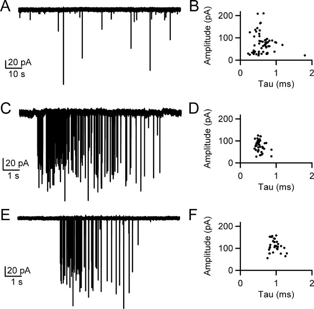
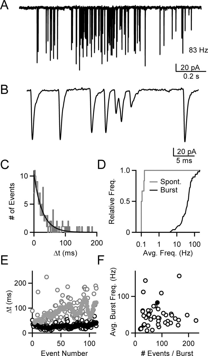
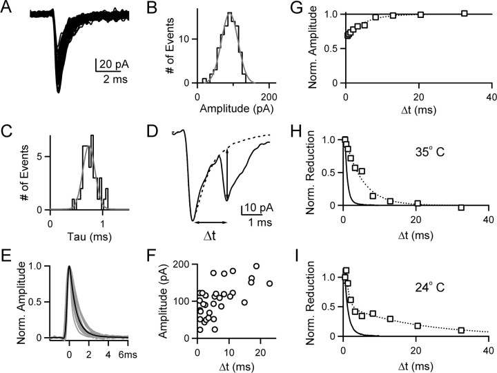
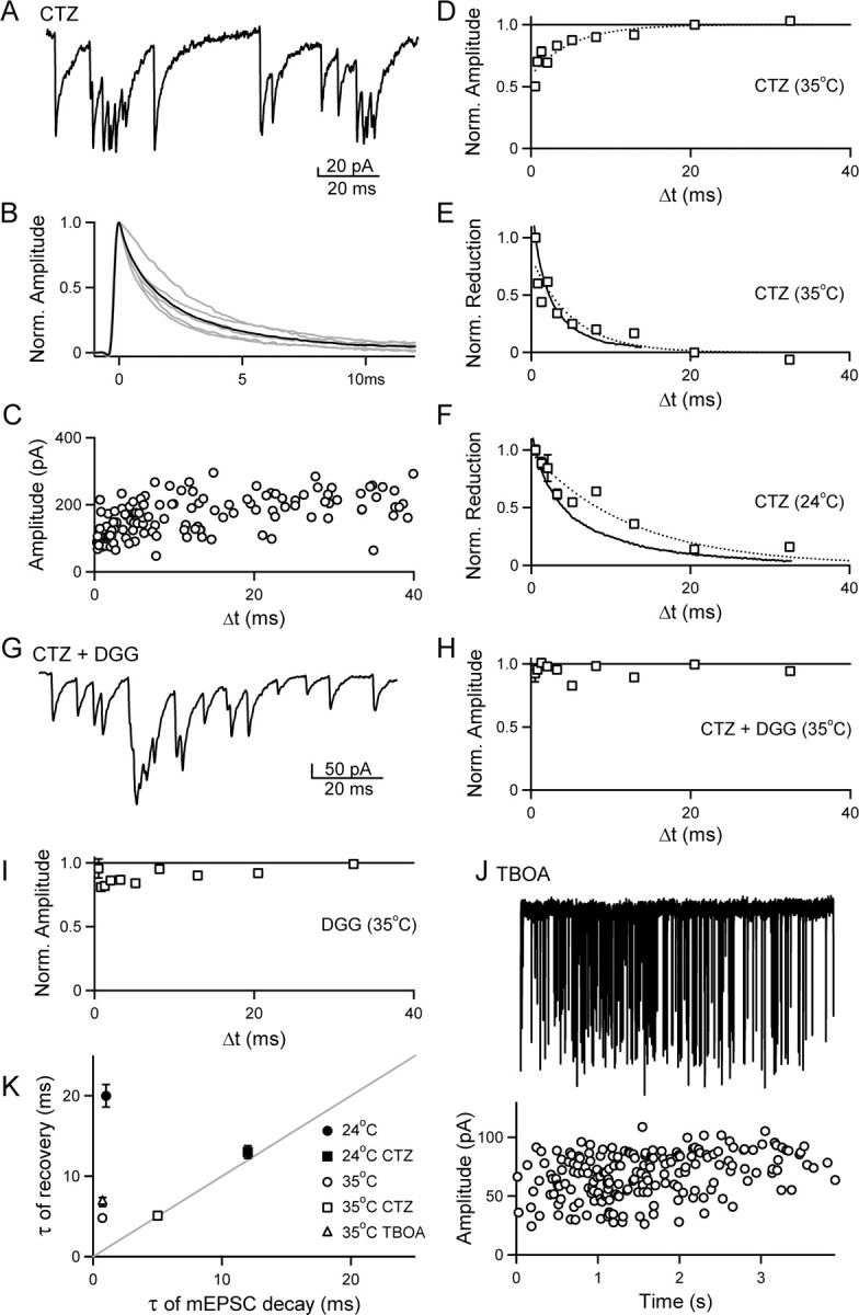
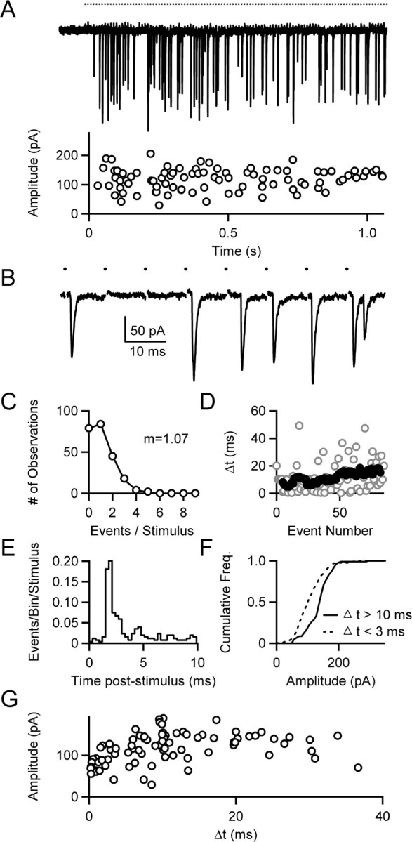
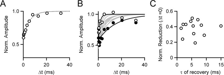
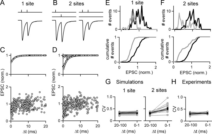
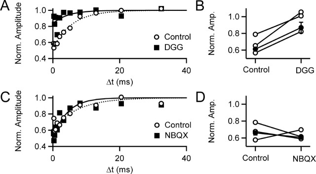
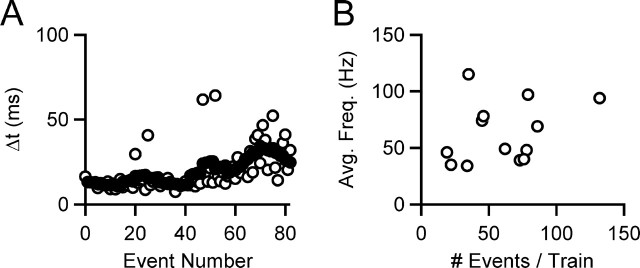
When the synaptic connection between two neurons consists of a small number of release sites, the ability to maintain transmission at high frequencies is limited by vesicle mobilization and by the response of postsynaptic receptors. These two properties were examined at single release sites between granule cells and stellate cells by triggering bursts of quantal events either with alpha-latrotoxin or with high-frequency trains of presynaptic activity. Bursts and evoked responses consisted of tens to hundreds of events with frequencies of up to hundreds per second. This indicates that single release sites can rapidly supply vesicles from a reserve pool to a release-ready pool. In addition, postsynaptic AMPA receptors recover from desensitization with a time constant of approximately 5 ms. Thus, even for synapses composed of a single release site, granule cells can effectively activate stellate cells during sustained high-frequency transmission because of rapid vesicle mobilization and fast recovery of AMPA receptors from desensitization.


Figures

Figure 1.
Figure 1.
α-LTX triggers high-frequency bursts of mEPSCs from single synaptic release sites. A, Representative recording of spontaneous mEPSCs recorded from a stellate cell (35°C). B, Plot of mEPSC amplitude versus decay time constant for events collected during a 10 min recording from the cell shown in A (58 events). C, Burst of mEPSCs triggered by α-LTX at 35°C (161 events; 7.6 s). D, Plot of mEPSC amplitude versus decay time constant for noninteracting events (Δt > 20 ms) from the burst (42 of 161 events), exhibiting lower variation than is evident for spontaneous mEPSCs. E, High-frequency burst of mEPSCs triggered by α-LTX at 24°C (75 events; 4.5 s). F, Plot of mEPSC amplitude versus decay time constant for noninteracting events (Δt > 20 ms) from the burst (32 of 75 events).
Figure 2.
Figure 2.
Single active zones can release large numbers of vesicles at high frequency. A, Representative recording of an α-LTX-triggered burst of mEPSCs recorded from a stellate cell (35°C, 4 Cae). B, Expanded view of the burst shown in A demonstrating that the instantaneous frequency can reach several hundred Hertz. C, Histogram of interevent intervals (Δt) within a representative burst, fit with a single exponential (τ = 27 ms). D, Cumulative histogram comparing the average frequency of spontaneous mEPSCs (gray line; n = 6 cells) and α-LTX bursts of mEPSCs (black line; n = 48 bursts). E, Plot of the average Δt (open circles) versus event number for bursts collected at 24°C (gray; n = 17 bursts) and at 35°C (black; n = 48 bursts). The running averages (10 point) for each are shown superimposed on the data (filled circles). F, Plot of the average burst frequency during a burst versus the number of events in a burst (35°C). Data from mEPSC bursts in several conditions are summarized: control (n = 17), 100 μm CTZ (n = 6), 100 μm CTZ plus 2 mm DGG (n = 10), 1 mm DGG (n = 6), and 100–300 μm TBOA (n = 8). The burst shown in A is denoted by the filled circle. There is no significant difference between groups in the event frequency of α-LTX-evoked bursts recorded under these conditions (ANOVA, p = 0.48).
Figure 3.
Figure 3.
Postsynaptic AMPAR responses are reduced as the interevent interval decreases, and the reduction outlasts the decay of the mEPSC. A–E, The analysis of α-LTX-induced bursts is illustrated for a representative burst at 35°C. Superimposed mEPSCs (90 of 137 events; A), and histograms of mEPSC amplitudes (B), and decay time constants (C) for events within the burst with an interevent interval (Δt) of at least 10 ms. D, Schematic illustrating the quantitation of mEPSC amplitude for short Δt intervals. When an mEPSC occurs on the decay of a previous event, the average mEPSC (calculated for each burst from the plot in A, dotted line) is superimposed, scaled, and subtracted from the trace. The arrows denote the approximate corrected amplitude and Δt. E, Average mEPSCs from 17 bursts for control conditions at 35°C are shown normalized and superimposed (n = 17; gray lines), along with the average for all bursts (black line). F, Plot of amplitude versus Δt for all events in a representative burst. G, Summary of amplitude versus Δt across all bursts recorded in control conditions at 35°C (n = 17 bursts; 1184 events). The amplitude for each mEPSC in the burst is normalized to the average mEPSC amplitude for events with a Δt ≥ 20 ms, and the events are binned logarithmically by Δt value. The average for each bin is plotted against Δt, using the Δt value at the center of each bin (open boxes). The data are fit (dotted line) to the following equation: normalized amplitude = Aexp(−Δt/τ), with fit parameters [A, τ (ms)] of [0.32, 4.8]. H, Plot comparing the time course for the reduction in mEPSC amplitude with Δt (open boxes), and corresponding exponential fit (dotted line) to the time course for the decay of the average mEPSC (solid line). The data from G are normalized to the first bin (Δt = 0.5 ms), inverted, and plotted against the decay of the average mEPSC (from E; normalized to 0.5 ms after the peak). The decay of the average mEPSC is well fit by the equation mEPSC decay = Aexp(−Δt/τ), with fit parameters [A, τ (ms)] of [1.1, 0.72]. I, Plot comparing the time constant for the reduction in mEPSC amplitude with Δt (open boxes) to the time course of the average mEPSC (solid line) at 24°C (n = 17 bursts; 1250 events). The decay of the average mEPSC is well approximated by a single exponential with fit parameters [1.0, 1.0]. The plot of mEPSC amplitude versus Δt is well approximated (dotted line) by the following equation: normalized amplitude = A1exp(−Δt1) + A2exp(−Δt2) with fit parameters [A1, A2, τ1 (ms), τ2 (ms)] of [0.30, 0.18, 0.80, 20].
Figure 4.
Figure 4.
Rapidly recovering AMPAR desensitization underlies the attenuation of mEPSC amplitude at small Δt values. A–F, α-LTX was used to induce bursts in the presence of 100 μm CTZ to prevent AMPA receptor desensitization. A, A portion of a representative burst in the presence of CTZ (35°C). B, Average mEPSCs from six bursts are superimposed (gray lines) and averaged (black line). C, Plot of amplitude versus Δt for all events in the burst shown in A. D, The amplitude versus Δt plot across all bursts (6 bursts; 600 events, open squares, binned as described in Fig. 3G) was well approximated (dotted line) by the following equation: normalized amplitude = Aexp(−Δt/τ) with fit parameters [A, τ (ms)] of [0.37, 5.1]. E, Plot comparing the time course for the reduction in mEPSC amplitude (open boxes, dotted line) with Δt to the time course for the decay of the average mEPSC (solid line), as described in Figure 3H. The average mEPSC is well approximated by the equation mEPSC decay = A1exp(−Δt1) + A2exp(−Δt2), with fit parameters [A1, A2, τ1 (ms), τ2 (ms)] of [0.54, 0.43, 1.1, 5.0]. F, Plot comparing the time course for the reduction in mEPSC amplitude with Δt (open boxes, dotted line) to the time course for the decay of the average mEPSC (solid line) at 24°C (5 bursts; 507 events). The average mEPSC is well approximated by the equation mEPSC decay = A1exp(−Δt1) + A2exp(−Δt2), with fit parameters [A1, A2, τ1 (ms), τ2 (ms)] of [0.50, 0.49, 2.3, 12]. The plot of mEPSC amplitude versus Δt is well approximated (dotted line) by the following equation: normalized amplitude = Aexp(−Δt/τ), with fit parameters [A, τ (ms)] of [0.42, 13]. G–I, The dependence of mEPSC amplitude on Δt is reduced by decreasing AMPAR occupancy. α-LTX-induced bursts were recorded at 35°C in the presence of CTZ and the low-affinity AMPA receptor antagonist DGG to reduce receptor occupancy. G, Time-expanded region of a burst in the presence of 100 μm CTZ and 2 mm DGG. H, Summary of amplitude versus Δt across all bursts recorded in 100 μm CTZ plus 2 mm DGG (10 bursts; 1074 events). I, Summary of amplitude versus Δt across all bursts recorded in 1 mm DGG (6 bursts; 680 events). J, Top, representative recording of a burst recorded in the presence of 300 μm TBOA. Below, the amplitude of each mEPSC is plotted against time on the aligned graph. K, Summary of the time constant of recovery of mEPSC amplitude with Δt is plotted against the mEPSC decay time constant for bursts under different experimental conditions. In 100 μm CTZ, the time constant for single-exponential fits of amplitude versus Δt is plotted against τ2 from the double-exponential fits of mEPSC decay. For the control data at 24°C, τ2 from the double-exponential fit of amplitude versus Δt is plotted against the single-exponential fit of the average mEPSC decay.
Figure 5.
Figure 5.
High-frequency trains of quantal events evoked by extracellular stimulation of single synaptic release sites at the granule cell to stellate cell synapse. A, Representative recording of AMPAR-mediated quantal events in response to a regular 100 Hz stimulus train (denoted by dots above the trace). An aligned plot of event amplitude versus time is shown below the trace. B, Time-expanded view of a section of the train shown in A. C, Histogram of the number of events per stimulus compiled for two regular trains from the representative example in A (open circles), along with a fit of the data using a Poisson function (solid line; m = 1.07). D, Plot of Δt versus event number for the train shown in A (open circles), along with the running average (10 point) (filled circles). E, Poststimulus time histogram for two regular 100 Hz trains from the site shown in A. F, Cumulative histogram exhibiting a shift toward smaller amplitudes for events that occur with a short Δt (<3 ms) relative to events with longer latencies (> 10 ms), compiled for three trains (regular and Poisson) of events from the cell shown in A. G, Plot of amplitude versus Δt for the representative train shown in A, exhibiting a reduction in amplitude at small values of Δt consistent with the behavior of a single synaptic release site.
Figure 6.
Figure 6.
Variation in the extent and recovery from desensitization at single release sites. A, Summary of amplitude versus Δt for trains of events evoked by extracellular stimulation. A plot of amplitude versus Δt is shown for the average of 13 sites (open circles). The amplitude versus Δt plot, normalized and binned as described in Figure 2G, was well approximated by the equation normalized amplitude = Aexp(−Δt/τ) with fit parameters [A = 0.38, τ = 5.4 ms] (dotted line). B, Plot of the individual fits (normalized amplitude = Aexp(−Δt/τ) across release sites (gray lines). The sites exhibiting the fastest (open circles, black line) and slowest (filled circles, black line) recovery time constants are highlighted. C, Plot of the extent of desensitization versus the time constant of recovery for 13 individual release sites.
Figure 7.
Figure 7.
A–F, Simulations of quantal responses during trains are shown for events arising from a single site (A, C, E) or from two sites (B, D, F). Example responses are shown with the timing of vesicle fusion shown above and the resulting currents shown below for a single site (A) and for two sites (B). B, If two sites are present no desensitization occurs if the quantal events reflect fusion at different sites (left), but significant desensitization occurs if two vesicles fuse in rapid succession at the same site (right). Simulations were conducted based on the measured probability of observing a quantal event at any time during the train, and on the observed dependence of event amplitude on the Δt value. In this example, there were a total of 300 stimuli at 100 Hz. (C, top) For a single site, the amplitude is given by EPSC = EPSC0(1 − 0.4 exp(−Δt/τ)), where Δt is the time between the given fusion and the preceding vesicle fusion. D, Top, For two sites, EPSC = EPSC0(1 − 0.8 exp(−Δt/τ)), where Δt is the time between the given fusion and the preceding vesicle fusion at that same site. C, D, Bottom, The variability of EPSC amplitude was included by determining EPSC amplitude stochastically using the observed coefficient of variation for widely spaced events (Δt >20 ms) that are unaffected by desensitization. The amplitude distributions for events arising from a single site (E, top) and two sites (F, top) are shown for Δt = 0–1 ms (gray) and Δt = 10–60 ms (black), along with the normalized cumulative histograms for a single site (E, bottom) and two sites (F, bottom). G, The predicted CV values are Δt = 20–100 ms and Δt = 0–1 ms for the single site and two site cases, respectively. H, The observed CV values conform to the prediction for a single site.
Figure 8.
Figure 8.
Reducing receptor occupancy with DGG, but not the high-affinity antagonist NBQX, minimizes the dependence of quantal event amplitude on Δt. A–D, Trains of extracellular stimuli were delivered to single release sites before and after washing in either the low-affinity antagonist DGG (1–2 mm) (A, B) or the high-affinity antagonist NBQX (125–200 nm) (C, D). In A and B, the normalized amplitude versus Δt is shown (as described in Fig. 3G) compiled from three sites before (open circles) and after (filled squares) wash in of the antagonist. In A, control data were well approximated by the equation normalized amplitude = Aexp(−Δt/τ), with [A = 0.46, τ = 5.7 ms] (dotted line), whereas in DGG (solid line), [A = 0.15 ± 0.05 τ = 4.4 ± 4.4 ms]. In C, fit parameters are [A = 0.38, τ = 7.4] (dotted line) for control, and [A = 0.47, τ = 4.2] (solid line) for NBQX. The normalized amplitude for Δt < 3 ms is shown for individual experiments (open circles) and for the average of the experiments (closed circles) for DGG (B) and NBQX experiments (D).
Figure 9.
Figure 9.
Single release sites rapidly mobilize vesicles for release in response to trains of extracellular stimuli. A, A plot of the Δt versus event number averaged across 13 individual release sites (open circles), along with the running average (10 point, filled circles). B, A plot of the average frequency of events in a train (defined as the number of events divided by the duration between first and last event) against the number of events in the mEPSC train demonstrates that parallel fiber boutons can release tens to hundreds of vesicles (average, 61 events), at frequencies of tens to hundreds of Hertz (average, 63 Hz).

Similar articles

Cited by

References

    1. Arai A, Lynch G. AMPA receptor desensitization modulates synaptic responses induced by repetitive afferent stimulation in hippocampal slices. Brain Res. 1998;799:235–242. - PubMed
    1. Atluri PP, Regehr WG. Delayed release of neurotransmitter from cerebellar granule cells. J Neurosci. 1998;18:8214–8227. - PMC - PubMed
    1. Auger C, Marty A. Heterogeneity of functional synaptic parameters among single release sites. Neuron. 1997;19:139–150. - PubMed
    1. Augustin I, Rosenmund C, Sudhof TC, Brose N. Munc13–1 is essential for fusion competence of glutamatergic synaptic vesicles. Nature. 1999;400:457–461. - PubMed
    1. Barbour B, Keller BU, Llano I, Marty A. Prolonged presence of glutamate during excitatory synaptic transmission to cerebellar Purkinje cells. Neuron. 1994;12:1331–1343. - PubMed

Publication types

LinkOut - more resources