Comparative Study
. 2007 May 16;27(20):5461-73.
doi: 10.1523/JNEUROSCI.4046-06.2007.

Zic1 and Zic3 regulate medial forebrain development through expansion of neuronal progenitors

Affiliations
Comparative Study

Zic1 and Zic3 regulate medial forebrain development through expansion of neuronal progenitors

Takashi Inoue et al. J Neurosci. .

Abstract

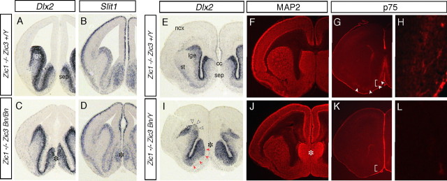
The medial telencephalon is a source of neurons that follow distinct tangential trajectories of migration to various structures such as the cerebral cortex, striatum, and olfactory bulb. In the present study, we characterized the forebrain anomalies in Zic1/Zic3 compound mutant mice. Zic1 and Zic3 were strongly expressed in the medial structures, including the septum, medial cerebral cortex, and choroid plexus. Mice homozygous for the Zic1 mutant allele together with the null Zic3 allele showed medial forebrain defects, which were not obvious in either Zic1 or Zic3 single mutants. Absence of both Zic1 and Zic3 caused hypoplasia of the hippocampus, septum, and olfactory bulb. Analysis of the cell cycle revealed that the cell cycle exit rate was increased in the septa of double mutants. Misexpression of Zic3 in the ventricular layer of the cerebral cortex inhibited neuronal differentiation. These results indicated that both Zic1 and Zic3 function in maintaining neural precursor cells in an undifferentiated state. The functions of these genes may be essential to increasing neural cell numbers regionally in the medial telencephalon and to proper mediolateral patterning of the telencephalon.


Figures

Figure 1.
Figure 1.
Locations of mouse Zic1, Zic2, and Zic3 mRNA, and Zic protein in the developing telencephalon. A–L, In situ hybridization for Zic1 (A–D), Zic2 (E–H), and Zic3 (I–L) mRNA was performed on coronal (A–C, E–G, I–K) and sagittal (D, H, L) sections at E12.5 (A, B, E, F, I, J) and E16.5 (C, D, G, H, K, L). A, E, I, B, F, J, Adjacent rostral (A, E, I) and caudal (B, F, J) parts of serial coronal sections. Zic1 and Zic2 mRNAs are broadly distributed in the septum, dorsomedial part of the telencephalon, and dorsal thalamus. Zic3 mRNA is in the VZ/SVZ of the ventral telencephalon (I, K, red arrowheads) and the medial marginal region of the cerebral cortex (cortical hem) with a clear boundary (J, arrow). Ventral expression of Zic1 and Zic3 extends laterally (A, C, I, K). M–P, Immunofluorescence staining of Zic proteins is shown in coronal (M–O) and sagittal (P) sections at E12.5 (M, N) and E16.5 (O, P). cc, Cingulate cortex-forming area; cpe, choroid plexus; dg, dentate gyrus of hippocampus; ep, entopeduncular area; fc, frontal cortex-forming area; fm, fimbria of hippocampus; he, cortical hem; lge, lateral ganglionic eminence; mge, medial ganglionic eminence; ncx, neocortex (cerebral cortex); ob, olfactory bulb; pm, primitive meninx; poa, preoptic area; th, thalamus; se, septum.
Figure 2.
Figure 2.
Expression patterns of Zic1 and Zic3 mRNA and pattern of production of Zic protein in the OB. A–G, Distributions of Zic1 (A), Zic3 (B), Dlx2 (C), GAD67 (D), and Reelin (E) mRNAs, and Zic protein (F, G). A–E are adjacent horizontal sections through the OB at P0. G, Higher magnification of the rectangle in F. Expression of Zic1 and Zic3 in the glomerular layer and granule cell layer is indicated by clear and solid arrows, respectively. Arrowheads, Rostral end of the presumptive rostral migratory stream; broken lines, remnant of olfactory vesicle. g, Glomeruli (some marked by broken circles); gcl, granule cell layer; gl, glomerular layer; mcl, mitral cell layer; vz, ventricular zone.
Figure 3.
Figure 3.
Forebrain defects in Zic1/3 embryos. A, Zic1/3 mutant (Zic1−/− Zic3 Bn/Y, right) is slightly smaller than Zic1−/− (indistinguishable from wild type, left) at E18.5, and shows dented hindbrain and kinky tail. B, C, Frontal views of the brains at E18.5. The forebrain of Zic1/3 (C) is smaller than that of Zic1−/− (B), and the left and right OBs of Zic1/3 are positioned with a wide interspace (arrowheads). D–O, Cresyl violet-stained coronal sections of E17.5 brains from wild type (Zic1+/+ Zic3 +/Y; D, H, L), Zic1 mutant (Zic1−/− Zic3 +/Y; E, I, M), Zic3 mutant (Zic1+/+ Zic3 Bn/Bn; F, J, N), and Zic1/3 mutant (Zic1−/− Zic3 Bn/Bn; G, K, O). D–G and H–K are comparable rostral and caudal sets of sections, respectively. L–O are higher-magnification views of the hippocampus in H–K, respectively. Comparison reveals hypoplasia or altered tissue architecture of the septum (G), corpus callosum (G), thalamus (K), cerebral cortex (G, K), hippocampus (K, O), and choroid plexus (G, K, O). III, Third ventricle; CA1, hippocampal field CA1; CA3, hippocampal field CA3; cc, corpus callosum; cpe, choroid plexus; dg, dentate gyrus; dt, dorsal thalamus; fm, fimbria; hip, hippocampus; ht, hypothalamus; lge, lateral ganglionic eminence; ncx, neocortex (cerebral cortex); s, subiculum; sep, septum; st, striatum.
Figure 4.
Figure 4.
Marker studies of dorsal forebrain in Zic1/3 mutant mice. A–O, Coronal sections of E14.5 brains from Zic1−/− (A, D, G, J, M), Zic1/3 (Zic1−/− Zic3 Bn/Bn; B, E, H, K, N), and Zic2 kd/kd (C, F, I, L, O) are shown, as well as in situ hybridization for Ngn2 (A–F), Tbr1 (G–I), Ttr (J–L), and Wnt3a (M–O). In Zic1/3 and Zic2 kd/kd, the defect is accompanied by altered expression patterns of Ngn2 and Tbr1 in the dorsal telencephalon. B and C are at the same rostrocaudal level as A. D–F are more caudal, comparable sections. Asterisks in E and F indicate hypoplastic thalamus. Arrowheads in H and I indicate thickening of the Tbr1-labeled layers, including the areas where Zic1, Zic2, and Zic3 are commonly expressed. Insets in K and L, and N and O are higher magnifications of choroid plexus, and cortical hem regions, respectively. cpe, Choroid plexus; he, cortical hem; lge, lateral ganglionic eminence; ncx, neocortex; th, thalamus.
Figure 5.
Figure 5.
Defective septal structures in the Zic1/3 mutant. A–L, Coronal sections of the Zic1 mutant (Zic1−/− Zic3 +/Y; A, B, E–H), and Zic1/3 double mutant (Zic1−/− Zic3 Bn/Y; C, D, I–L) at E14.5 (A–D), E15.5 (G, H, K, L), and E18.5 (E, F, I, J) are shown. In situ hybridization for Dlx2 (A, C, E, I) and Slit1 (B, D), and immunofluorescence staining for MAP2 (F, J) and p75 (G, H, K, L) were performed on brain coronal sections. Asterisks in C, D, I, and J indicate hypoplastic septum. Red arrowheads in I indicate unusual expansion of Dlx2 in the septal region of the Zic1/3 mutant. Open arrowheads in I indicate area of Dlx2 signal reduction of unknown cause (e.g., change secondary to ventricular collapse). White arrowheads in G indicate p75-positive neurons. H and L are higher-magnification views of areas indicated in G and K, respectively. p75-positive cholinergic neurons are reduced in number (or not detected) in the medial septum. cc, Corpus callosum, lge, lateral ganglionic eminence; ncx, neocortex; se, septum. st, striatum.
Figure 6.
Figure 6.
Proliferation and differentiation of neural progenitors in the Zic1/3 mutant. A–H, BrdU-labeling index analysis. Coronal sections through septum of Zic3 Bn/Y (A–D) and Zic1/3 (Zic1−/− Zic3 Bn/Bn) (E–H) E13.5 embryos after 1 h pulse labeling of BrdU are shown. Immunofluorescence staining for BrdU (green; A, B, E, F) and Ki67 (red; C, G) of the embryo is shown. B–D and F–H are higher magnifications of the areas indicated in A and E, respectively. D and H are merged views of B and C, and F and G, respectively. The results were quantified and used to calculate the BrdU-labeling index in Q. I–P, Cell cycle exit fraction analysis. Animals were exposed to a single-pulse label of BrdU at E11.5; 24 h later (at E12.5), the animals were killed and subjected to immunofluorescence staining for BrdU (green) and Ki67 (red). K and O are merged views of I and J, and M and N, respectively. The results were quantified and used to calculate cell fractions exiting the cell cycle (R). Neighboring sections were stained with class-III β-tubulin (L, P), which showed a thicker layer in Zic1/3 than in Zic1−/− (P, arrowheads). Q, Quantification of progenitor BrdU-labeling index. The percentage of BrdU-labeled cells in Ki67-positive proliferating cells was not altered in Zic1−/−, Zic3 Bn/Y, or Zic1/3. Results represent mean numbers of BrdU-labeled cells as percentages of total numbers of Ki67-labeled cells. Error bars indicate SEMs. R, Quantification of cell fractions exiting cell cycle. The ratio of the number of cells labeled only with BrdU (BrdU+/Ki67−, no longer dividing) to the number of double-labeled BrdU+/Ki67+ cells (yellow, re-entered cell cycle) was compared. Zic1/3 showed a significantly higher ratio than did the wild type, Zic1−/−, or Zic3 Bn/Y (or Bn/Bn). The results represent the mean ratios of the numbers of cells no longer dividing to the numbers of cells that had re-entered the cycle in the septum. *p < 0.05 by t test.
Figure 7.
Figure 7.
Misexpression of Zic3 inhibited neuronal differentiation in cerebral cortex. A–H, A Zic3 expression plasmid vector, pCAG-myc-tagged-Zic3-IRES EGFP (CAG-Zic3-IE) (E–H) or its control vector, pCAG-IRES-EGFP (CAG-EGFP) (A–D) was transfected by in utero electroporation into the dorsolateral cerebral cortex, where Zic3 is not expressed. Electroporation was performed at E14.5, and the fates of the transfected cells were examined at E17.5 by examining the EGFP signals. Transfected cells were detected with immunofluorescence staining for anti-GFP (A–H), anti-myc epitope tag (A, E), the neural progenitor marker Nestin (B, F), neuronal marker MAP2 (C, G), and proliferation marker Ki67 (D, H). Whereas many CAG-EGFP-transfected cells (EGFP+) migrated out of the VZ and were located in the intermediate zone (iz) and cortical plate (cp) (A), CAG-Zic3-transfected cells (EGFP+) remained in the VZ (E). CAG-Zic3-transfected cells were generally negative for MAP2 production (G), but produced Nestin (insets in F). Nestin staining outside the VZ was also observed in the blood vessels. I, Quantification of the percentage of MAP2-expressing neurons or Nestin-expressing precursor cells in the transfected (EGFP+) cells. The results represented in A–H were examined as in Materials and Methods. The results are shown as the mean percentage of four independent experiments. Error bars indicate SDs.
Figure 8.
Figure 8.
Defects in Zic1/3 OB. A, D, Dorsal views of the Zic1−/− (A), and Zic1/3 (D) head at E18.5. In Zic1/3, unusual structures (asterisks) are seen in the interspace between the OBs. B, E, Immunofluorescence staining for olfactory marker protein (OMP), which is located on the neuronal processes from the olfactory neurons, in Zic1−/− (B) and Zic1/3 (E). In Zic1−/−, the olfactory nerve layer surrounded the whole surface of the OB. In contrast, in the Zic1/3 mutant, no apparent olfactory nerve layer was present and most of the olfactory axons terminated in fibrocellular mass-like structures (asterisks in E). C, F, Immunofluorescence staining for BrdU at E15.5. The embryo was labeled with BrdU for 1 h before being killed. G–N, Neuronal subsets in Zic1/3 OB. Cresyl violet staining (G, K) and immunofluorescence staining for Tbx21 (H, L), TH (I, M), and GABA (J, N) in horizontal sections of E18.5 OBs of Zic1−/− (G–J) and Zic1/3 (K–N) are shown. O, Quantification of OB neurons examined in sections. The number of neurons positive for Tbx21, TH, and GABA in the OB were counted in each section. The mean numbers of immunoreactive cells in each OB section, including the maximum diameter of the OB, were compared. Cells were counted in wild type (n = 6), Zic1−/− (n = 5), Zic3 Bn/Y or Zic3 Bn/Bn (n = 5), and Zic1/3 (black bars; n = 4) OBs in comparable sections. Error bars indicate SDs. **p < 0.01 by t test. mcl, mitral cell layer; gl, glomerular layer; gcl, granule cell layer.
Figure 9.
Figure 9.
Focal transfection by in utero electroporation into the medial forebrain specifically targets OB interneurons. A, Electroporation of pCAG-EGFP into the lateral ganglionic eminence (left) and medial wall of the lateral ventricle (right) were performed at E13.5, and brains were fixed at P4. Dorsal views of the whole brains. Arrows, EGFP-labeled OB; arrowheads, the transfection sites. B, C, Higher magnification of the OBs in A. Control electroporation into the ganglionic eminences labeled the neuronal progenitors that migrated into the OB (B). Introduction of EGFP into the medial wall of the lateral ventricle also labeled the neuronal progenitors that migrated into the OB (C). D, E, Coronal sections of the forebrain from a brain electroporated with pCAG-EGFP into the medial wall of the lateral ventricle were stained with stained with DAPI (blue, D) or anti-GFP antibody (red, E). EGFP was introduced into the septum, and clusters of EGFP+ cells are seen in the ventral RMS (arrowheads, D, E). The transfected site was confirmed to be more caudally located. F, G, Coronal sections of the OB from the medially electroporated brain were stained with DAPI (blue) and anti-GFP antibody (green). A higher magnification of the region indicated in F is shown in G, where the EGFP+ cells have the typical shape of periglomerular interneurons (arborized within glomeruli; dashed circles) in the glomerular layer and granule interneurons in the granule cell layer. g, Glomeruli; gcl, granule cell layer; gl, glomerular layer; lv, lateral ventricle; mcl, mitral cell layer; ncx, neocortex; st, striatum.

Similar articles

Cited by

References

    1. Anderson SA, Eisenstat DD, Shi L, Rubenstein JL. Interneuron migration from basal forebrain to neocortex: dependence on Dlx genes. Science. 1997a;278:474–476. - PubMed
    1. Anderson SA, Qiu M, Bulfore A, Eisenstat DD, Meneses, Pederson S, Rubenstein JL. Mutations of the homeobox genes Dlx-1 and Dlx-2 disrupt the striatal subventricular zone and differentiation of late born striatal neurons. Neuron. 1997b;19:27–37. - PubMed
    1. Anderson SA, Marin O, Horn C, Jennings K, Rubenstein JL. Distinct cortical migrations from the medial and lateral ganglionic eminences. Development. 2001;128:353–363. - PubMed
    1. Aruga J. The role of Zic genes in neural development. Mol Cell Neurosci. 2004;26:205–221. - PubMed
    1. Aruga J, Minowa O, Yaginuma H, Kuno J, Nagai T, Noda T, Mikoshiba K. Mouse Zic1 is involved in cerebellar development. J Neurosci. 1998;18:284–293. - PMC - PubMed

Publication types

MeSH terms