Comparative Study
. 2007 May 16;27(20):5495-505.
doi: 10.1523/JNEUROSCI.1384-07.2007.

Prolonged glial expression of Sox4 in the CNS leads to architectural cerebellar defects and ataxia

Affiliations
Comparative Study

Prolonged glial expression of Sox4 in the CNS leads to architectural cerebellar defects and ataxia

Melanie Hoser et al. J Neurosci. .

Abstract

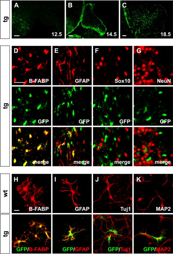
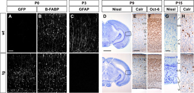
Sox proteins of group C are strongly expressed in the developing nervous system and have been associated with maturation of neurons and glia. Here, we overexpressed the group C protein Sox4 in transgenic mice under the control of the human GFAP promoter. Transgene expression was detected in radial glia and astrocytes throughout the CNS. The transgenic mice were ataxic and exhibited hydrocephaly as well as cerebellar malformations. In the cerebellum, fissures were not formed and neuronal layering was dramatically disturbed. Nevertheless, all neuronal cell types of the cerebellum were present as well as cells with characteristics of early radial glia, astrocytes, and oligodendrocytes. However, radial glia failed to migrate into the position normally taken by Bergmann glia and did not extend radial fibers toward the pial surface. The cerebellar malformations can therefore be explained by the absence of functional Bergmann glia. We conclude that Sox4 expression counteracts differentiation of radial glia and has to be downregulated before full maturation can occur.


Figures

Figure 1.
Figure 1.
Generation of GFAP–Sox4 transgenic mice. A, Schematic representation of the GFAP–Sox4 transgene consisting of human GFAP (hGFAP) promoter (positions −2163 to +47), rat Sox4 open reading frame, and IRES-EGFP cassette. Landmark restriction sites in the construct are indicated by letters N (NheI), E (EcoRI), and A (AscI). B, Southern blot analysis of genomic DNA from wild-type and transgenic mice derived from female founders (#1, #2, #3) after EcoRI digestion. The 3.4 kb band is indicative of the wild-type Sox4 gene; the 2.0 kb band represents the Sox4 transgene. Copy numbers were determined using a phosphorimager and are given below the lanes. C, Quantitative RT-PCR. Amounts of Sox4 transcripts were determined in total RNA prepared from forebrain and cerebellum of wild-type mice and progeny of transgenic founders at P15. Values were normalized to β-actin for each sample and expressed as multiples of the wild-type values. Error bars indicate SD. D, Gross morphology of the brain from wild-type (top) and GFAP–Sox4 transgenic (bottom) mice at P19. The right panels focus on the area of the cerebellum. Note the hydrocephalus and the strongly reduced foliation of the transgenic cerebellum. E, Nissl staining of sections from the mesencephalic region of wild-type and GFAP–Sox4 transgenic brains at P3. Inlays show a magnification of the boxed areas for better visualization of the mesencephalic aqueduct. F, Immunohistochemical staining of the choroid plexus of wild-type and GFAP–Sox4 transgenic mice at P15 using antibodies directed against Sox9. G, Confocal imaging of ependymal cilia in wild-type and GFAP–Sox4 transgenic mice at P9 after immunohistochemical staining with antibodies directed against acetylated α tubulin. The same magnifications were used for pictures of wild-type and transgenic cerebella. Scale bars: E, 1 mm (overview), 50 μm (inlays); F, 50 μm; G, 10 μm. wt, Wild type; tg, transgenic.
Figure 2.
Figure 2.
Embryonic expression pattern of the GFAP–Sox4 transgene. AH, EGFP autofluorescence was used to determine transgene expression in the spinal cord of GFAP–Sox4 transgenic embryos at 10.5 dpc (A), 11.5 dpc (B), 12.5 dpc (C, F, G, H), 14.5 dpc (D), and 18.5 dpc (E). The region boxed in C is magnified in F–H. F–H, Using immunohistochemistry with antibodies directed against SoxB1 (F), Sox11 (G), and NeuN (H) (all in red), EGFP autofluorescence (in green) was detected in ventricular zone cells at 12.5 dpc but not in neuronal progenitors and neurons. Top, Staining for the stage-specific marker. Middle, Corresponding EGFP staining. Bottom, Merged pictures. Scale bars, 100 μm. The scale bar in F also applies to G and H. The pattern is indicative of expression in radial glia and astrocytes.
Figure 3.
Figure 3.
Expression of the GFAP–Sox4 transgene in the cerebellum and in cerebellar cultures. A–C, EGFP autofluorescence was used to determine transgene expression in the developing cerebellum of GFAP–Sox4 transgenic embryos at 12.5 dpc (A), 14.5 dpc (B), and 18.5 dpc (C) as indicated. D–G, Antibodies directed against B-FABP (D), GFAP (E), Sox10 (F), and NeuN (G) as markers for specific cell types (all in red) were combined with antibodies directed against EGFP (in green) in coimmunohistochemistry on sections of transgenic cerebella at P9. Top, Staining for the cell-type specific marker. Middle, Corresponding EGFP staining. Bottom, Merged pictures. H–K, Antibodies directed against B-FABP (H), GFAP (I), Tuj1 (J), and MAP2 (K) as markers for specific cell types (all in red) were combined with antibodies directed against EGFP (in green) in coimmunocytochemistry on primary cell cultures prepared from wild-type (top) or GFAP–Sox4 transgenic (bottom) cerebella. Note that expression of the transgene overlaps both in vivo and in culture with B-FABP and GFAP staining, confirming expression in radial glia and astrocytes. Scale bars: A–C, 100 μm; (in D) D–G, 25 μm; (in H) H–K, 25 μm. wt, Wild type; tg, transgenic.
Figure 4.
Figure 4.
Cerebellar histology in GFAP–Sox4 transgenic mice. AF, Paraffin-embedded sections of wild-type (wt; A, C, E) and GFAP–Sox4 transgenic (tg; B, D, F) cerebella were stained with hematoxylin–eosin at P3 (A, B), P9 (C, D), and P15 (E, F). Roman numerals depict vermal lobules. Higher magnifications are derived from the area around lobule IV/V in wild-type and from the central lobe in transgenic cerebella (boxed in the low magnifications). Arrows depict the external granule cell layer, arrowheads depict the molecular layer and areas of Purkinje cell clusters, and double arrows depict the internal granule cell layer and granule cell clusters. Scale bars: low magnification, 200 μm (A), 500 μm (C), 1 mm (E); high magnification, 100 μm (A), 100 μm (C), 100 μm (E). The same magnifications were used for pictures of wild-type and transgenic cerebella. fc, Culminate fissure; fp, primary fissure; fs, secondary fissure; fl, flocculonodular fissure.
Figure 5.
Figure 5.
Apoptosis and proliferation in cerebella of GFAP–Sox4 transgenic mice. A, B, Immunohistochemistry with antibodies specific for the proliferation marker Ki67 was performed on wild-type and transgenic cerebella at P3. C, D, Adjacent sections were used for TUNEL assays. E, F, Quantification of all Ki67-expressing cells outside the external granule cell layer (E) and of TUNEL-positive cells (F) in the cerebellum. At least 12 separate 10 μm sections from two independent cerebella were counted for each genotype. Counted cells were normalized to cerebellar size and are presented per area unit as mean ± SD. Differences to the wild type were statistically significant for proliferation rates in the mutant genotype (p ≤ 0.001 in E) as determined by the Student's t test. Scale bar: (in A) A–D, 100 μm. wt, Wild type; tg, transgenic.
Figure 6.
Figure 6.
Analysis of neuronal subtypes in cerebella of GFAP–Sox4 transgenic mice. AJ, Immunohistochemistry was performed on paraffin sections of cerebella from wild-type (wt) and GFAP–Sox4 transgenic (tg) mice at P3 (A–E) and P15 (F–J) using antibodies directed against Ki67 (A, F), NeuN (B, G), calbindin D28K (C, H), RORα (D, I), and Pax2 (E, J). All pictures depict areas around the central lobe. Scale bar, (in A) 50 μm. Antibody staining is visible as a brown diaminobenzidine precipitate. Hematoxylin was used for counterstaining. Arrows point toward Golgi neurons. Arrowheads depict undifferentiated and differentiated basket/stellate cells within the white matter and molecular layer. All major neuronal subtypes are specified and mature in the transgenic cerebellum.
Figure 7.
Figure 7.
Analysis of glial cells in cerebella of GFAP–Sox4 transgenic mice. AI, Immunohistochemistry was performed on sections of cerebella from wild-type (wt) and GFAP–Sox4 transgenic (tg) mice at P3 (A–C), P9 (D–F), and P15 (G–I) using antibodies directed against EGFP (A, D, G), the radial and Bergmann glia marker B-FABP (B, E, H), and the Bergmann glia and astrocyte marker GFAP (C, F, I). Scale bar, (in A) 50 μm. Development of Bergmann glia was selectively disturbed.
Figure 8.
Figure 8.
Analysis of the cortex of GFAP–Sox4 transgenic mice. AH, Immunohistochemistry was performed on cortical sections (temporal lobe immediately dorsal to the prepiriform cortex) of wild-type (wt) and GFAP–Sox4 transgenic (tg) mice at the day of birth (A, B), at P3 (C), P9 (DF), and P15 (G, H) using antibodies directed against EGFP (A), B-FABP (B), GFAP (C), calretinin (Calr; E, H), and Oct-6 (F). D, G, Nissl stainings at P9 (D) and P15 (G). The area in which higher magnifications were taken is boxed in D. Scale bars: A–C, 50 μm; D, 1 mm; E, F, 100 μm; G, H, 25 μm. Development of radial glia and layering in the forebrain were both disrupted with ongoing development.

Similar articles

Cited by

References

    1. Baader SL, Schilling K. Glutamate receptors mediate dynamic regulation of nitric oxide synthase expression in cerebellar granule cells. J Neurosci. 1996;16:1440–1449. - PMC - PubMed
    1. Baader SL, Bergmann M, Mertz KD, Fox PA, Gerdes J, Oberdick J, Schilling K. The differentiation of cerebellar interneurons is independent of their mitotic history. Neuroscience. 1999;90:1243–1254. - PubMed
    1. Bayer SA, Altman J, Chaper I. San Diego: Academic; 2004. Development in the rat nervous system.
    1. Bergsland M, Werme M, Malewicz M, Perlmann T, Muhr J. The establishment of neuronal properties is controlled by Sox4 and Sox11. Genes Dev. 2006;20:3475–3486. - PMC - PubMed
    1. Bermingham JR, Scherer SS, O'Connell S, Arroyo E, Kalla KA, Powell FL, Rosenfeld MG. Tst-1/Oct-6/SCIP regulates a unique step in peripheral myelination and is required for normal respiration. Genes Dev. 1996;10:1751–1762. - PubMed

Publication types

MeSH terms

LinkOut - more resources