. 2007 Aug;18(8):2873-82.
doi: 10.1091/mbc.e07-02-0166. Epub 2007 May 16.

Mesenchymal stem cells use integrin beta1 not CXC chemokine receptor 4 for myocardial migration and engraftment

Affiliations

Mesenchymal stem cells use integrin beta1 not CXC chemokine receptor 4 for myocardial migration and engraftment

James E Ip et al. Mol Biol Cell. 2007 Aug.

Abstract

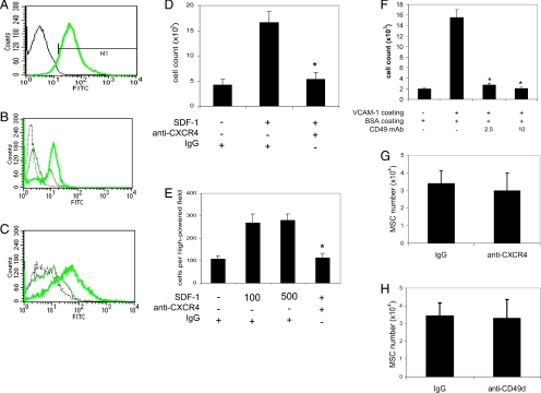
Recent evidence has demonstrated the importance of bone marrow-derived mesenchymal stem cells (BM-MSCs) in the repair of damaged myocardium. The molecular mechanisms of engraftment and migration of BM-MSCs in the ischemic myocardium are unknown. In this study, we developed a functional genomics approach toward the identification of mediators of engraftment and migration of BM-MSCs within the ischemic myocardium. Our strategy involves microarray profiling (>22,000 probes) of ischemic hearts, complemented by reverse transcription-polymerase chain reaction and fluorescence-activated cell sorting of corresponding adhesion molecule and cytokine receptors in BM-MSCs to focus on the coexpressed pairs only. Our data revealed nine complementary adhesion molecules and cytokine receptors, including integrin beta1, integrin alpha4, and CXC chemokine receptor 4 (CXCR4). To examine their functional contributions, we first blocked selectively these receptors by preincubation of BM-MSCs with specific neutralizing antibodies, and then we administered these cells intramyocardially. A significant reduction in the total number of BM-MSC in the infarcted myocardium was observed after integrin beta1 blockade but not integrin alpha4 or CXCR4 blockade. The latter observation is distinctively different from that reported for hematopoietic stem cells (HSCs). Thus, our data show that BM-MSCs use a different pathway from HSCs for intramyocardial trafficking and engraftment.


Figures

Figure 1.
Figure 1.
(A) Strategy using genomics to identify potential receptor/ligand pairs involved in stem cell homing and trafficking. (B) Real-time PCR showing increased expression of numerous cytokines and adhesion molecules in MI versus sham hearts after 24 h (p < 0.05 except VEGF-α). Sele, endothelial selectin; TNFRII, tumor necrosis factor receptor II; CC, chemokine (C-C motif); CXC, chemokine (C-X-C motif); FN, fibronectin; Lam, laminin.
Figure 2.
Figure 2.
(A) RT-PCR showing expression of receptors/ligands in BM-MSCs, PBMCs, JGCs, and VSMCs. Items displayed in red were shown to be up-regulated in ischemic myocardium compared with sham at 24 h by RT-PCR. Items displayed in blue were shown to be counterreceptors/ligands expressed by MSCs. IL6RA, IL-6 receptor, α; IL6ST, IL-6 signal transducer; CCR, CC receptor; Selel, E-selectin-1 ligand; VN, vitronectin; Tnc, tenascin-C; Itg, integrin. The other abbreviations were donated in the legend of Figure 1. (B) RT-PCR (30 cycles) showing expression of integrin β1 isoforms (A, B, C, and D) in BM-MSCs and dermal keratinocytes.
Figure 3.
Figure 3.
Protein expression of receptor/ligand pairs. (A) Flow cytometric analysis of BM-MSC surface receptors. Aliquots of cultured BM-MSCs were incubated with FITC- or PE-conjugated monoclonal antibodies against CD45, CD14, CD29, CD49d, CD105, CXCR4, IL-6 receptor α chain, or Sca-1. Cells stained with isotype control IgG conjugated to FITC served as a negative controls (gray peak). Representative results from one of three individual experiments were shown. Values represent percentages of positive cells. (B–D) Immunohistochemical staining for ICAM-1 (B), VCAM-1 (C), and tenascin-C (D). Murine heart sections, 48 h (B and C) and 72 h (D) after MI, were stained with anti-ICAM-1, VCAM-1, or tenascin-C (green) mAb. Myocytes were stained red except in D, and nuclei were stained with blue.
Figure 4.
Figure 4.
Effect of CD29 blockade on BM-MSC adhesion, migration, and engraftment. (A and B) Blocking mAb against CD29 (B) reduced BM-MSCs attachment and spreading onto the fibronectin-coated plates compared with control IgM (A). (C) Real-time PCR assessment of BM-MSC migration and engraftment into the infarcted myocardium. BM-MSCs derived from male mice were incubated with anti-CD29 mAb or control IgM, and then they were injected into the myocardium of female mice after MI above the ligation. Seventy-two hours later, the BM-MSCs in the apical region of the heart below the ligation was assessed by real-time PCR assay of the Y-chromosome–specific DNA sequence. BM-MSCs incubated with antibody against CD29 had reduced accumulation in the apical region compared with the cells treated with control IgM (n = 5; **p = 0.012).
Figure 5.
Figure 5.
CD29 blockade reduced the accumulation of BM-MSCs in the infarcted myocardium. BM-MSCs incubated with control IgM (A, C, and E) or anti-CD29 mAb (B, D, and F) were injected into the myocardium at one site above the ligation. Seventy-two hours later, sections of the heart below the ligation were immunostained for GPF-positive BM-MSCs (green). BM-MSCs treated with anti-CD29 blocking mAb (B and D) had reduced accumulation in the heart than BM-MSCs incubated with control IgM (A and C). BM-MSCs incubated with control IgM (E) were found to have migrated from the injection site, and they homed to the entire left ventricular wall infarct, whereas reduced BM-MSC migration and accumulation were seen in the BM-MSCs incubated with anti-CD29 (F). Myocytes (red) were detected by anti-sarcomeric α-actin, and nuclei (blue) were stained with Hoechst. (G) The area of GFP-positive BM-MSCs in each section was quantified. Treatment of BM-MSCs with CD29 blocking mAb reduced BM-MSC volume in the apical region of the hearts compared with incubation of the cells with control IgM (n = 6; **p = 0.004).
Figure 6.
Figure 6.
Effect of CXCR4 or CD49d blockade on BM-MSC intramyocardial homing and engraftment to the infarcted myocardium. (A) FACS analysis indicated that >90% of EL4 cells expressed CXCR4. (B) EL4 cells were preincubated with anti-CXCR4 (peak in middle) or control IgG (peak on right) at a concentration of 10 μg/ml, and then they were incubated with FITC-labeled SDF-1. EL4 cells with SDF-1 binding were determined by FACS. Cell incubated with FITC-labeled nonimmune IgG were used as a negative control (gray peak). (C) Passage 0 adherent cells from culture of mouse bone marrow-nucleated cells were first incubated with anti-CD49d (peak in middle) or control IgG (peak on right) at a concentration of 10 μg/ml, and then they were incubated with FITC-labeled VCAM-1. Cells with VCAM-1 binding were determined by FACS. Cell incubated with FITC-labeled nonimmune IgG were used as a negative control (gray peak). (D and E) Anti-CXCR4 (10 μg/ml) reduced SDF-1–mediated migration of EL4 cell (D) and passage 0 adherent mouse bone marrow-nucleated cells (E). Each experiment was performed twice in six replicate wells, p < 0.00001 in D and E. (F) Anti-CD49d (2.5 and 10 μg/ml) inhibited attachment of passage 0 adherent mouse bone marrow-nucleated cells. The experiment was performed twice in quadruplet wells for each variable (p < 0.0001 for both antibody doses). (G and H) A similar procedure as described in Figure 5 was used for BM-MSC injection and assessment by real-time PCR. Treatment of BM-MSCs with anti-CXCR4 (G; n = 6, p = 0.83) or anti-CD49d (H; n = 5, p = 0.31) had no significant effect on the amount of BM-MSCs accumulated in the infarcted myocardium compared with treatment with control IgG.

References

    1. Abbott J. D., Huang Y., Liu D., Hickey R., Krause D. S., Giordano F. J. Stromal cell-derived factor-1alpha plays a critical role in stem cell recruitment to the heart after myocardial infarction but is not sufficient to induce homing in the absence of injury. Circulation. 2004;110:3300–3305. - PubMed
    1. Amado L. C., et al. Cardiac repair with intramyocardial injection of allogeneic mesenchymal stem cells after myocardial infarction. Proc. Natl. Acad. Sci. USA. 2005;102:11474–11479. - PMC - PubMed
    1. Anversa P., Nadal-Ginard B. Myocyte renewal and ventricular remodelling. Nature. 2002;415:240–243. - PubMed
    1. Askari A. T., et al. Effect of stromal-cell-derived factor 1 on stem-cell homing and tissue regeneration in ischaemic cardiomyopathy. Lancet. 2003;362:697–703. - PubMed
    1. Assmus B., et al. Transplantation of progenitor cells and regeneration enhancement in acute myocardial infarction (TOPCARE-AMI) Circulation. 2002;106:3009–3017. - PubMed

Publication types

MeSH terms

LinkOut - more resources