. 2007 Jul;18(7):2728-34.
doi: 10.1091/mbc.e07-02-0117. Epub 2007 May 16.

The signal recognition particle (SRP) RNA links conformational changes in the SRP to protein targeting

Affiliations

The signal recognition particle (SRP) RNA links conformational changes in the SRP to protein targeting

Niels Bradshaw et al. Mol Biol Cell. 2007 Jul.

Abstract

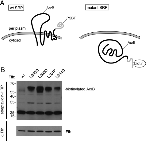
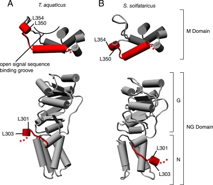
The RNA component of the signal recognition particle (SRP) is universally required for cotranslational protein targeting. Biochemical studies have shown that SRP RNA participates in the central step of protein targeting by catalyzing the interaction of the SRP with the SRP receptor (SR). SRP RNA also accelerates GTP hydrolysis in the SRP.SR complex once formed. Using a reverse-genetic and biochemical analysis, we identified mutations in the E. coli SRP protein, Ffh, that abrogate the activity of the SRP RNA and cause corresponding targeting defects in vivo. The mutations in Ffh that disrupt SRP RNA activity map to regions that undergo dramatic conformational changes during the targeting reaction, suggesting that the activity of the SRP RNA is linked to the major conformational changes in the signal sequence-binding subunit of the SRP. In this way, the SRP RNA may coordinate the interaction of the SRP and the SR with ribosome recruitment and transfer to the translocon, explaining why the SRP RNA is an indispensable component of the protein targeting machinery.


Figures

Figure 1.
Figure 1.
Removal of the M domain does not alter the interaction kinetics of Ffh and FtsY in the absence of the 4.5S RNA. (A) The NG domain was severed from Ffh by limited proteolysis with V8 protease and purified as described in Materials and Methods. A Coomassie blue–stained SDS polyacrylamide gel displaying selected fractions from the purification procedure is shown. The lane labeled SP FT contains the flow through fraction from an SP Sepharose column, which binds to the liberated M domain, and the lane labeled NG contains purified Ffh-NG after gel filtration. This fraction was used in the subsequent assays. (B) Binding of the purified Ffh-NG fragment to FtsY produces a change of the tryptophan fluorescence of FtsY. •, the intensity of fluorescence from the complex of Ffh-NG and FtsY when excited with 290-nm light; ○, the fluorescence spectrum of unbound Ffh-NG and FtsY. Complex formation was initiated by addition of Mg2+ in excess over EDTA as previously described (Shan and Walter, 2003). (C) Representative data monitoring the increase in tryptophan fluorescence intensity as a function of time. The time course shown was obtained at 0.85 μM Ffh-NG and 0.1 μM FtsY and no 4.5S RNA. Data points represent intensity measurements taken when the sample was excited at 290 nm and emission was measured at 340 nm. (D) Ffh-NG binds to FtsY with rates indistinguishable from full-length Ffh in the absence of the 4.5S RNA. Observed rate constants from data such as in C are plotted as a function of concentration of Ffh (•: full-length Ffh + 4.5S RNA; ○: full-length Ffh − 4.5S RNA; ▴: Ffh NG − 4.5S RNA). The inset graph is magnified to show the slow reactions. (E) Stimulated GTP hydrolysis reactions for Ffh·FtsY and Ffh-NG·FtsY. Hydrolysis rates (per complex, per second) are plotted as a function of concentration of FtsY (•: full-length Ffh + 4.5S RNA, ○: full-length Ffh − 4.5S RNA, ▴: Ffh NG − 4.5S RNA, ▵: Ffh NG + 4.5S RNA).
Figure 2.
Figure 2.
Binding to 4.5S RNA and basal GTPase activity is not affected by mutations L301P, L303D, L350D, and L354D. (A) Gel shift analysis using 0.25 μM Ffh and 0.25 μM 32P-labeled 4.5S RNA shows that all mutant variants quantitatively shift the RNA. (B) Single-turnover GTP hydrolysis assays of wild-type (•), Ffh(L301P) (○), Ffh(L303D) (□), Ffh(L350D) (◇), and Ffh(L354D) (▿). Reactions were performed with trace [32P]GTP and varying amounts of Ffh. Curves (solid line Ffh(wt), dashed lines for Ffh mutants) were fit to the equation kobs = kcat ∗ [Ffh]/(KM + [Ffh]). Values of kcat are as follows: Ffh(wt), 0.070 min−1; Ffh(L301P), 0.077 min−1; Ffh(L303D), 0.092 min−1; Ffh(L350D), 0.10 min−1; and Ffh(L354D), 0.10 min−1.
Figure 3.
Figure 3.
Ffh mutations of L303D, L350D, and L354D abrogate the activity of the 4.5S RNA to catalyze association of Ffh and FtsY. (A) Observed rate constants for binding of wild-type Ffh (•, ○) or Ffh(L303D) (▴, ▵) in the presence (filled symbols) or absence (open symbols) of 4.5S RNA are plotted as a function of Ffh concentration. Lines represent fits to the equation kobs = kon ∗ [Ffh] + koff [solid lines for wt Ffh, dashed lines for Ffh(L303D)]. (B) The mutations selectively affect the binding rate in the presence but not absence of the 4.5S RNA. The binding rates relative to wt Ffh are plotted (note log-scaled Y axis). (C) Dissociation of wild-type (•) or Ffh(L303D) (▴) from FtsY was measured in the presence of the 4.5S RNA by adding GDP to trap dissociated complexes and monitored by changes in tryptophan fluorescence. Samples were excited at 290 nm, and fluorescence emission at 340 nm was recorded. The x-axis in the inset is expanded to show the curve for wild-type Ffh. Data were fit to a single exponential equation to calculate the koff. (D) Plot of the koff or KD of Ffh mutants relative to wild-type Ffh in the presence of the 4.5S RNA. kon, koff, and KD values are summarized in Table 2.
Figure 4.
Figure 4.
Mutations L301P and L303D abrogate the activity of the 4.5S RNA to enhance the stimulated GTPase activity of the SRP and FtsY. Multiple turnover GTP hydrolysis reactions were carried out in which wild-type Ffh (•) or Ffh mutants (▴) were mixed with varying concentrations of FtsY in the presence (filled symbols) or absence (open symbols) of the 4.5S RNA. The Ffh mutants are shown in (A) Ffh(L301P), (B) Ffh(L303D), and (C) Ffh(L350D). Solid lines are curve fits to reactions containing wild-type Ffh and dashed lines to reactions containing Ffh mutants. (D) Plot of kmax in the presence and absence of 4.5S RNA. The values for kmax and K1/2 are summarized in Table 1. Error bars represent the accuracy of the fits to the data.
Figure 5.
Figure 5.
Ffh mutations L301P, L303D, L350D, and L354D show membrane protein integration defects in vivo. (A) The multispanning transmembrane protein AcrB fused to the PSBT biotinylation domain is efficiently targeted and integrated in cells expressing wild-type SRP, locating the PSBT biotinylation domain to the periplasmic space, where it is not biotinylated. If SRP-dependent protein targeting is impaired, AcrB accumulates in the cytoplasm, where it is biotinylated. (B) E. coli cells containing a genomic deletion of Ffh and a genomically inserted copy of wt Ffh that is exclusively expressed in the presence of arabinose and bearing two plasmids: one containing wild-type or mutant Ffh, and the other containing AcrB fused to the PSBT biotinylation domain were grown in the absence of arabinose. Cell extracts were fractionated by SDS polyacrylamide electrophoresis, and gels were blotted using a streptavidin-HRP conjugate (top panel) or anti-Ffh antibodies followed by HRP-conjugated secondary antibodies (bottom panel). HRP was visualized by chemiluminescence.
Figure 6.
Figure 6.
Mutations in Ffh that lead to defects in 4.5S RNA activity map to conformationally dynamic regions. Comparison of x-ray crystal structures from T. aquaticus (A) and S. solfataricus (B) reveals that the linker between the NG and M domains (bottom) and the finger loop of the M domain (top) are mobile. The corresponding amino acid positions of the mutations in E. coli Ffh described in this article are indicated: L301 (298 in T. aquaticus and 298 in S. solfataricus) and L303 (300,300) in the linker and L350(341,348) and L354(345,352) lead to defects in 4.5S RNA activity. Molecular graphics images were produced using the UCSF Chimera package from the Resource for Biocomputing, Visualization, and Informatics at the University of California, San Francisco (supported by NIH P41 RR-01081) (Pettersen et al., 2004).

Similar articles

Cited by

References

    1. Batey R. T., Rambo R. P., Lucast L., Rha B., Doudna J. A. Crystal structure of the ribonucleoprotein core of the signal recognition particle. Science. 2000;287:1232–1239. - PubMed
    1. Bernstein H. D., Hyndman J. B. Physiological basis for conservation of the signal recognition particle targeting pathway in Escherichia coli. J. Bacteriol. 2001;183:2187–2197. - PMC - PubMed
    1. Buskiewicz I., Kubarenko A., Peske F., Rodnina M. V., Wintermeyer W. Domain rearrangement of SRP protein Ffh upon binding 4.5S RNA and the SRP receptor FtsY. RNA. 2005a;11:947–957. - PMC - PubMed
    1. Buskiewicz I., Peske F., Wieden H. J., Gryczynski I., Rodnina M. V., Wintermeyer W. Conformations of the signal recognition particle protein Ffh from Escherichia coli as determined by FRET. J. Mol. Biol. 2005b;351:417–430. - PubMed
    1. Connolly T., Rapiejko P. J., Gilmore R. Requirement of GTP hydrolysis for dissociation of the signal recognition particle from its receptor. Science. 1991;252:1171–1173. - PubMed

Publication types

MeSH terms