. 2007 Aug;18(8):2805-16.
doi: 10.1091/mbc.e06-09-0882. Epub 2007 May 16.

Spc24 and Stu2 promote spindle integrity when DNA replication is stalled

Affiliations

Spc24 and Stu2 promote spindle integrity when DNA replication is stalled

Lina Ma et al. Mol Biol Cell. 2007 Aug.

Abstract

The kinetochore, a protein complex that links chromosomes to microtubules (MTs), is required to prevent spindle expansion during S phase in budding yeast, but the mechanism of how the kinetochore maintains integrity of the bipolar spindle before mitosis is not well understood. Here, we demonstrate that a mutation of Spc24, a component of the conserved Ndc80 kinetochore complex, causes lethality when cells are exposed to the DNA replication inhibitor hydroxyurea (HU) due to premature spindle expansion and segregation of incompletely replicated DNA. Overexpression of Stu1, a CLASP-related MT-associated protein or a truncated form of the XMAP215 orthologue Stu2 rescues spc24-9 HU lethality and prevents spindle expansion. Truncated Stu2 likely acts in a dominant-negative manner, because overexpression of full-length STU2 does not rescue spc24-9 HU lethality, and spindle expansion in spc24-9 HU-treated cells requires active Stu2. Stu1 and Stu2 localize to the kinetochore early in the cell cycle and Stu2 kinetochore localization depends on Spc24. We propose that mislocalization of Stu2 results in premature spindle expansion in S phase stalled spc24-9 mutants. Identifying factors that restrain spindle expansion upon inhibition of DNA replication is likely applicable to the mechanism by which spindle elongation is regulated during a normal cell cycle.


Figures
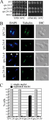
Figure 1.
Figure 1.
spc24-9 mutants are sensitive to HU due to inappropriate spindle expansion. (A) Cell dilution assay of indicated strains grown on YPD and 0.05 M HU at 30°C for 3 d. (B) Immunofluorescence analysis of wild-type (SPC24), spc24-8, spc24-9, and spc24-10 cells synchronized in G1 phase with α-factor and then released into 0.2 M HU for 3 h at 30°C. Shown are representative cells after 3-h HU treatment imaged for DNA (4,6-diamidino-2-phenylindole [DAPI]), MTs (tubulin) and cell morphology (differential interference contrast [DIC]). White arrowheads point to separated nuclei in the spc24-9 mutant. (C) Percentage of cells (100 cells counted) described in B displaying single (gray bars) or separated (black bars) nuclei.
Figure 2.
Figure 2.
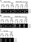
Ndc80 CEN association is disrupted in spc24-9 mutants. Multiplex PCR analysis of CEN1, PGK1, and CEN3 loci was performed with total chromatin (T) or immunoprecipitate (IP) as PCR templates. Strains were grown at 25°C to log phase and then either shifted to 37°C for 3 h or incubated in 0.2 M HU at 30°C for 3 h. (A) Ndc10-Myc wild type (lanes 1–6) or spc24-9 cells (lanes 7–12). (B) Ndc80-Myc wild-type (lanes 1–6) or spc24-9 cells (lanes 7–12). (C) Wild-type strain carrying no epitope tag (No Tag, lanes 1–6) shown as a control. An untagged spc24-9 mutant was also used as a control and showed similar results (data not shown).
Figure 3.
Figure 3.
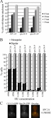
spc24-9 mutants are capable of establishing bipolar attachment. (A) Wild-type (SPC24), spc24-9, and spc24-10 strains carrying LacO repeats integrated 1.8 kb from CEN15, LacI-GFP, and Spc29-CFP were synchronized in G1 phase with α-factor and released at 30°C. Samples were taken every 15 min and imaged using fluorescence microscopy for the presence of the CEN15-GFP and Spc29-CFP signal. Shown are the time points (105 min onward) at which the cells began to display bipolar attachment (separation of CEN15-GFP signals). Duplicate experiments were performed with similar results. Shown is the result of one experiment in which 100 cells containing a spindle of 0.5 μm or larger were counted for each time point. (B) Wild-type (SPC24) and spc24-9 CEN15-GFP Spc29-CFP strains were synchronized in G1 phase with α-factor, split into four cultures, and indicated concentrations of HU were added for 3 h at 30°C. Similar results were seen with duplicate experiments; thus, data from one experiment are shown (100 cells counted). (C) Example of a wild-type (SPC24) cell from B after 3 h of 0.3 M HU treatment that displays monopolar CEN15 attachment. In the overlay, CEN15-GFP is green and Spc29-CFP is red.
Figure 4.
Figure 4.
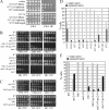
Spindle expansion in spc24-9 mutants depends on active Stu2. (A) Cell dilution assay of spc24-9 mutants carrying the 2μ plasmid vector (pRS202), STU1ΔN-library (HCS clone identified in screen), STU1ΔN-subclone (subclone of STU1ΔN-library containing only the STU1 gene), STU1 full-length (gift from T. Huffaker), full-length SPC24, STU2ΔN-library (HCS clone identified in screen), STU2ΔN-subclone (subclone of STU2ΔN-library containing only the STU2 gene), and STU2 full-length (gift from T. Huffaker) were grown on −URA plates at 30°C for 4 d or −URA 0.05 M HU at 30°C for 5 d. (B and C) Cell dilution assay of indicated strains grown on YPD at 25°C (2 d), 30 and 33°C (3 d), and 0.05 M HU at 25, 30, and 33°C (3 d). (D) Immunofluorescence analysis of spc24-9 mutants carrying the indicated HCS plasmids synchronized in G1 phase with α-factor and then released into 0.2 M HU for 3 h at 30°C. Cells were counted (100 per sample) for single nuclei (gray bars) or separated nuclei (black bars) by DAPI staining. Duplicate experiments were performed with similar data, and shown is the result of one experiment. (E) Immunofluorescence analysis of indicated strains treated and analyzed as described in D.
Figure 5.
Figure 5.
Stu1 localizes to kinetochores and the spindle midzone. Wild-type Stu1-VFP Spc29-CFP cells were synchronized in G1 phase with α-factor and released into the cell cycle at 30°C. Cells were fixed in 70% ethanol every 15 min for 90 min, and they were imaged as described in the Materials and Methods. In the overlay, green is VFP signal and red is CFP. Bar, 2 μm for all images.
Figure 6.
Figure 6.
Stu2 CEN binding is abolished in spc24-9 mutants, whereas Stu1 is still able to associate with CEN DNA. Multiplex PCR analysis of CEN1, PGK1, and CEN3 loci was performed with total chromatin (T) or immunoprecipitate (IP) as PCR templates. Strains were grown at 25°C to log phase and then either kept at 25°C or incubated at 30 or 37°C for 3 h. (A) Wild-type log phase cells grown at 30°C and carrying no epitope tag (No Tag; lanes 1 and 2), Stu1-Myc (lanes 3 and 4), and Stu2-Myc (lanes 5 and 6). (B) spc24-9 mutants carrying Stu1-Myc (lanes 1–4) and a wild-type strain with no tag at 37°C (lanes 5 and 6). (C) Stu2-Myc in a wild-type (lanes 1 and 2), spc24-9 (lanes 3 and 4), and an untagged wild-type strain (No Tag; lanes 5 and 6). Strains were grown to log phase at 25°C, HU was added to a final concentration of 0.2 M HU, and cells were shifted to 30°C for 3 h. (D) Stu2-Myc in a wild-type (lanes 1–6) and spc24-9 (lanes 7–12) strains. No Tag strain (lanes 13–18) is an untagged wild-type strain. For all ChIP assays where the spc24-9 mutant was used, we included both a wild-type and spc24-9 untagged control, and we saw similar results; thus, only the wild-type untagged control is presented.
Figure 7.
Figure 7.
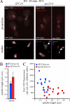
Increased spindle length correlates with Stu2 mislocalization and reduction. Wild-type (SPC24) and spc24-9 cells expressing Stu2-VFP Spc29-CFP were synchronized in G1 phase with pheromone, released into 0.2 M HU at 25°C for 1.5 h, and shifted to 30°C in HU for 60 min, and then fixed. (A) Representative images (extended focus and three-dimensional render) of Stu2-VFP (gray scale) and Spc29-CFP (red) fluorescence in wild-type and spc24-9 cells at 60 min after shift to 30°C are shown; mp indicates monopolar localization (near the SPB), whereas cs indicates localization to the central spindle. (B) Average (ave) spindle length of wild-type (SPC24) and spc24-9 cells after a 60-min incubation in 0.2 M HU at 30°C (n = 15). (C) Quantitative analysis of Stu2-VFP fluorescence per micrometer plotted as a function of spindle length.
Figure 8.
Figure 8.
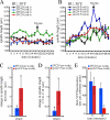
Decreased Stu2 in the spc24-9 mutant results in oscillation of spindle length. Wild-type (SPC24) and spc24-9 cells carrying Stu2-VFP Spc29-CFP were synchronized in G1 phase with pheromone, released into 0.2 M HU at 25°C for 1.5 h, mounted in FP supplemented with HU, shifted to 30°C on a heated stage, and time-lapse microscopy was performed. Time zero is time in HU at 30°C after equilibration on the stage at 30°C for 15 min. Spindle length is plotted for four cells of each strain (A, SPC24; B, spc24-9) as a function of time, each depicted with a different color, and shows oscillation with a net increase in length observed in all four spc24-9 cells at 18 min. (C) In contrast with wild-type cells, spindle length increases in spc24-9 cells. (D) Spindle length is significantly increased in all four spc24-9 cells at 18 min relative to length at t = 0. (E) At 18 min, Stu2-VFP fluorescence on the spindle is significantly decreased in all spc24-9 mutant cells relative to wild-type cells.

Similar articles

Cited by

References

    1. Adams I. R., Kilmartin J. V. Localization of core spindle pole body (SPB) components during SPB duplication in Saccharomyces cerevisiae. J. Cell Biol. 1999;145:809–823. - PMC - PubMed
    1. Al-Bassam J., Larsen N. A., Hyman A. A., Harrison S. C. Crystal structure of a TOG domain: conserved features of XMAP215/Dis1-family TOG domains and implications for tubulin binding. Structure. 2007;15:355–362. - PubMed
    1. Al-Bassam J., van Breugel M., Harrison S. C., Hyman A. Stu2p binds tubulin and undergoes an open-to-closed conformational change. J. Cell Biol. 2006;172:1009–1022. - PMC - PubMed
    1. Allen J. B., Zhou Z., Siede W., Friedberg E. C., Elledge S. J. The SAD1/RAD53 protein kinase controls multiple checkpoints and DNA damage-induced transcription in yeast. Genes Dev. 1994;8:2401–2415. - PubMed
    1. Bachant J., Jessen S. R., Kavanaugh S. E., Fielding C. S. The yeast S phase checkpoint enables replicating chromosomes to bi-orient and restrain spindle extension during S phase distress. J. Cell Biol. 2005;168:999–1012. - PMC - PubMed

Publication types

MeSH terms

Substances

LinkOut - more resources