. 2007 Jul;13(7):1103-15.
doi: 10.1261/rna.577707. Epub 2007 May 16.

Specific trans-acting proteins interact with auxiliary RNA polyadenylation elements in the COX-2 3'-UTR

Affiliations

Specific trans-acting proteins interact with auxiliary RNA polyadenylation elements in the COX-2 3'-UTR

Tyra Hall-Pogar et al. RNA. 2007 Jul.

Abstract

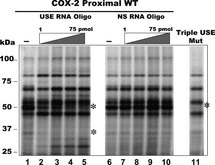
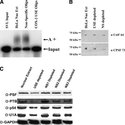
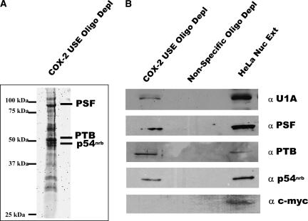
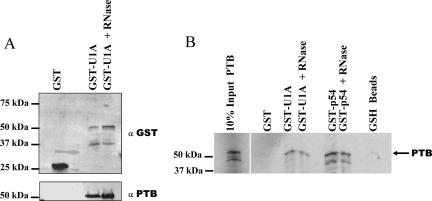
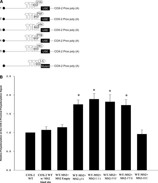
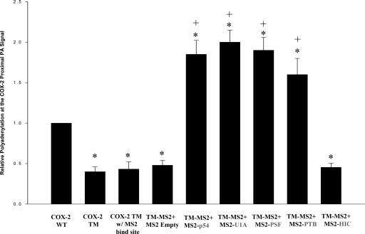
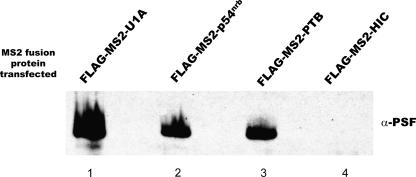
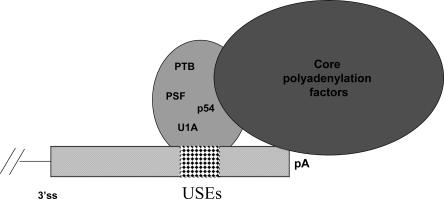
Two cyclooxygenase (COX) enzymes, COX-1 and COX-2, are present in human cells. While COX-1 is constitutively expressed, COX-2 is inducible and up-regulated in response to many signals. Since increased transcriptional activity accounts for only part of COX-2 up-regulation, we chose to explore other RNA processing mechanisms in the regulation of this gene. Previously, we showed that COX-2 is regulated by alternative polyadenylation, and that the COX-2 proximal polyadenylation signal contains auxiliary upstream sequence elements (USEs) that are very important in efficient polyadenylation. To explore trans-acting protein factors interacting with these cis-acting RNA elements, we performed pull-down assays with HeLa nuclear extract and biotinylated RNA oligonucleotides representing COX-2 USEs. We identified PSF, p54(nrb), PTB, and U1A as proteins specifically bound to the COX-2 USEs. We further explored their participation in polyadenylation using MS2 phage coat protein-MS2 RNA binding site tethering assays, and found that tethering any of these four proteins to the COX-2 USE mutant RNA can compensate for these cis-acting elements. Finally, we suggest that these proteins (p54(nrb), PTB, PSF, and U1A) may interact as a complex since immunoprecipitations of the transfected MS2 fusion proteins coprecipitate the other proteins.


Figures

FIGURE 1.
FIGURE 1.
Analysis of proteins which are UV-cross-linked and competed by specific USE oligoribonucleotides. Radiolabeled RNA transcript representing wild-type COX-2 proximal polyadenylation signal was incubated with HeLa nuclear extract and UV cross-linked in the absence of any oligoribonucleotides (lanes 1,6), in the presence of increasing amounts of a COX-2 USE oligoribonucleotide (1, 10, 25, 50 nmol oligo, lanes 2–5, respectively), or in the presence of increasing amounts of a nonspecific oligoribonucleotide (1, 10, 25, 50 nmol oligo, lanes 7–10, respectively). (Lane 11) RNA representing the COX-2 proximal polyadenylation signal with mutations in all three COX-2 USEs was used in a UV cross-linking reaction, called Triple USE mut (Hall-Pogar et al. 2005).
FIGURE 2.
FIGURE 2.
COX-2 USE depleted HeLa nuclear extracts are unable to polyadenylate substrate RNAs in vitro, yet the core polyadenylation factors remain. Biotinylated COX-2 USE or nonspecific oligoribonucleotides were used to deplete HeLa nuclear extract. Depleted supernatants were then used in in vitro polyadenylation reactions with SVL substrate RNA. (A) Results of in vitro polyadenylation reactions. SVL input, unreacted substrate RNA; HeLa Nuc Ext, regular undepleted nuclear extract; nonspecific oligo, nonspecific oligo depleted extract; COX-2 USE oligo, COX-2 USE oligo depleted extract; A+, polyadenylated product. (B) Western blot of depleted and undepleted extracts using antibodies to two core polyadenylation factors, CstF-64 and CPSF-73, as indicated at right. HeLa Nuc Ext, regular undepleted nuclear extract; USE depleted, COX-2 USE oligo depleted extract; NS depleted, nonspecific oligo depleted extract. Molecular weight markers are indicated at left. (C) Western blot of depleted and nondepleted extracts using specific antibodies as indicated to the left. Nuclear extract, HeLa undepleted nuclear extract; USE depleted, supernatant of biotinylated COX-2 USE oligo depleted HeLa nuclear extract; NS1 depleted, supernatant of biotinylated NS1 oligo depleted HeLa nuclear extract; NS2 depleted, supernatant of biotinylated NS2 oligo depleted HeLa nuclear extract; NS3 depleted, supernatant of biotinylated NS3 oligo depleted HeLa nuclear extract. See Materials and Methods for sequences of the oligos.
FIGURE 3.
FIGURE 3.
Specific proteins interact with the COX-2 USE oligoribonucleotide. (A) Sypro Ruby stained gel from which protein bands were excised and submitted for MALDI TOF/TOF analysis. Identified proteins are indicated on the right, molecular weight markers at left. (B) Western blot analyses confirm that the identified proteins do interact specifically with the COX-2 USE oligoribonucleotide and not a nonspecific oligo. Proteins bound to biotinylated COX-2 USE oligo or a nonspecific oligo were isolated using streptavidin agarose followed by extensive washing, then bound proteins were separated on a 12% SDS-PAGE gel and Western blotted with antibodies as indicated to the right. HeLa nuclear extract (HeLa Nuc Ext) and anti-c-myc antibody were used as controls.
FIGURE 4.
FIGURE 4.
PTB interacts directly with U1A protein. (A) HeLa nuclear extracts were incubated with the GST moiety alone (GST) or with GST-full length U1A protein. Bound proteins were separated on a 12% SDS-PAGE gel and were assayed using Western blotting with anti-GST antibodies (top) or with anti-PTB antibodies (bottom). Addition of RNase A did not affect the U1A-PTB interaction (GST U1A + RNase). Molecular weight markers are indicated to the left. (B) GST pull-down assays were performed in the presence of in vitro transcribed and translated 35S methionine labeled PTB protein. GST fusion proteins used in this experiment were the GST moiety alone (GST), GST full-length U1A (GST-U1A), or GST-p54nrb (GST-p54). After incubation of the GST fusion proteins to the 35S PTB and subsequently to the glutathione beads, extensive washing took place, and the bound proteins were separated on a 12% SDS-PAGE gel. Any proteins bound to the glutathione-sepharose beads when incubated alone with the 35S PTB were shown in the last lane (GSH beads). Proteins were visualized by autoradiography.
FIGURE 5.
FIGURE 5.
MS2 tethering assays show that each protein can enhance polyadenylation. (A) Schematic of each protein fused to MS2 coat protein and cotransfected with a reporter vector containing wild-type COX-2 proximal polyadenylation signal with MS2 stem-loop structures inserted upstream. Only three stem-loops are depicted; actually six were inserted. (Bottom) Each individual MS2 coat protein fusion was also cotransfected with a similar COX-2 construct that had the three USE elements mutated. (B) Quantification of RNase protection assays using cotransfections of COX-2 wild-type reporter vector containing MS2 binding sites and individual MS2 coat-protein fusion proteins. Results are shown as relative polyadenylation at the COX-2 polyadenylation signal, with usage of the COX-2 proximal polyadenylation signal set as a value of one. COX-2 WT w/MS2 bind site, COX-2 proximal polyadenylation signal with six MS2 stem–loops inserted; WT-MS2 + MS2 empty, cotransfected with MS2 coat protein vector alone; WT-MS2 + MS2 p54, cotransfected with MS2-p54nrb; WT-MS2 + U1A, cotransfected with MS2-U1A; WT-MS2 + PSF, cotransfected with MS2-PSF; WT + MS2 PTB, cotransfected with MS2-PTB; WT-MS2 + HIC, cotransfected with MS2-HIC as a negative control. Bars represent means ±SD, n = 7. Values were significantly different (two-tailed, two-sample t-test assuming unequal variances) from COX-2 proximal WT. *P < 0.001467.
FIGURE 6.
FIGURE 6.
MS2 tethering abrogates the need for the cis-acting USE elements. Same as Figure 4B but the COX-2 proximal polyadenylation signal reporter contained mutations in all three COX-2 USEs (Triple mutation [TM]) so that each USE was mutated (n = 6; Hall-Pogar et al. 2005). Values were significantly different (two-tailed, two-sample t-test assuming unequal variances) from COX-2 proximal WT. *P < 0.02915 and from COX-2 TM, +P < 0.001076.
FIGURE 7.
FIGURE 7.
Immunoprecipitation of individual transfected FLAG-tagged proteins coprecipitate PSF. Individual transfections were performed using FLAG-tagged MS2 protein constructs as indicated at the top. Immunoprecipitations were performed using a FLAG-tag antibody, and following separation of precipitated proteins on a 12% SDS-PAGE gel, the gel was Western blotted using anti-PSF antibodies.
FIGURE 8.
FIGURE 8.
A model: proteins bound to USEs at the COX-2 proximal polyadenylation signal may enhance polyadenylation at that signal by recruiting or stabilizing core polyadenylation factors on the polyadenylation signal. The proteins we have found associated with the COX-2 USEs are listed inside a large oval; since each protein has been found to associate with each other and with USEs it is not clear which one(s) may be in direct association with the polyadenylation machinery.

Similar articles

Cited by

References

    1. Appleby, S.B., Ristimaki, A., Neilson, K., Narko, K., Hla, T. Structure of the human cyclo-oxygenase-2 gene. Biochem. J. 1994;302:723–727. - PMC - PubMed
    1. Bazan, N.G. COX-2 as a multifunctional neuronal modulator. Nat. Med. 2001;7:414–415. - PubMed
    1. Bazan, N.G., Fletcher, B.S., Herschmann, H.R., Mukherjee, P.K. Platelet-activating factor and retinoic acid synergistically activate the inducible prostaglandin synthase gene. Proc. Natl. Acad. Sci. 1994;91:5252–5256. - PMC - PubMed
    1. Bentley, D.L. Rules of engagement: Co-transcriptional recruitment of pre-mRNA processing factors. Curr. Opin. Cell Biol. 2005;17:251–256. - PubMed
    1. Bishop-Bailey, D., Calatayud, S., Warner, T.D., Hla, T., Mitchell, J.A. Prostaglandins and the regulation of tumor growth. J. Environ. Pathol. Toxicol. Oncol. 2002;21:93–101. - PubMed

Publication types

MeSH terms