. 2007 Jul;176(3):1609-24.
doi: 10.1534/genetics.107.073569. Epub 2007 May 16.

Defective decapentaplegic signaling results in heart overgrowth and reduced cardiac output in Drosophila

Affiliations

Defective decapentaplegic signaling results in heart overgrowth and reduced cardiac output in Drosophila

Aaron N Johnson et al. Genetics. 2007 Jul.

Abstract

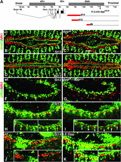
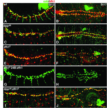
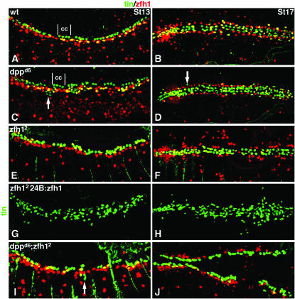
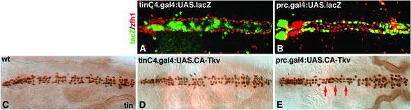
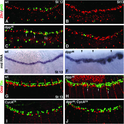
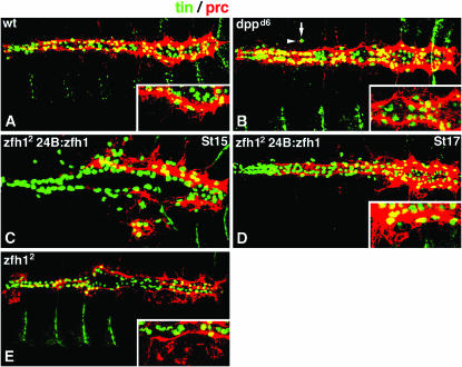
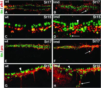
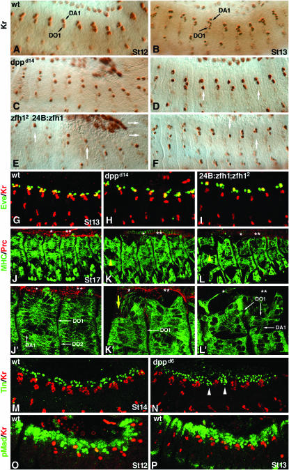
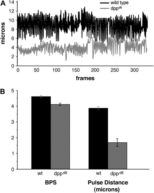
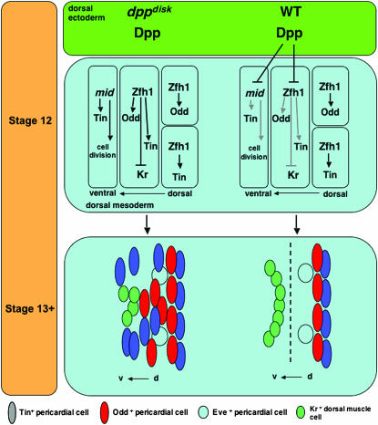
During germ-band extension, Decapentaplegic (Dpp) signals from the dorsal ectoderm to maintain Tinman (Tin) expression in the underlying mesoderm. This signal specifies the cardiac field, and homologous genes (BMP2/4 and Nkx2.5) perform this function in mammals. We showed previously that a second Dpp signal from the dorsal ectoderm restricts the number of pericardial cells expressing the transcription factor Zfh1. Here we report that, via Zfh1, the second Dpp signal restricts the number of Odd-skipped-expressing and the number of Tin-expressing pericardial cells. Dpp also represses Tin expression independently of Zfh1, implicating a feed-forward mechanism in the regulation of Tin pericardial cell number. In the adjacent dorsal muscles, Dpp has the opposite effect. Dpp maintains Krüppel and Even-skipped expression required for muscle development. Our data show that Dpp refines the cardiac field by limiting the number of pericardial cells. This maintains the boundary between pericardial and dorsal muscle cells and defines the size of the heart. In the absence of the second Dpp signal, pericardial cells overgrow and this significantly reduces larval cardiac output. Our study suggests the existence of a second round of BMP signaling in mammalian heart development and that perhaps defects in this signal play a role in congenital heart defects.


Figures

F<sc>igure</sc> 1.—
Figure 1.—
dpp mutant embryos display pericardial but not cardiac cell overgrowth and have reduced pMad accumulation in the dorsal mesoderm. (A) Map of the dpp locus showing three genetically defined regions: shortvein (shv), Hin, and disk. The structures of dpp transcripts are shown with black boxes representing the open reading frame and open boxes corresponding to untranslated regions. dpp disk region mutations used in this study are the deficiency dppd14 and the inversions dppd12 and dppd6. Red boxes represent the degree of uncertainty in the position of the distal breakpoint associated with each aberration. The dpp151H enhancer trap is also shown (Johnson et al. 2003). (B–E) Embryos double labeled for dMef2 (green) and Zfh1 (red). (B and C) Stage 15. (B) Wild type. (C) dppd6 embryos contain a significantly greater number of Zfh1-expressing pericardial cells (see Johnson et al. 2003 for statistics) but show no change in the number of dMef2-expressing cells. Note some Zfh1-expressing cells are visible in the dorsal muscle domain (white arrow). (D and E). Stage 17. (D) Wild type. (E) dppd6 embryos continue to display pericardial cell hyperplasia. (F–K) Merged scans of embryos double labeled for pMad (green) and Zfh1 (red). Insets are single mesodermal scans to exclude ectodermal pMad accumulation. (F and G) Stage 12. (F) Wild type. (G) dppd12/dppd6. pMad is detected in all Zfh1-expressing pericardial precursor cells in both genotypes. (H and I) Stage 13. (H) Wild type. pMad is detected in a majority of the Zfh1-expressing cells (yellow arrows) as well as in non-Zfh1-expressing dorsal mesoderm cells (yellow arrowheads). (I) dppd6/dppd12. pMad is not detected in a subset of Zfh1-expressing cells (white arrows). pMad is detected in fewer nonpericardial dorsal mesoderm cells (white arrowheads). (J and K) Stage 14. (J) Wild type. pMad is largely undetectable in Zfh1-expressing cells. (K) LE.Gal4:UAS.Dpp. Ectopic pMad accumulates in Zfh1-expressing and non-Zfh1-expressing dorsal mesoderm cells.
F<sc>igure</sc> 2.—
Figure 2.—
Dpp restricts the number of Odd-skipped pericardial cells via zfh1. Stage 13 embryos in lateral view (A, C, E, G, and I) and stage 17 in dorsal view (B, D, F, H, and J) double labeled for Odd (green) and Zfh1 (red). See Table 1 for statistics. (A and B) Wild type. Odd is expressed in a subset of Zfh1-expressing pericardial cells. (C and D) dppd6. The number of Odd-expressing pericardial cells is significantly increased. All Odd pericardial cells co-express Zfh1. (E and F) zfh12. The number of Odd-expressing cells is comparable to wild type at stage 13 (E) but is significantly less than wild type by stage 17 (F). (G and H) 24B.Gal4:UAS.Zfh1; zfh12. Pan-mesodermal Zfh1 expression in zfh12 mutants not only rescues Odd expression in pericardial cells but also induces ectopic Odd-expressing cells in lateral regions of the mesoderm. (I and J) dppd6; zfh12. The number of Odd-expressing cells is comparable to wild type at stage 13 (I) but is significantly decreased by stage 17 (J), a phenocopy of zfh12 single mutants.
F<sc>igure</sc> 3.—
Figure 3.—
Dpp restricts the number of Tin pericardial cells via zfh1 and independently of zfh1. Stage and view as in Figure 2 for embryos double labeled for Tin (green) and Zfh1 (red). See Table 1 for statistics. (A and B) Wild type. Tin is expressed in a subset of Zfh1-expressing pericardial cells (yellow) and in a subset of cardial cells (cc). (C and D) dppd6. The number of Tin-expressing pericardial cells is significantly increased. Note that Tin-positive cells are located ventral to the Zfh1-expressing pericardial cells but they do not co-express Zfh1 (white arrows). (E and F) zfh12. The number of Tin-expressing cells is significantly fewer than wild type. (G and H) 24B.Gal4:UAS.Zfh1; zfh12. Pan-mesodermal Zfh1 expression in zfh12 mutants not only rescues Tin expression in pericardial cells but also induces ectopic Tin-expressing cells in lateral regions of the mesoderm. (I and J) dppd6; zfh12. The number of Tin-expressing cells is significantly decreased compared to wild type—a phenocopy of zfh12 single mutants. Some Tin-expressing cells positioned ventral to the Zfh1-expressing pericardial cells do not co-express Zfh1 in double-mutant embryos (white arrow in I).
F<sc>igure</sc> 4.—
Figure 4.—
Expressing activated Tkv in pericardial cells, but not in cardiac cells, reduces the number of cells expressing Tin. Dorsal view of stage 17 embryos. (A) tinCΔ4.Gal4 drives lacZ expression (green) in a majority of cardiac cells but not in Zfh1-expressing pericardial cells (red). (B) prc.gal4 drives lacZ expression in a subset of Zfh1-expressing pericardial cells. (C) Wild type Tin expression. (D) tinCΔ4.Gal4:UAS.CA-Tkv embryos do not show a significant change in the number of Tin-expressing cells. (E) prc.Gal4:UAS.CA-Tkv embryos show a significant decrease in the number of Tin-expressing cells (see Table 1 for statistics). Note that, in the region indicated by red arrows, only two rows (of presumably cardiac cells) instead of three or four rows in the comparable region of wild-type embryos are present.
F<sc>igure</sc> 5.—
Figure 5.—
Dpp signaling reduces cell proliferation in the dorsal mesoderm by restricting mid expression. (A–D) Merged mesodermal scans of embryos double labeled for Zfh1 (red) and the mitosis marker phospho-histone3 (pH3, green). (A) Wild-type stage 12. Note the limited cell proliferation (pH3 staining) dorsal to the Zfh1-expressing pericardial cells. (B) Wild-type stage 13. No cell proliferation in the dorsal mesoderm. (C) dppd6 stage 12. Cell proliferation is apparent ventrally and medially to the Zfh1-expressing cells. White arrows identify ectopic pH3 staining. Note that apparent colocalization of Zfh1 and pH3 (yellow cells) is an artifact of merging scans. (D) dppd6 stage 13. Cell proliferation persists. (E and F) mid RNA expression stage 13. (E) Wild type. mid expression is restricted to cardiac cells. (F) dppd6. mid expression expands laterally, specifically in the posterior dorsal mesoderm. This image is a composite of four images. Black arrowheads denote points of overlay. The embryo shown is an extreme example. (G–I) Stage 13 embryos double labeled for Tin (green) and Odd (red). (G) Wild type. (H) dppd6. (I) CycAC8. (J) dppd6; CycAC8. The number of Odd-expressing cells in CycAC8 embryos is approximately half the number of wild type. However, the number of Odd-expressing cells in dppd6; CycAC8 double mutants is greater than that observed in CycAC8 embryos. In addition, none of these embryos co-express Tin and Odd. Numerous laterally displaced Tin-expressing cells (arrows in H) are observed in dppd6 embryos.
F<sc>igure</sc> 6.—
Figure 6.—
Modifying Zfh1 expression alters Prc expression. Embryos double labeled for Prc (red) and Tin (green). Insets are high-magnification merged scans from the posterior regions of the heart. (A) Wild type stage 17. Prc is broadly expressed throughout the pericardial cell domain, most prominently in the posterior. (B) dppd6 stage 17. Prc expression is expanded. Ectopic Tin-expressing cells (arrow) co-express Prc (arrowhead). (C) 24B.Gal4:UAS.Zfh1; zfh12 stage 15 and (D) stage 17. Ventrally positioned Tin-expressing cells co-express Prc. (E) zfh12 stage 17. Prc expression domain is greatly reduced.
F<sc>igure</sc> 7.—
Figure 7.—
Mutations in lame duck also display pericardial cell hyperplasia. (A–D) Embryos double labeled for Tin (green) and Zfh1 (red) at the indicated stages. (A and B) Dorsal (bilateral) view of stage 17 embryonic hearts at low magnification. (C and D) Lateral (unilateral) view of stage 15 embryonic hearts at high magnification. (A and C) In wild-type embryos, all Tin-positive pericardial cells co-express Zfh1 (yellow cells lateral to the green Tin-expressing cardiac cells). (B and D) In lmd1 embryos, Tin-expressing cardiac cells look normal (cells indicated by an arrowhead in D). There are an excessive number of Zfh1-expressing cells and an excess of Tin-expressing cells, including many quite lateral to their normal position that do not co-express Zfh1 (cells indicated by an arrow in B and D). (E–H) Embryos double labeled for Tin (green) and Prc (red). (E and F) Dorsal (bilateral) view of stage 17 embryonic hearts at low magnification. (G and H) Lateral (unilateral) view of stage 15 embryonic hearts at intermediate magnification. (E and G) In wild-type embryos, all Tin-expressing pericardial cells express Prc but Tin-expressing cardiac cells do not (cardiac cells are indicated by an arrowhead in G and H). (F and H) In lmd1 embryos, all Tin-expressing cells, except for Tin cardiac cells (arrowhead), also express Prc (cells indicated by an arrow).
F<sc>igure</sc> 8.—
Figure 8.—
Dpp indirectly maintains dorsal muscle cell fates. (A–F) Lateral views of embryos stained for Kr expression. See Table 2 for statistics. (A) Wild-type stage 12. Kr-expressing DA1 and DO1 muscle founder cells are shown. (B) Wild-type stage 13. An increased number of Kr-expressing nuclei are evident. (C) dppd14 stage12 and (D) stage 13. Roughly half of the Kr-expressing cells per segment are present. White arrows in D identify segments with reduced Kr expression. (E) 24B.Gal4:UAS.Zfh1; zfh12 stage 12 and (F) stage 13. Kr expression is dramatically reduced. (G–I) Stage 13 embryos double labeled for Kr (red) and Eve (green). (G) Wild type. (H) dppd14. (I) 24B.Gal4:UAS.Zfh1; zfh12. Eve pericardial cells, two per segment, are present in all genotypes, but the number of Kr and Eve co-expressing dorsal muscle cells (in yellow) is considerably reduced in dpp and zfh mutants. (J–L) Stage 17 embryos double labeled for myosin heavy chain (green) and Prc (red). The DO1, DO2, and DA1 muscles are indicated with white arrows. Asterisks indicate the two segments viewed at high magnification in J′–L′. (J and J′) Wild type. The DO1 and DO2 muscles are tightly associated. The DO1 and DA1 muscles abut the pericardial cells. (K and K′) dppd14. The DO1 and DO2 muscles are loosely associated (yellow arrowhead in K) and the size of the DO1 muscles is reduced (yellow arrow in K′). (L and L′) 24B.Gal4:UAS.Zfh1; zfh12. The DO1 and DO2 muscles are loosely associated (yellow arrowhead in L), a subset of DA1 muscles is absent, and the DO1 muscles are reduced. (M and N) Stage 14 embryos labeled for Tin (green) and Kr (red). (M) Wild type. Tin-expressing cells are exclusively positioned dorsal to the Kr-expressing cells. (N) dppd6. There are Tin-expressing cells positioned ventral to the dorsal-most Kr-expressing cells (white arrowheads). (O and P) Stage 12 and 13 embryos labeled for pMad (green) and Kr (red). (O) Wild type. (P) dppd6. Colabeling of Kr and pMad is not observed in either embryo, indicating that the effect of Dpp on Kr is indirect.
F<sc>igure</sc> 9.—
Figure 9.—
Reduced cardiac output in dpp mutant larvae. (A) Representative traces showing the distance between a pair of cardiac cells in the posterior region of the heart over time in wild type (top) and dppd6 (bottom) first instar larvae. One second corresponds to 12 frames. (B) Average number of beats per second (BPS) and pulse distance for wild type (n = 3) and dppd6 (n = 4) first instar larvae. Pulse distance is the difference between the maximum diastolic position and the minimum systolic position for each heartbeat. Error bars signify the standard error of the mean.
F<sc>igure</sc> 10.—
Figure 10.—
Dpp signals pattern the embryonic dorsal mesoderm during germ-band retraction. dppdisk at stage 12: in the absence of Dpp, we observed that (1) mid expression expands ventrally, drives cell proliferation, and induces ectopic Tin expression; (2) Zfh1 expression expands ventrally, inducing ectopic Tin and Odd expression and repressing Kr (and Eve) expression; and (3) pericardial cell specification of Odd and Tin by Zfh is unaffected. Wild type at stage 12: Dpp signals from the dorsal ectoderm restrict the expression of mid and Zfh1. dppdisk at stage 13+: in the absence of Dpp, we observed that (1) pericardial cells populate ventral regions of the dorsal mesoderm normally occupied exclusively by dorsal muscle cells and (2) the inappropriate presence of pericardial cells reduces the expression of the dorsal muscle genes Kr (and Eve). It appears that there is a failure to maintain the pericardial cell–dorsal muscle cell boundary and that this failure results in an increase in heart size. Wild type (WT): Dpp maintains the boundary between pericardial and dorsal muscle cells (dashed line) by restricting the number of Tin- and Odd-expressing pericardial cells and by reducing cell division via the repression of mid.

Similar articles

Cited by

References

    1. Alvarez, A., W. Shi, B. Wilson and J. Skeath, 2003. pannier and pointedP2 act sequentially to regulate Drosophila heart development. Development 130: 3015–3026. - PubMed
    1. Bour, B., M. O'Brien, W. Lockwood, E. Goldstein, R. Bodmer et al., 1995. dMef2, a transcription factor that is essential for myogenesis. Genes Dev. 9: 730–741. - PubMed
    1. Brand, A., and N. Perrimon, 1993. Targeted gene expression as a means of altering cell fates and generating dominant phenotypes. Development 118: 401–415. - PubMed
    1. Carmena, A., S. Gisselbrecht, J. Harrison, F. Jimenez and A. Michelson, 1998. a Combinatorial signaling codes for the progressive determination of cell fates in the Drosophila embryonic mesoderm. Genes Dev. 12: 3910–3922. - PMC - PubMed
    1. Carmena, A., B. Murugasu-Oei, D. Menon, F. Jimenez and W. Chia, 1998. b Inscuteable and Numb mediate asymmetric muscle progenitor cell divisions during Drosophila myogenesis. Genes Dev. 12: 304–315. - PMC - PubMed

Publication types

LinkOut - more resources