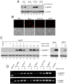
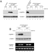
. 2007 Jun 12;46(23):6844-58.
doi: 10.1021/bi6024484. Epub 2007 May 18.

Erythroid-specific expression of beta-globin by the sleeping beauty transposon for Sickle cell disease

Affiliations

Erythroid-specific expression of beta-globin by the sleeping beauty transposon for Sickle cell disease

Jianhui Zhu et al. Biochemistry. .

Abstract

Sickle cell disease (SCD) results predominately from a single monogenic mutation that affects thousands of individuals worldwide. Gene therapy approaches have focused on using viral vectors to transfer wild-type beta- or gamma-globin transgenes into hematopoietic stem cells for long-term expression of the recombinant globins. In this study, we investigated the use of a novel nonviral vector system, the Sleeping Beauty (SB) transposon (Tn) to insert a wild-type beta-globin expression cassette into the human genome for sustained expression of beta-globin. We initially constructed a beta-globin expression vector composed of the hybrid cytomegalovirus (CMV) enhancer chicken beta-actin promoter (CAGGS) and full-length beta-globin cDNA, as well as truncated forms lacking either the 3' or 3' and 5' untranslated regions (UTRs), to optimize expression of beta-globin. Beta-globin with its 5' UTR was efficiently expressed from its cDNA in K-562 cells induced with hemin. However, expression was constitutive and not erythroid-specific. We then constructed cis SB-Tn-beta-globin plasmids using a minimal beta-globin gene driven by hybrid promoter IHK (human ALAS2 intron 8 erythroid-specific enhancer, HS40 core element from human alphaLCR, ankyrin-1 promoter), IHbetap (human ALAS2 intron 8 erythroid-specific enhancer, HS40 core element from human alphaLCR, beta-globin promoter), or HS3betap (HS3 core element from human betaLCR, beta-globin promoter) to establish erythroid-specific expression of beta-globin. Stable genomic insertion of the minimal gene and expression of the beta-globin transgene for >5 months at a level comparable to that of the endogenous gamma-globin gene were achieved using a SB-Tn beta-globin cis construct. Interestingly, erythroid-specific expression of beta-globin driven by IHK was regulated primarily at the translational level, in contrast to post-transcriptional regulation in non-erythroid cells. The SB-Tn system is a promising nonviral vector for efficient genomic insertion conferring stable, persistent erythroid-specific expression of beta-globin.


Figures
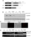
Figure 1
Figure 1
Expression of β-globin from cDNA in K-562 and HuH-7 cells. (A) Structure of the β-globin cDNA expression cassettes. CAGGS, cytomegalovirus (CMV) enhancer:chicken β-actin promoter; 5′UTR, 5′-untranslated region; CDS, β-globin coding region; 3′UTR, 3′-untranslated region; poly A, rabbit globin poly (A) signal from the pCAGGS vector. (B) Western blot analysis of cell extracts from K-562 and HuH-7 cells transiently transfected with pCAGGS-β, pCAGGS-β3′Δ or pCAGGS-βCDS constructs and induced to express β-globin with hemin (20 μM). The cells were transfected as described in the “Materials and Methods” section. After 36 h, hemin was added to the culture media, and the cells were harvested following an additional 48 h of incubation. Lane loading was normalized to 50 μg protein using β-actin as an internal control. The addition of hemin (+) is indicated for each construct at top. β, pCAGGS-β-globin; β3′Δ, pCAGGS-β3′Δ; βCDS, pCAGGS-β-CDS. (C) Transfection efficiency with pCAGGS-DsRed2 plasmid in K-562 and HuH-7 cells. The cells were transfected in parallel with the β-globin constructs described in (A). The live cell cultures were photographed at 48 h post-transfection. The phase contrast panels are shown at left for each pair and the same field using red fluorescent microscopy is shown at right. (D) Reverse-transcribed (RT)PCR analysis of β-globin mRNA levels from pCAGGS-β, pCAGGS-β3′Δ and pCAGGS-βCDS in the presence or absence of hemin in K-562 (left panel) and HuH-7 (right panel) cells. The construct and addition of hemin (+) to the cultures is indicated at top, and glyceraldehyde-3-phosphate dehydogenase (GAPDH) transcript levels were used as an internal standard to verify RNA quality and lane loading. The smaller size of the amplicon observed using the pCAGGS-βCDS construct reflects the predicted size due to the deletion of the 51 nt 5′UTR.
Figure 2
Figure 2
Expression of β-globin by the CAGGS promoter in non-erythroid cells. (A) Western blot analysis of cell extracts from U87, Daoy, HCT116 and MCF7 cells transiently transfected with pCAGGS-β3′Δ plasmid. The cells transfected with the β-globin construct were collected 48 h after the addition of hemin (20 μM) and the western blot analysis was performed as described in “Material and Methods.” β-actin was used to control for lane loading. (B) The transfection efficiency in U87, Daoy, HCT117, and MCF7 cells was determined by parallel transfection with the pCAGGS-DsRed2 plasmid. (C) β-globin protein undergoes proteasomal degradation in non-erythroid cells. Western blot analysis of HuH-7 (left), or Daoy and MCF7 cells (right) transiently transfected with pCAGGS-β3′Δ plasmid. Twenty-four h post-transfection, the cells were first induced with hemin (20 μM) and then MG132 was added at the indicated concentrations (top) after an additional 24 h. The cells were harvested and processed for western blot analysis 72 h after transfection. Control, no transfection control for HuH-7. (D) β-globin transcript levels in erythroid and non-erythroid cell lines. Reverse-transcribed (RT) PCR analysis of β-globin mRNA levels from pCAGGS-β3′Δ in the presence or absence of hemin. The cell line is indicated above and β-actin transcript levels were used as an internal standard to verify RNA quality. The 500 bp DNA maker is shown at left with the transcript and its respective size indicated at right.
Figure 3
Figure 3
β-globin expression from different cis pT2-β-globin//eIF-SB10 constructs in K-562 cells. (A) Structure of cis pT2-promoter-β-gene//eIF-SB10 constructs (left), and schematic of the cut-and-paste transposition mechanism used by the SB-Tns (right). The obligate SB transposase is expressed from the transgene located in the vector backbone, external to the IR/DRs flanking the Tn IHK-βgene cargo. Following binding of the transposase to the IR/DRs, the Tn is cut from the plasmid vector and pasted into a TA dinucleotide in the host genomic DNA which is duplicated during the insertion process. i8, Human ALAS2 intron 8 erythroid-specific enhancer element; HS40, core element of αLCR; HS3, core element of βLCR; βp, human β-globin promoter; Ank-1p, human ankyrin-1 promoter; I, II and III are β-globin exons; poly A, rabbit globin poly(A) signal from the pCAGGS vector; IR/DR, transposon inverted repeat/direct repeat; eIF, mouse initiation factor 4A1 promoter; SB10, Sleeping Beauty transposase gene, version 10. (B) Western blot (left) and RT-PCR (right) analysis of β-globin protein and transcript levels in K-562 cells transfected with pT2-IHK-β-gene//eIF-SB10 (lanes 7,8) or pT2-IHβp-β-gene//eIF-SB10 (lane 5,6), or pT2-HS3βp-β-gene//eIF-SB10 (lanes 3,4), respectively. Lanes 1 and 2 are non-transfected controls. Twenty-four h after transfection, hemin (20 μM) was added to induce β-globin expression. The cells were harvested for western blot and RT-PCR analysis after additional 48 h incubation. The proteins and transcripts are indicated at left. The hybrid promoter used to drive the β-gene in the construct and addition (+) of hemin to the cultures is shown at the top. HS3βp, HS3 core element of βLCR and human β-globin promoter; IHβp, human ALAS2 intron 8 erythroid-specific enhancer element, HS40 core element of αLCR and human β-globin promoter; IHK, human ALAS2 intron 8 erythroid-specific enhancer element, HS40 core element of αLCR and human ankyrin-1 promoter. β-actin and GAPDH served as normalization controls.
Figure 4
Figure 4
Erythroid-specific expression of β-globin by the hybrid IHK promoter. (A) Western blot analysis of cell extracts from blood origin cells, K-562 and Jurkat, transfected with pT2-IHK-β-gene//eIF-SB10, induced to express β-globin with 20 μM hemin (left), and from non-erythrocyte HuH-7, MCF7, and HCT116 cells transfected with pT2-IHK-β-gene//eIF-SB10 (lanes 2-7) or pT2-pCAGGS-β3′Δ//eIF-SB10 (lane 1) (right). MG132 (5 μM) was added 48 h post-transfection to inhibit proteasome activity and the non-erythrocyte cells incubated for additional 18 h before harvesting. Total protein extracts (50 μg) from the collected cells were subjected to western blot analysis using β-actin as an internal control for equal lane loading. The proteins detected are indicated in the middle, and the addition (+) of hemin or MG132 is shown above the blots. (B) Parallel western blot (upper panel) and RT-PCR analysis (lower panel) of cell samples for K-562 and HuH-7 cells transfected with pT2-IHK-β-gene//eIF-SB10. Hemin (20 μM) was added to induce β-globin synthesis in K-562 cells and MG132 (5 μM) to inhibit proteasome activity in HuH-7 cells. Lanes 1,4 are non-transfected controls for K-562 and HuH-7, respectively. An RT-PCR negative control without reverse transcriptase using RNA sample from lane 6 is included (lane 7). Ten μl of the RT-PCR (25 cycles) products were analyzed by agarose gel electrophoresis. Glyceraldehyde-3-phosphate dehydrogenase (GAPDH) transcript was amplified in parallel and served to normalize RNA sample loading. The cell line and the addition of hemin (+) or MG132 (+) to the cultures are indicated at top.
Figure 5
Figure 5
Long Term expression of β-globin mediated by SB10 and HSB3. Western blot analysis of β-globin expression in K-562 cells transfected with pT2-IHK-β-gene//eIF-HSB3 (right) and pT2-IHK-β-gene//eIF-SB10 or pT2-IHK-β-gene (left). The cells were passaged every 3 days by plating 10% of cells from the previous passage. Hemin (20 μM) was included in the media 48 h prior to harvesting cells at each time point. Fifty μg of total protein was subjected to western blot analysis; and β-actin was used to normalize sample loading. The time (days) to harvest after transfection is indicated at top. Cell extracts from pT2-IHK-β-globin (left, lanes 1-4); pT2-IHK-β-globin//eIF-SB10 transfected cells (left, lanes 5-12); and pT2-IHK-β-globin//eIF-HSB3 (right) are shown. The protein levels determined by western blot are shown in the middle and the addition (+) of hemin to the culture is indicated above the panels. SB, Sleeping Beauty transposase, version 10 (left) and hyperactive version HSB3 (right).
Figure 6
Figure 6
SB10 mediated β-globin transposition and identification of the insertion sites in K-562 cells transfected with pT2-IHK-β-gene//eIF-SB10 or pT2-IHK-β-gene. (A) Schematic design for inverted nested PCR analysis. The genomic DNA samples were digested by KpnI, and subjected to self-ligation. The ligated products were used as template for the initial inverted PCR amplification with primer pair LP1/RP1. The second inverted PCR amplification step was performed using an internal nested primer pair LP2/RP2. The consequent PCR products were analyzed by agarose gel electrophoresis and visualized by UV light after ethidium bromide staining. (B) Inverted nested PCR analysis of the genomic DNA samples purified from K-562 cells 4 weeks post-transfection with an unrelated plasmid (control); pT2-IHK-β-gene; and pT2-IHK-β-gene//eIF-SB10. Amplified PCR bands from either episomal plasmid or random integration are indicated by open circles; and those from canonical SB insertion footprints by filled circles. ?, the amplified band of unknown identity. M, DNA marker lane with the size of the bands indicated in kb at left.
Figure 7
Figure 7
Identification of the insertion sites in K-562 cells transfected with pT2-IHK-β-gene//eIF-SB10. The amplicons resulting from the second inverted PCR reactions were isolated from the agarose gel and sequenced directly using the RP2 or LP2 primer as described in “Materials and Methods.” The identified flanking genomic sequences of the insertion sites are displayed. The regions in the ID/DR of the transposon and the requisite duplicated TA are shown in bold letters. The chromosomal location established by BLAST analysis is shown at right and the intronic insertion sites are marked (*).
Figure 8
Figure 8
Expression of the different cis β-gene//eIF-SB10 constructs in adult erythroid cells. Proerythroid MEL cells were differentiated to adult erythroid cells by addition of DMSO to media 56 h prior to transfection. After induction with hemin 24 h post-transfection, the cells were harvested 48 h later and processed for western blot and RT-PCR analysis as described in “Materials and Methods.” Non-induced “proerythroid” MEL cell (left panel) western blots of β-globin and β-actin are shown in the upper two panels, while the agarose gels of RT-PCR analysis of β-globin and GAPDH transcript levels are shown below. Glyceraldehyde-3-phosphate dehydrogenase (GAPDH) transcript was amplified in parallel and served to normalize RNA sample loading. β-actin was used as a lane loading control for the western blots. The expression levels in the adult erythroid cells of the same constructs by western blot (upper right panels) and RT-PCR analysis (lower right panels) demonstrated robust expression by all three promoter constructs. The erythroid promoter used to drive the β-gene and addition (+) of hemin to the cultures is indicated at top. The protein and transcript are shown at right with the predicted size of the β-globin amplicon indicated in parentheses. HS3βp, HS3 core element of βLCR and human β-globin promoter; IHBp, human ALAS2 intron 8 erythroid-specific enhancer element, HS40 core element of αLCR and human β-globin promoter; IHK, human ALAS2 intron 8 erythroid-specific enhancer element, HS40 core element of αLCR and human ankyrin-1 promoter.

Similar articles

Cited by

References

    1. Thomas CE, Ehrhardt A, Kay MA. Progress and problems with the use of viral vectors for gene therapy. Nat Rev Genet. 2003;4:346–358. - PubMed
    1. Pawliuk R, Westerman KA, Fabry ME, Payen E, Tighe R, Bouhassira EE, Acharya SA, Ellis J, London IM, Eaves CJ, Humphries RK, Beuzard Y, Nagel RL, Leboulch P. Correction of sickle cell disease in transgenic mouse models by gene therapy. Science. 2001;294:2368–2371. - PubMed
    1. Mohamedali A, Moreau-Gaudry F, Richard E, Xia P, Nolta J, Malik p. Self-inactivating lentiviral vectors resist proviral methylation but do not confer position-independent expression in hematopoietic stem cells. Mol Ther. 2004;10:249–259. - PubMed
    1. Hacein-Bey-Abina S, Von Kalle C, Schmidt M, McCormack MP, Wulffraat N, Leboulch P, Lim A, Osborne CS, Pawliuk R, Morillon E, Sorensen R, Forster A, Fraser P, Cohen JI, de Saint Basile G, Alexander I, Wintergerst U, Frebourg T, Aurias A, Stoppa-Lyonnet D, Romana S, Radford-Weiss I, Gross F, Valensi F, Delabesse E, Macintyre E, Sigaux F, Soulier J, Leiva LE, Wissler M, Prinz C, Rabbitts TH, Le Deist F, Fischer A, Cavazzana-Calvo M. LMO2-associated clonal T cell proliferation in two patients after gene therapy for SCID-X1. Science. 2003;302:415–419. - PubMed
    1. Moreau-Gaudry F, Xia P, Jiang G, Perelman NP, Bauer G, Ellis J, Surinya KH, Mavilio F, Shen CK, Malik P. High-level erythroid-specific gene expression in primary human and murine hematopoietic cells with self-inactivating lentiviral vectors. Blood. 2001;98:2664–2672. - PubMed

Publication types