. 2007;9(3):R49.
doi: 10.1186/ar2204.

Pro-apoptotic Bid is required for the resolution of the effector phase of inflammatory arthritis

Affiliations

Pro-apoptotic Bid is required for the resolution of the effector phase of inflammatory arthritis

John C Scatizzi et al. Arthritis Res Ther. 2007.

Abstract

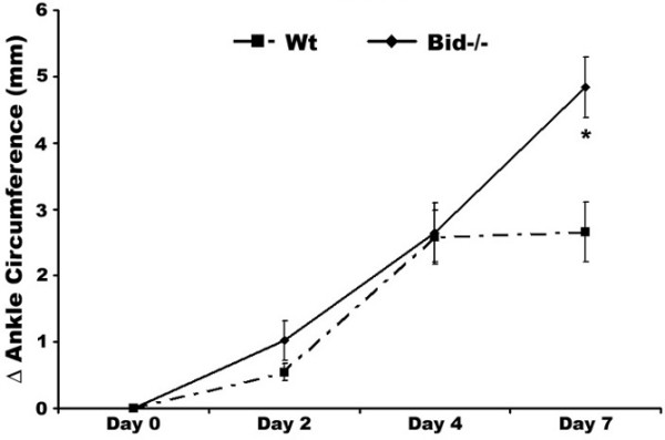
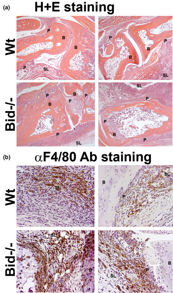
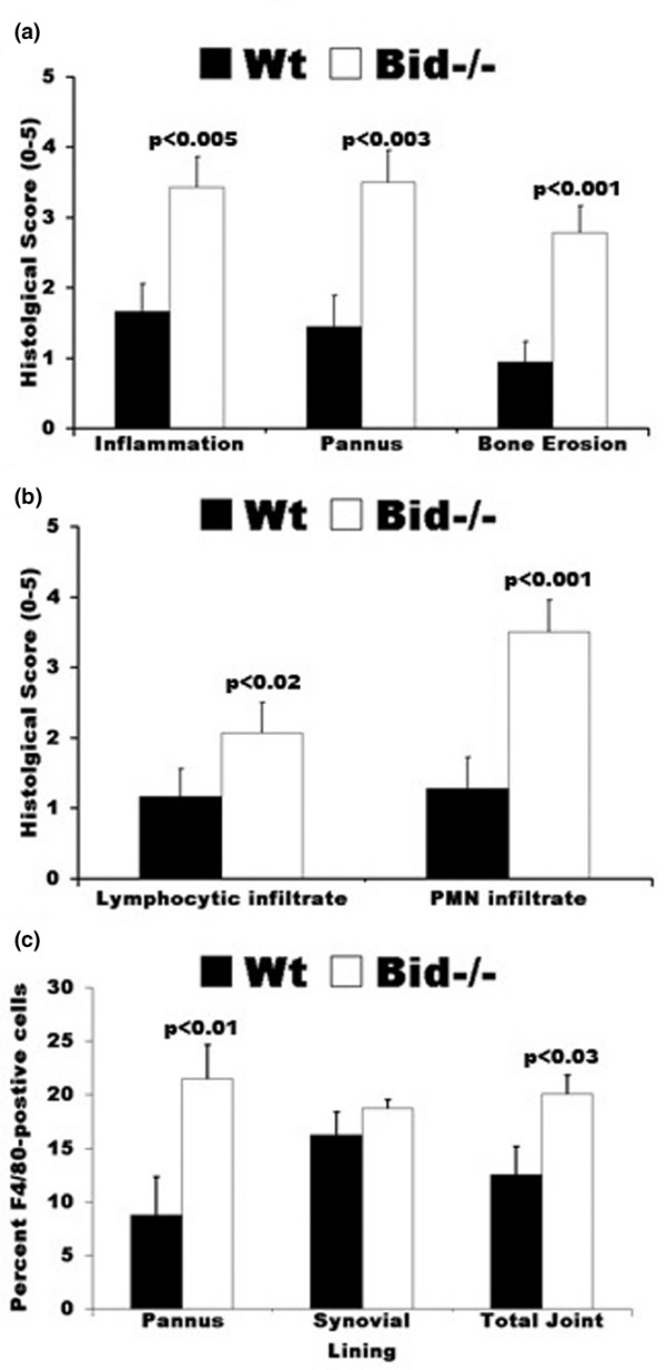
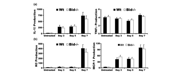
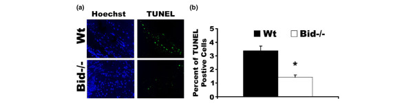
Rheumatoid arthritis is an autoimmune disease characterized by hyperplasia of the synovial lining and destruction of cartilage and bone. Recent studies have suggested that a lack of apoptosis contributes to the hyperplasia of the synovial lining and to the failure in eliminating autoreactive cells. Mice lacking Fas or Bim, two pro-apoptotic proteins that mediate the extrinsic and intrinsic death cascades, respectively, develop enhanced K/BxN serum transfer-induced arthritis. Since the pro-apoptotic protein Bid functions as an intermediate between the extrinsic and intrinsic apoptotic pathways, we examined the role that it plays in inflammatory arthritis. Mice deficient in Bid (Bid-/-) show a delay in the resolution of K/BxN serum transfer-induced arthritis. Bid-/- mice display increased inflammation, bone destruction, and pannus formation compared to wild-type mice. Furthermore, Bid-/- mice have elevated levels of CXC chemokine and IL-1beta in serum, which are associated with more inflammatory cells throughout the arthritic joint. In addition, there are fewer apoptotic cells in the synovium of Bid-/- compared to Wt mice. These data suggest that extrinsic and intrinsic apoptotic pathways cooperate through Bid to limit development of inflammatory arthritis.


Figures

Figure 1
Figure 1
Bid-deficient mice develop a sustained and prolonged edema of the ankles following transfer of K/BxN serum. Pooled serum (300 μl) from K/BxN mice was injected intra-peritoneally (IP) into Bid-/- (n = 32) and wild-type (Wt) (n = 42) mice. Ankle joints were examined for arthritis by measuring two perpendicular diameters of both joints (anterior-posterior; medio-lateral) by calipers. The change in (Δ) ankle circumference at each time point is defined as the difference between the ankle circumference and the measurement at day 0. values represent the mean ± standard error of ankles/time point, which were compared by Student's t-test to Wt mice under parallel conditions. The asterisk denotes p < 0.002 compared to Wt under parallel conditions.
Figure 2
Figure 2
Increased inflammation and destruction of the joint is associated with more macrophages in Bid-/- compared to wild-type (Wt) mice following transfer of serum. Mice (Wt, n = 9; Bid-/-, n = 7) underwent K/BxN serum transfer as described in Figure 1 and were euthanized at seven days post-serum transfer. Both ankles from each mouse were harvested, fixed, embedded in paraffin, sectioned, and stained with either (a) hematoxylin (blue) and eosin (pink) (H&E) or (b) F4/80 antigen (macrophage specific marker). Shown are representative photomicrographs of the synovium and pannus formation from Wt and Bid-/- mice. B, bone; SL, synovial lining; P, pannus.
Figure 3
Figure 3
Histological scores of ankle sections from wild-type (Wt) and Bid-/- mice. (a) Bid-/- mice have increased inflammation and joint destruction compared to Wt mice. Ankles isolated from mice (Wt, n = 9; Bid-/- n = 7) were prepared as described in Figure 2. Ankle sections were evaluated and scored by a pathologist blinded to the study as described in the Materials and methods section. Values represent the mean ± standard error of ankles/time point, which were compared by Student's t-test. (b) Increased numbers of lymphocytes and polymorphonuclear (PMNs) cells in inflamed Bid-/- joints. Ankles were prepared as described above. Values represent the mean ± standard error of ankles/time point, which were compared by Student's t-test. (c) Arthritic Bid-/- mice have more macrophages in the pannus and in the whole joint. Ankles were examined for F4/80 antigen as described in Materials and methods. The number of positive cells for F4/80 in pannus, synovial lining, and whole joint was determined by a pathologist blinded to the study. Values represent the mean ± standard error of ankles/time point, which were compared by Student's t-test.
Figure 4
Figure 4
Loss of Bid does not alter the cytokine and chemokine milieu of the joint. (a) Pro-inflammatory cytokine production in ankle joints following transfer of K/BxN serum. Untreated wild-type (Wt) and Bid-/- mice were euthanized at three, five, or seven days post-serum transfer. Ankles from each mouse (days 3, n = 6 (Wt) and n= 8 (Bid-/-); day 5, n = 10; day 7, n = 12 (Wt) and n = 8 (Bid-/-)) were isolated, snap frozen, ground into a fine powder, lysed, and examined for production of tumor necrosis factor (TNF)α and IL-1β using sandwich ELISAs. (b) Chemokine production in ankle joints following transfer of K/BxN serum. Ankles lysates as described above were examined for production of CXC chemokine (KC) and monocyte chemoattractant protein (MCP)-1 using ELISA. Data are shown as μg/μl per joint. Values represent the mean ± standard error, which were compared by Student's t-test.
Figure 5
Figure 5
Bid-/- mice have fewer TUNEL positive cells following serum transfer. (a) Decreased numbers of apoptotic cells in Bid-/- ankle joints following transfer of K/BxN serum. Ankle sections from Bid-/- mice and wild-type (Wt) mice as described in Figure 2 were stained with Hoechst (blue) and TUNEL (green) as detailed in the Materials and methods section. Shown are representative photomicrographs (200 × magnification) of ankle joints (n = 14 for Bid-/- and n = 18 for Wt) isolated seven days post-serum transfer. (b) Percentage of TUNEL-positive cells in the joint. Ankle joints as described above were stained with TUNEL and Hoechst. Three areas of TUNEL positive cells were examined (400 × magnification) and percentages of TUNEL-positive cells were determined as described in Materials and methods. The values represent the mean ± standard error, which were compared by Student's t-test. The asterisk denotes p < 0.001 compared to Wt under parallel conditions.

References

    1. Catrina AI, Trollmo C, af Klint E, Engstrom M, Lampa J, Hermansson Y, Klareskog L, Ulfgren AK. Evidence that anti-tumor necrosis factor therapy with both etanercept and infliximab induces apoptosis in macrophages, but not lymphocytes, in rheumatoid arthritis joints: extended report. Arthritis Rheum. 2005;52:61–72. doi: 10.1002/art.20764. - DOI - PubMed
    1. Yanni G, Whelan A, Feighery C, Bresnihan B. Synovial tissue macrophages and joint erosion in rheumatoid arthritis. Ann Rheum Dis. 1994;53:39–44. - PMC - PubMed
    1. Mulherin D, Fitzgerald O, Bresnihan B. Synovial tissue macrophage populations and articular damage in rheumatoid arthritis. Arthritis Rheum. 1996;39:115–124. doi: 10.1002/art.1780390116. - DOI - PubMed
    1. Liu H, Pope RM. Apoptosis in rheumatoid arthritis: friend or foe. Rheum Dis Clin North Am. 2004;30:603–625. doi: 10.1016/j.rdc.2004.04.010. - DOI - PubMed
    1. Matsumoto S, Muller-Ladner U, Gay RE, Nishioka K, Gay S. Ultrastructural demonstration of apoptosis, Fas and Bcl-2 expression of rheumatoid synovial fibroblasts. J Rheum. 1996;23:1345–1352. - PubMed

Publication types