. 2007 May 17:4:33.
doi: 10.1186/1742-4690-4-33.

Immunogenicity of the outer domain of a HIV-1 clade C gp120

Affiliations

Immunogenicity of the outer domain of a HIV-1 clade C gp120

Hongying Chen et al. Retrovirology. .

Abstract

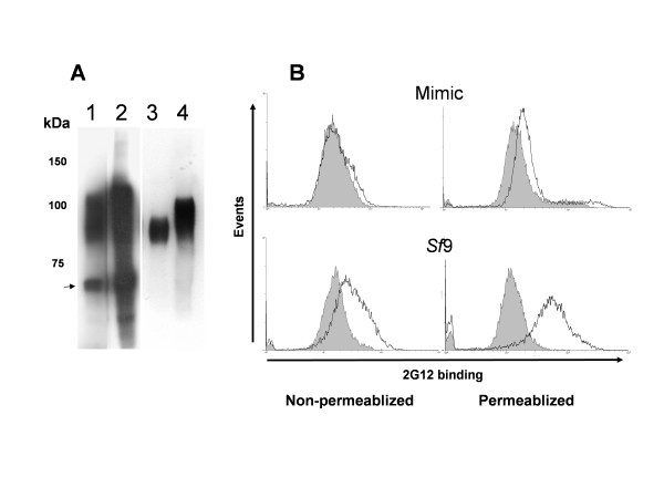
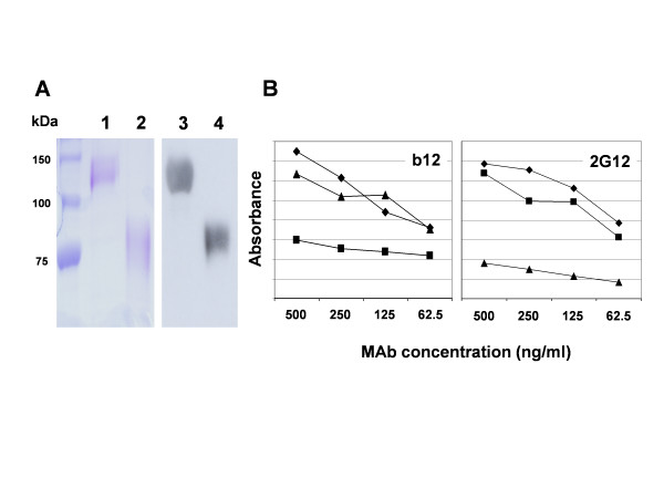
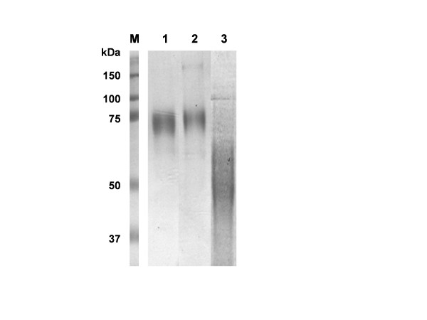
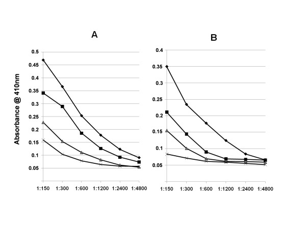
Background: The possibility that a sub domain of a C clade HIV-1 gp120 could act as an effective immunogen was investigated. To do this, the outer domain (OD) of gp120CN54 was expressed and characterized in a construct marked by a re-introduced conformational epitope for MAb 2G12. The expressed sequence showed efficient epitope retention on the isolated ODCN54 suggesting authentic folding. To facilitate purification and subsequent immunogenicity ODCN54 was fused to the Fc domain of human IgG1. Mice were immunised with the resulting fusion proteins and also with gp120CN54-Fc and gp120 alone.

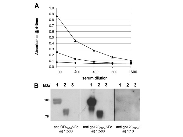
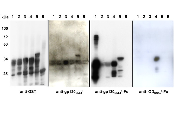
Results: Fusion to Fc was found to stimulate antibody titre and Fc tagged ODCN54 was substantially more immunogenic than non-tagged gp120. Immunogenicity appeared the result of Fc facilitated antigen processing as immunisation with an Fc domain mutant that reduced binding to the FcR lead to a reduction in antibody titre when compared to the parental sequence. The breadth of the antibody response was assessed by serum reaction with five overlapping fragments of gp120CN54 expressed as GST fusion proteins in bacteria. A predominant anti-inner domain and anti-V3C3 response was observed following immunisation with gp120CN54-Fc and an anti-V3C3 response to the ODCN54-Fc fusion.

Conclusion: The outer domain of gp120CN54 is correctly folded following expression as a C terminal fusion protein. Immunogenicity is substantial when targeted to antigen presenting cells but shows V3 dominance in the polyvalent response. The gp120 outer domain has potential as a candidate vaccine component.


Figures

Figure 1
Figure 1
Characterisation of HIV-1 gp120CN54 (V295N+A394T) expressed as a transmembrane bound protein in both Spodoptera frugiperda (Sf9) and Mimic cells (Invitrogen). A. Western blot of cell extracts (tracks 1 and 2) and supernatant (tracks 3 and 4) showing the increase in size associated with gp120 with modified glycans. Track 1 and 3 – Sf9 cells, Track 2 and 4 – Mimic cells. Note the presence of the unglycosylated apoprotein (arrowed) and glycosylation intermediates in the cell extracts which are absent in the supernatant. The gp120 present in the supernatant is due to pickup by budding baculovirus particles (see [53]). Markers to the left are in kilodaltons. B. Flow cytometry of permeablized or non-permeablized Sf9 and Mimic cells following staining with 2G12. Cells were permeablized by incubation in 0.1% Triton X-100 followed by fixing in 3% paraformaldehyde. Control cell profiles are shaded, infected cell profiles are unshaded.
Figure 2
Figure 2
Purification of gp120CN54+-Fc and ODCN54+-Fc and reaction with MAbs b12 and 2G12. A. Protein present in the supernatant of recombinant baculovirus infected insect cells was purified by lectin and protein A chromatography and analysed by SDS-PAGE (tracks 1 and 2) and western blot (tracks 3 and 4) using an anti-human Ig conjugate. Tracks 1 and 3 – gp120CN54+-Fc;Tracks 2 and 4 – ODCN54+-Fc. Protein size markers indicated to the left are in kilodaltons (kDa). B. Purified protein was coated onto plastic at 10 μg/ml and used to assess the binding of monoclonal antibodies b12 or 2G12 as shown. The samples were (▲) – gp120CN54+-Fc; (■) – ODCN54+-Fc; (◆) – gp120CN54+. Binding was detected by an anti-human Ig light chain conjugate.
Figure 3
Figure 3
Analysis of serum response to various CN54 gp120s by ELISA and Western blot. A. Pooled mouse sera were assayed on highly purified CN54 gp120 in a reciprocal dilution series starting at 1:100 and binding detected with an anti-mouse conjugate. The immunogens were: (▲) – gp120CN54+-Fc;(■) – ODCN54+-Fc;(◆) – gp120CN54+. B. Western blot analysis of each serum on gp120CN54+ (track 1) ODCN54+-Fc (track 2) and gp120Bal (track3 – to assess cross clade reactivity). The panels were blotted with the sera raised to the proteins shown and were used at the dilutions indicated. Anti-Fc reactivity in the sera was blocked by pre-incubation with excess HCV E2-Fc (not described).
Figure 4
Figure 4
Analysis of serum response to GST-gp120CN54 fragments by Western blot. Expression of each GST fusion protein was induced for 3 hrs and the equivalent of 50 μl of the culture fractionated on 10% SDS-PAGE before the transfer. Lanes 1–5 are the GST-gp120CN54 fusion proteins 1 to 5 with the origin of the gp120CN54 fragments as shown in additional file 2. Lane 6 is GST only (western blot reaction at 26 kDa and 54 kDa in panel A are monomer and dimer of GST respectively). The GST serum (Sigma) was used at 1:5000, serum dilutions for blots B-D were as described for figure 3.
Figure 5
Figure 5
Purification of ODCN54+-Fc, ODCN54+-Fc(VA) and ODCN54+-His. Protein present in the supernatant of recombinant baculovirus infected insect cells was purified by a combination of lectin and protein A chromatography and analysed by 10% SDS-PAGE. Track M – markers with molecular weights as shown; track 1 – ODCN54+-Fc; track 2 – ODCN54+-Fc(VA) and track 3 – ODCN54+-His. Note the slightly slower migration of the VA mutant as a result of the hydrophobicity change in the loss of two leucines within the Fc coding region. The smear of ODCN54+-His was typical and reflects the high level of glycosylation on a fairly small protein fragment. The yield for all constructs was ~1 mg/L of culture.
Figure 6
Figure 6
Analysis of serum responses to gp120CN54 (A) and ODCN54 (B) assessed by ELISA following immunization with the various ODCN54constructs described. Protein immunogens, as shown in figure 5 plus the complex between ODCN54+-Fc and GG7, were used to immunise groups of mice as before and terminal serum titres obtained following incubation on immobilised gp120CN54 and ODCN54 followed by an anti mouse conjugate. The sera were generated to: ODCN54+-Fc (◆); ODCN54+-Fc + GG7 (■); ODCN54+-Fc(VA) (▲) and ODCN54+-His (X).

References

    1. Burton DR, Desrosiers RC, Doms RW, Koff WC, Kwong PD, Moore JP, Nabel GJ, Sodroski J, Wilson IA, Wyatt RT. HIV vaccine design and the neutralizing antibody problem. Nat Immunol. 2004;5:233–236. doi: 10.1038/ni0304-233. - DOI - PubMed
    1. Burton DR, Stanfield RL, Wilson IA. Antibody vs. HIV in a clash of evolutionary titans. Proc Natl Acad Sci U S A. 2005;102:14943–14948. doi: 10.1073/pnas.0505126102. - DOI - PMC - PubMed
    1. Burton DR, Moore JP. Why do we not have an HIV vaccine and how can we make one? Nat Med. 1998;4:495–498. doi: 10.1038/nm0598supp-495. - DOI - PubMed
    1. Wei X, Decker JM, Wang S, Hui H, Kappes JC, Wu X, Salazar-Gonzalez JF, Salazar MG, Kilby JM, Saag MS, Komarova NL, Nowak MA, Hahn BH, Kwong PD, Shaw GM. Antibody neutralization and escape by HIV-1. Nature. 2003;422:307–312. doi: 10.1038/nature01470. - DOI - PubMed
    1. Moore PL, Crooks ET, Porter L, Zhu P, Cayanan CS, Grise H, Corcoran P, Zwick MB, Franti M, Morris L, Roux KH, Burton DR, Binley JM. Nature of nonfunctional envelope proteins on the surface of human immunodeficiency virus type 1. J Virol. 2006;80:2515–2528. doi: 10.1128/JVI.80.5.2515-2528.2006. - DOI - PMC - PubMed

Publication types

MeSH terms