. 2007 Jul;206(1):95-100.
doi: 10.1016/j.expneurol.2007.04.002. Epub 2007 Apr 18.

Estradiol augments peri-infarct cerebral vascular density in experimental stroke

Affiliations

Estradiol augments peri-infarct cerebral vascular density in experimental stroke

Agnieszka A Ardelt et al. Exp Neurol. 2007 Jul.

Abstract

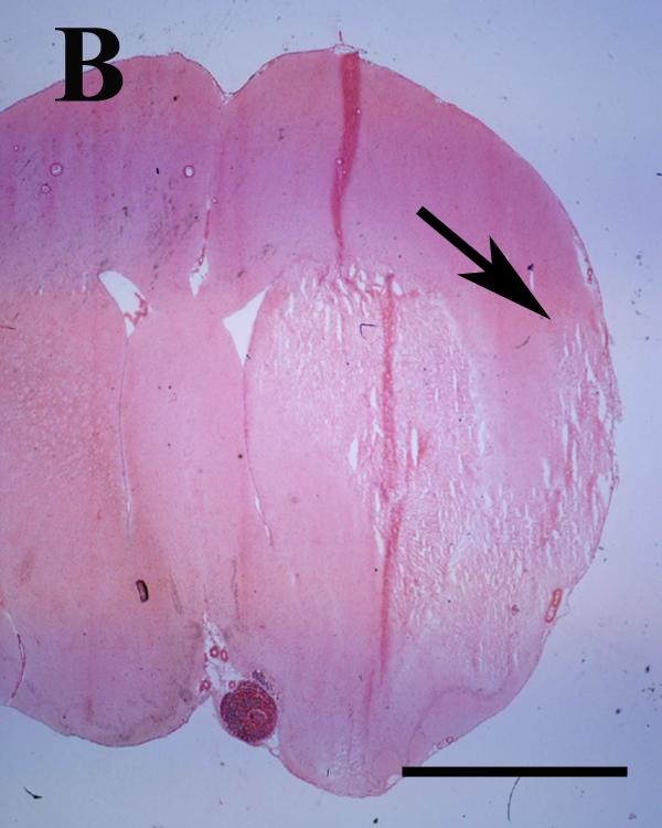
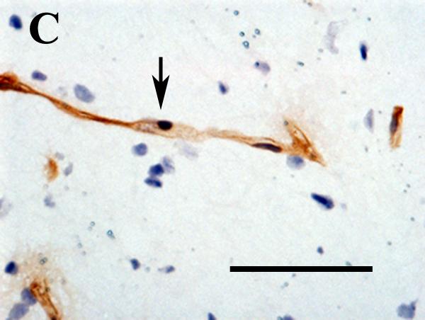
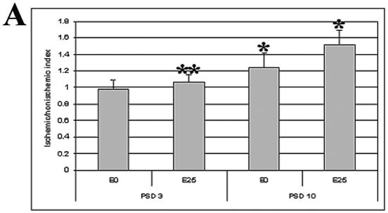
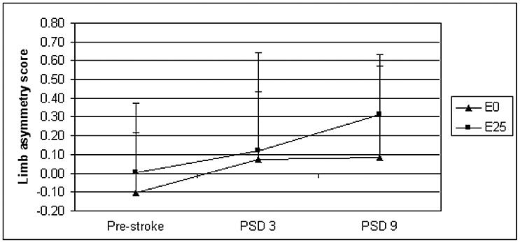
Peri-infarct increase of vascular density has been observed in animals and in humans with ischemic stroke. Increased peri-infarct vascular density correlates with improved functional outcome after stroke. We hypothesized that pre-treatment with estradiol will increase post-ischemic peri-infarct capillary density in a rat model of transient ischemic stroke. Estradiol, compared to placebo, augmented post-ischemic peri-infarct vascular density by 22% 10 days after stroke. Recovery of forelimb function was not improved with estradiol treatment on day three and nine post-stroke. Loss of estradiol may limit repair in the peri-infarct region by limiting angiogenesis, but functional significance in stroke recovery requires further investigation.


Figures

Figure 1
Figure 1
A and B. Eosin-stained coronal rat brain section photographed at 1.25X illustrating the approximate peri-infarct region analyzed (arrows) in rats with the largest (A) and smallest (B) infarcts on post stroke day three. Infarct dimensions and sub-regional localization of the cortical portion of the lesion were heterogeneous within and between cohorts. Scale bar = 5 mm.
Figure 1
Figure 1
A and B. Eosin-stained coronal rat brain section photographed at 1.25X illustrating the approximate peri-infarct region analyzed (arrows) in rats with the largest (A) and smallest (B) infarcts on post stroke day three. Infarct dimensions and sub-regional localization of the cortical portion of the lesion were heterogeneous within and between cohorts. Scale bar = 5 mm.
Figure 2
Figure 2
A and B. Low magnification views of CD31-labeled structures in the non-ischemic cortex (A) and peri-infarct region (B) photographed 10 days after experimental ischemic stroke. Counts of these structures in the peri-infarct and contralateral mirror-image regions were used to derive ischemic/nonischemic vascular indices. The structures located in the peri-ischemic region were more numerous, larger, and torturous. Scale bar = 200μm. C and D. High magnification views of CD31-labeled structures (arrows) in intact brain (C) and in the peri-infarct region (D) 10 days after experimental stroke. In intact brain, the morphology is typical of a normal parenchymal capillary: the endothelial cells form a narrow lumen, with relatively few nuclei oriented along the axis of the vessel. In the injured area, the morphology somewhat resembles that of angiogenic blood vessels: the lumen is wide, and there are numerous endothelial cell nuclei clustered around the lumen. CD31 outlines the surface membranes. Nuclei are labeled with hematoxylin. Scale bar = 50μm.
Figure 2
Figure 2
A and B. Low magnification views of CD31-labeled structures in the non-ischemic cortex (A) and peri-infarct region (B) photographed 10 days after experimental ischemic stroke. Counts of these structures in the peri-infarct and contralateral mirror-image regions were used to derive ischemic/nonischemic vascular indices. The structures located in the peri-ischemic region were more numerous, larger, and torturous. Scale bar = 200μm. C and D. High magnification views of CD31-labeled structures (arrows) in intact brain (C) and in the peri-infarct region (D) 10 days after experimental stroke. In intact brain, the morphology is typical of a normal parenchymal capillary: the endothelial cells form a narrow lumen, with relatively few nuclei oriented along the axis of the vessel. In the injured area, the morphology somewhat resembles that of angiogenic blood vessels: the lumen is wide, and there are numerous endothelial cell nuclei clustered around the lumen. CD31 outlines the surface membranes. Nuclei are labeled with hematoxylin. Scale bar = 50μm.
Figure 3
Figure 3
A. Peri-infarct vascular density indices were determined in brain sections labeled with anti-CD31 antibody from placebo (E0) and estradiol-treated (E25) animals on post stroke day three and 10 (PSD 3 and PSD 10, respectively). *p=0.011 for E0 vs. E25 on PSD 10; **p=0.010 for E25 on PSD 3 vs. E25 on PSD 10. One-way ANOVA. Sample size: E0, PSD 3, n=4; E25, PSD 3, n=4; E0, PSD 10, n=6; E25, PSD 10, n=5. B. Ischemic/nonischemic difference in the proportions of CD31-positive structures of different sizes was derived 10 days post stroke. CD31-positive structures were counted in placebo (E0) and estradiol (E25)-treated rats and sorted into five bins by pixel area. The percent difference in proportion of structures within each bin between the peri-infarct area and the contralateral matched region was determined. *p<0.017 for E0 vs. E25 in >1000 pixel bin; **p<0.005 for >1000 versus 50-250 pixel bin for estradiol-treated animals. Two-way ANOVA.
Figure 3
Figure 3
A. Peri-infarct vascular density indices were determined in brain sections labeled with anti-CD31 antibody from placebo (E0) and estradiol-treated (E25) animals on post stroke day three and 10 (PSD 3 and PSD 10, respectively). *p=0.011 for E0 vs. E25 on PSD 10; **p=0.010 for E25 on PSD 3 vs. E25 on PSD 10. One-way ANOVA. Sample size: E0, PSD 3, n=4; E25, PSD 3, n=4; E0, PSD 10, n=6; E25, PSD 10, n=5. B. Ischemic/nonischemic difference in the proportions of CD31-positive structures of different sizes was derived 10 days post stroke. CD31-positive structures were counted in placebo (E0) and estradiol (E25)-treated rats and sorted into five bins by pixel area. The percent difference in proportion of structures within each bin between the peri-infarct area and the contralateral matched region was determined. *p<0.017 for E0 vs. E25 in >1000 pixel bin; **p<0.005 for >1000 versus 50-250 pixel bin for estradiol-treated animals. Two-way ANOVA.
Figure 4
Figure 4
Forelimb use asymmetry scores were derived for estradiol (E2) and placebo (E0)-treated animals using the cylinder test pre stroke and on post stroke day three (PSD 3) and 10 (PSD 10). Limb asymmetry score = (right - left) / (right + left + both); thus, positive scores reflect decreased left forelimb use (as expected with a right hemispheric stroke).

Similar articles

Cited by

References

    1. Alkayed NJ, Harukuni I, Kimes AS, London ED, Traystman RJ, Hurn PD.Gender-linked brain injury in experimental stroke Stroke 199829159–165. discussion 166 - PubMed
    1. Ardelt AA, McCullough LD, Korach KS, Wang MM, Munzenmaier DH, Hurn PD. Estradiol regulates angiopoietin-1 mRNA expression through estrogen receptor-alpha in a rodent experimental stroke model. Stroke. 2005;36:337–341. - PubMed
    1. Chen J, Li Y, Zhang R, Katakowski M, Gautam SC, Xu Y, Lu M, Zhang Z, Chopp M. Combination therapy of stroke in rats with a nitric oxide donor and human bone marrow stromal cells enhances angiogenesis and neurogenesis. Brain Res. 2004;1005:21–28. - PubMed
    1. Chen J, Zhang C, Jiang H, Li Y, Zhang L, Robin A, Katakowski M, Lu M, Chopp M. Atorvastatin induction of VEGF and BDNF promotes brain plasticity after stroke in mice. J Cereb Blood Flow Metab. 2005;25:281–290. - PMC - PubMed
    1. Chen J, Zhang ZG, Li Y, Wang Y, Wang L, Jiang H, Zhang C, Lu M, Katakowski M, Feldkamp CS, Chopp M. Statins induce angiogenesis, neurogenesis, and synaptogenesis after stroke. Ann Neurol. 2003;53:743–751. - PubMed

MeSH terms