

Cone-based vision in the aging mouse
- PMID: 17509638
- PMCID: PMC2049007
- DOI: 10.1016/j.visres.2007.03.023
Cone-based vision in the aging mouse
Abstract
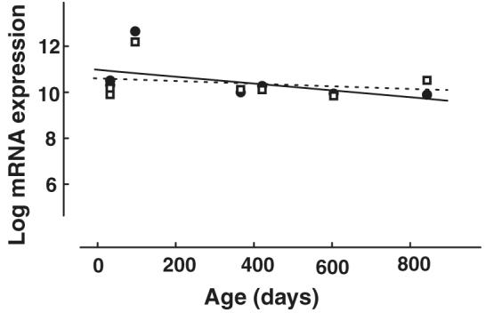
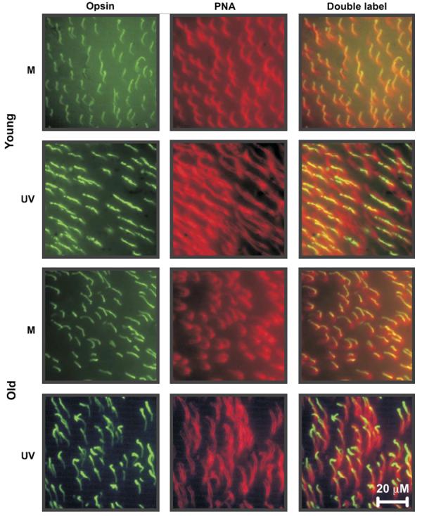
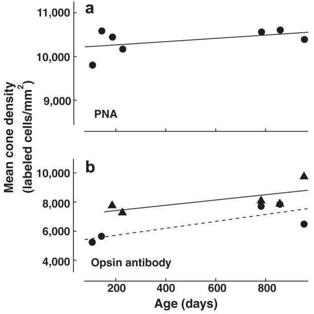
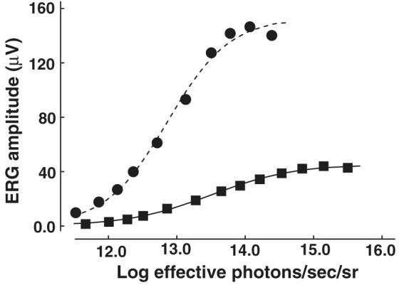
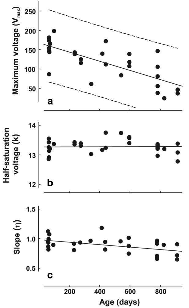
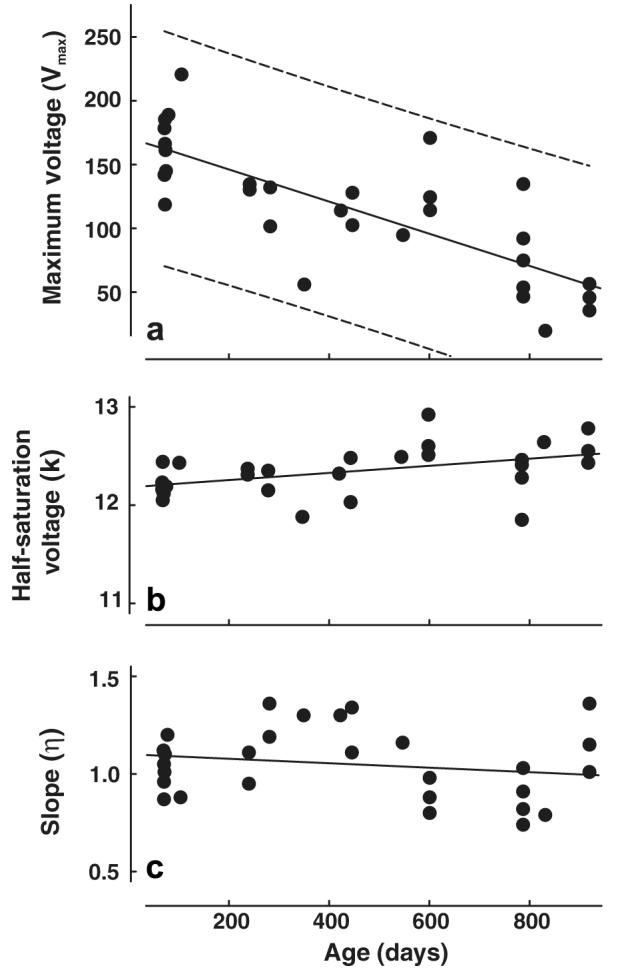
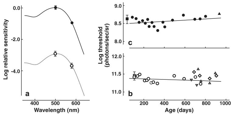
People often experience age-related declines in cone-based visual capacities despite an absence of apparent visual pathology. Although mice are used as models of human visual pathologies associated with aging, little is known about how age impacts vision in animals with disease-free retinas since most studies have heretofore examined relatively young mice. We examined the effects of age on cone-based vision by assessing opsin gene transcription, cone densities, the flicker electroretinogram (ERG), and behavioral increment thresholds in mice. ERG measurements of cone function showed age-related declines in maximum voltage (Vmax), while opsin gene transcription, cone density, and increment thresholds were unchanged even in extremely old mice. The age-related decline in Vmax seen in mice is qualitatively similar to that documented for human subjects. It is notable that Vmax, a commonly used index of ERG activity, does not predict behavioral performance in the mouse.
Figures
Similar articles
-
Transgenic mice expressing a functional human photopigment.Invest Ophthalmol Vis Sci. 1998 May;39(6):1036-43. Invest Ophthalmol Vis Sci. 1998. PMID: 9579484
-
Cone receptor variations and their functional consequences in two species of hamster.Vis Neurosci. 1999 Jan-Feb;16(1):53-63. doi: 10.1017/s0952523899161029. Vis Neurosci. 1999. PMID: 10022478
-
Cone opsin mislocalization in Rpe65-/- mice: a defect that can be corrected by 11-cis retinal.Invest Ophthalmol Vis Sci. 2005 Oct;46(10):3876-82. doi: 10.1167/iovs.05-0533. Invest Ophthalmol Vis Sci. 2005. PMID: 16186377
-
Human retinal dark adaptation tracked in vivo with the electroretinogram: insights into processes underlying recovery of cone- and rod-mediated vision.J Physiol. 2022 Nov;600(21):4603-4621. doi: 10.1113/JP283105. Epub 2022 Jun 7. J Physiol. 2022. PMID: 35612091 Free PMC article. Review.
-
The visual cycle of the cone photoreceptors of the retina.Nutr Rev. 2004 Jul;62(7 Pt 1):283-6. doi: 10.1111/j.1753-4887.2004.tb00053.x. Nutr Rev. 2004. PMID: 15384919 Review.
Cited by
-
Validating Fluorescent Chrnb4.EGFP Mouse Models for the Study of Cone Photoreceptor Degeneration.Transl Vis Sci Technol. 2020 Aug 18;9(9):28. doi: 10.1167/tvst.9.9.28. eCollection 2020 Aug. Transl Vis Sci Technol. 2020. PMID: 32879784 Free PMC article.
-
Effects of aging and sensory loss on glial cells in mouse visual and auditory cortices.Glia. 2012 Apr;60(4):541-58. doi: 10.1002/glia.22287. Epub 2012 Jan 5. Glia. 2012. PMID: 22223464 Free PMC article.
-
Color and contrast vision in mouse models of aging and Alzheimer's disease using a novel visual-stimuli four-arm maze.Sci Rep. 2021 Jan 13;11(1):1255. doi: 10.1038/s41598-021-80988-0. Sci Rep. 2021. PMID: 33441984 Free PMC article.
-
Amyloid Precursor-Like Protein 2 deletion-induced retinal synaptopathy related to congenital stationary night blindness: structural, functional and molecular characteristics.Mol Brain. 2016 Jun 8;9(1):64. doi: 10.1186/s13041-016-0245-z. Mol Brain. 2016. PMID: 27267879 Free PMC article.
-
The first synapse in vision in the aging mouse retina.Front Cell Neurosci. 2023 Nov 3;17:1291054. doi: 10.3389/fncel.2023.1291054. eCollection 2023. Front Cell Neurosci. 2023. PMID: 38026697 Free PMC article.
References
-
- Applebury ML, Antoch MP, Baxter LC, Chun LLY, Falk JD, Farhangfar F, et al. The murine cone photoreceptor: a single cone type expresses both S and M opsins with retinal spatial patterning. Neuron. 2000;27:513–523. - PubMed
-
- Betts LR, Taylor CP, Sekuler AB, Bennett PJ. Aging reduces center-surround antagonism in visual motion procession. Neuron. 2005;45:361–366. - PubMed
-
- Birch DG, Anderson JL. Standardized full-field electroretinography: Normal values and their variation with age. Archives of Ophthalmology. 1992;110:1571–1576. - PubMed
-
- Birch DG, Hood DC, Locke KG, Hoffman DR, Tzekov RT. Quantitative electroretinogram measures of phototransduction in cone and rod photoreceptors. Archives of Ophthalmology. 2002;120:1045–1051. - PubMed
-
- Blanks JC, Johnson LV. Specific binding of peanut lectin to a class of retinal photoreceptor cells. Investigative Ophthalmology and Visual Science. 1984;25:546–557. - PubMed
Publication types
MeSH terms
Substances
Grants and funding
LinkOut - more resources
Full Text Sources
Other Literature Sources
Medical
