

MUC1 is involved in trophoblast transendothelial migration
- PMID: 17509701
- PMCID: PMC1986703
- DOI: 10.1016/j.bbamcr.2007.04.006
MUC1 is involved in trophoblast transendothelial migration
Abstract
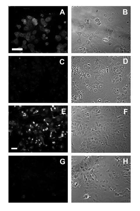
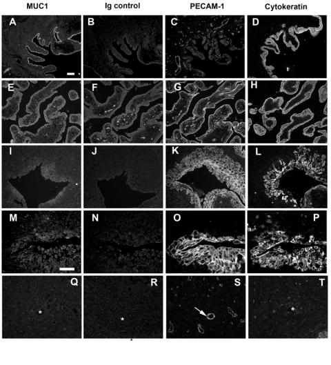
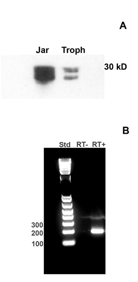
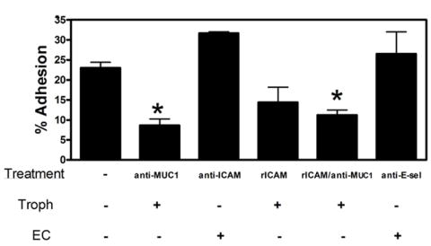
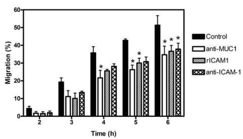
The factors that regulate trophoblast invasion of the uterine vasculature are incompletely understood. In this paper we show that macaque trophoblasts express the mucin, MUC1, and that it is involved in trophoblast-endothelial interaction. Immunocytochemistry, Western blotting and RT-PCR analyses confirmed that MUC1 was expressed by isolated early gestation macaque trophoblasts. MUC1 was also detected in endovascular trophoblasts in sections of placental-decidual tissue during early gestation. A blocking antibody against MUC1 reduced trophoblast adhesion to uterine endothelial cells and also blocked trophoblast transendothelial migration. MUC1 is known to bind to Intercellular Adhesion Molecule-1 (ICAM-1) in other systems. Incubation in the presence of a blocking antibody against Intercellular Adhesion Molecule-1 (ICAM-1) or recombinant ICAM-1 modestly, but significantly, reduced transendothelial trophoblast migration. These results are consistent with the idea that MUC1 is involved in trophoblast adhesion to uterine endothelial cells and in trophoblast transendothelial migration.
Figures
Similar articles
-
Expression of adhesion molecules by endovascular trophoblast and decidual endothelial cells: implications for vascular invasion during implantation.Placenta. 1994 Jan;15(1):21-33. doi: 10.1016/s0143-4004(05)80233-4. Placenta. 1994. PMID: 7516077
-
Platelet-endothelial cell adhesion molecule-1 is expressed by a subpopulation of human trophoblasts: a possible mechanism for trophoblast-endothelial interaction during haemochorial placentation.Mol Hum Reprod. 1998 Apr;4(4):357-67. doi: 10.1093/molehr/4.4.357. Mol Hum Reprod. 1998. PMID: 9620836
-
MUC1 expression is increased during human placental development and suppresses trophoblast-like cell invasion in vitro.Biol Reprod. 2008 Aug;79(2):233-9. doi: 10.1095/biolreprod.108.067629. Epub 2008 Apr 16. Biol Reprod. 2008. PMID: 18417712
-
[Molecular mechanisms of regulation of cytotrophoblastic invasion in uteroplacental region].Arkh Patol. 2001 Sep-Oct;63(5):3-8. Arkh Patol. 2001. PMID: 11765411 Review. Russian.
-
IFPA meeting 2016 workshop report III: Decidua-trophoblast interactions; trophoblast implantation and invasion; immunology at the maternal-fetal interface; placental inflammation.Placenta. 2017 Dec;60 Suppl 1:S15-S19. doi: 10.1016/j.placenta.2017.04.019. Epub 2017 Apr 25. Placenta. 2017. PMID: 28456431 Review.
Cited by
-
Blockade of extracellular NM23 or its endothelial target slows breast cancer growth and metastasis.Integr Cancer Sci Ther. 2015;2(4):192-200. doi: 10.15761/icst.1000139. Integr Cancer Sci Ther. 2015. PMID: 26413311 Free PMC article.
-
Adrenomedullin 2 (ADM2) Regulates Mucin 1 at the Maternal-Fetal Interface in Human Pregnancy.Biol Reprod. 2015 Dec;93(6):136. doi: 10.1095/biolreprod.115.134296. Epub 2015 Oct 28. Biol Reprod. 2015. PMID: 26510869 Free PMC article.
-
Distinct genomic signatures of adaptation in pre- and postnatal environments during human evolution.Proc Natl Acad Sci U S A. 2008 Mar 4;105(9):3215-20. doi: 10.1073/pnas.0712400105. Epub 2008 Feb 27. Proc Natl Acad Sci U S A. 2008. PMID: 18305157 Free PMC article.
-
AP-1 transcription factors, mucin-type molecules and MMPs regulate the IL-11 mediated invasiveness of JEG-3 and HTR-8/SVneo trophoblastic cells.PLoS One. 2012;7(1):e29745. doi: 10.1371/journal.pone.0029745. Epub 2012 Jan 3. PLoS One. 2012. PMID: 22235337 Free PMC article.
-
The MUC1 extracellular domain subunit is found in nuclear speckles and associates with spliceosomes.PLoS One. 2012;7(8):e42712. doi: 10.1371/journal.pone.0042712. Epub 2012 Aug 8. PLoS One. 2012. PMID: 22905162 Free PMC article.
References
-
- Gendler SJ. MUC1, the renaissance molecule. J Mammary Gland Biol Neoplasia. 2001;6:339–53. - PubMed
-
- Cloosen S, Thio M, Vanclee A, van Leeuwen EB, Senden-Gijsbers BL, Oving EB, Germeraad WT, Bos GM. Mucin-1 is expressed on dendritic cells, both in vitro and in vivo. Int Immunol. 2004;16:1561–71. - PubMed
-
- Wykes M, MacDonald KP, Tran M, Quin RJ, Xing PX, Gendler SJ, Hart DN, McGuckin MA. MUC1 epithelial mucin (CD227) is expressed by activated dendritic cells. J Leukoc Biol. 2002;72:692–701. - PubMed
-
- Hilkens J, Wesseling J, Vos HL, Storm J, Boer B, van der Valk SW, Maas MC. Involvement of the cell surface-bound mucin, episialin/MUC1, in progression of human carcinomas. Biochem Soc Trans. 1995;23:822–6. - PubMed
Publication types
MeSH terms
Substances
Grants and funding
LinkOut - more resources
Full Text Sources
Medical
Research Materials
Miscellaneous
