Clinical Trial
. 2007 Sep 1;110(5):1656-63.
doi: 10.1182/blood-2007-03-081240. Epub 2007 May 17.

Targeting MEK induces myeloma-cell cytotoxicity and inhibits osteoclastogenesis

Affiliations
Clinical Trial

Targeting MEK induces myeloma-cell cytotoxicity and inhibits osteoclastogenesis

Yu-Tzu Tai et al. Blood. .

Abstract

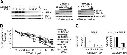
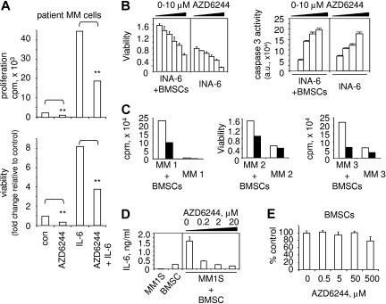
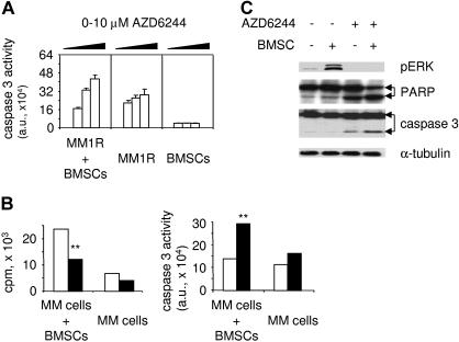
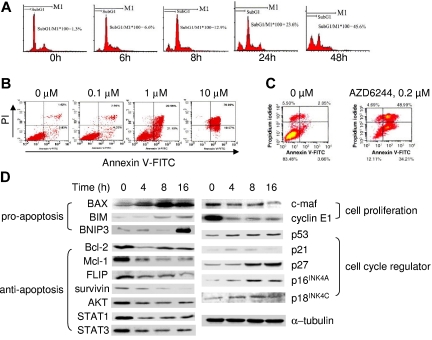
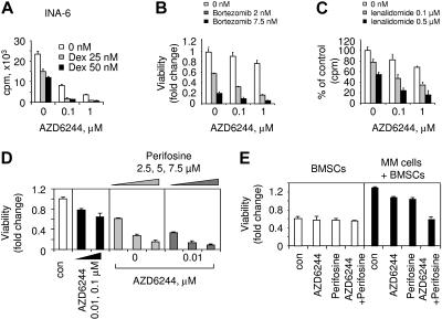
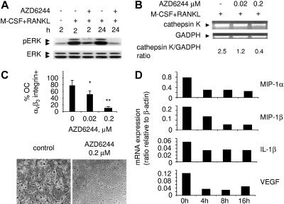
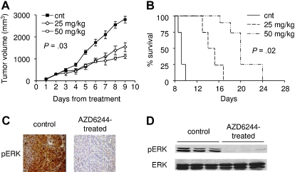
Activation of the extracellular signal-regulated kinase1/2 (ERK1/2) signaling cascade mediates human multiple myeloma (MM) growth and survival triggered by cytokines and adhesion to bone marrow stromal cells (BMSCs). Here, we examined the effect of AZD6244 (ARRY-142886), a novel and specific MEK1/2 inhibitor, on human MM cell growth in the bone marrow (BM) milieu. AZD6244 blocks constitutive and cytokine-stimulated ERK1/2 phosphorylation and inhibits proliferation and survival of human MM cell lines and patient MM cells, regardless of sensitivity to conventional chemotherapy. Importantly, AZD6244 (200 nM) induces apoptosis in patient MM cells, even in the presence of exogenous interleukin-6 or BMSCs associated with triggering of caspase 3 activity. AZD6244 sensitizes MM cells to both conventional (dexamethasone) and novel (perifosine, lenalidomide, and bortezomib) therapies. AZD6244 down-regulates the expression/secretion of osteoclast (OC)-activating factors from MM cells and inhibits in vitro differentiation of MM patient PBMCs to OCs induced by ligand for receptor activator of NF-kappaB (RANKL) and macrophage-colony stimulating factor (M-CSF). Finally, AZD6244 inhibits tumor growth and prolongs survival in vivo in a human plasmacytoma xenograft model. Taken together, these results show that AZD6244 targets both MM cells and OCs in the BM microenvironment, providing the preclinical framework for clinical trials to improve patient outcome in MM.


Figures

Figure 1
Figure 1
AZD6244 specifically inhibits ERK phosphorylation and induces cytotoxicity in MM lines resistant to conventional chemotherapy, as well as in primary patient MM cells. (A) Serum-starved MM1S MM cells were pretreated with either AZD6244 (10 nM) or control medium for 1 hour and then stimulated with IGF-1 (100 ng/mL) for indicated time intervals (left panel). Similarly, MM1S cells were pretreated with serial dilutions of AZD6244 (0-10 μM) and stimulated with IL-6 (50 ng/mL, middle panel) or sCD40L (5 μg/mL, right panel). Immunoblotting was performed using anti-pAKT and pERK Abs, as well as anti–β-actin or anti–α-tubulin mAbs as loading controls. (B) Ten MM lines, including drug-sensitive RPMI8226, Dox-resistant RPMI8226 (Dox40), melphalan-resistant RPMI8226 (LR5), Dex-sensitive MM1S and -resistant MM1R, IL-6–dependent INA-6, and MCCAR, 28PE, 28BM, and OPM1 cells, were cultured with AZD6244 for 2 days and then pulsed with [3H]thymidine for the last 8 hours for measurement of DNA synthesis. Data represent mean (+ SE) of quadruplicate cultures; cpm indicates counts per minute. (C) Freshly isolated tumor cells from 3 MM patients (MM 1, MM 2, MM3) were cultured with AZD6244 (0.02-20 μM) for 2 days, and cytotoxicity was assessed by MTT assay.
Figure 2
Figure 2
AZD6244 induces MM-cell cytotoxicity in the presence of IL-6 or BMSCs. (A) CD138-purified patient MM cells were treated with AZD6244 (20 nM) in the presence or absence of IL-6 (10 ng/mL). Cytotoxicity (growth and viability) was measured by [3H]thymidine uptake and MTT assay. **P < .01 compared with control. (B) INA-6 MM cells were treated for 2 days with serial dilutions of AZD6244 (0-10 μM) in the presence or absence of BMSCs, followed by MTT assay (left panel) and caspase 3 activity assay (right panel). Data represent mean (+ SE) of triplicate cultures; au indicates arbitrary unit. (C) Freshly purified CD138+ tumor cells from 3 MM patients were cultured with AZD6244 (200 nM, ■) or control medium (□), alone or with BMSCs. Cytotoxicity was determined by [3H]thymidine incorporation assay (MM1 and MM3) or MTT assay (MM2). (D) MM1S cells were treated with AZD6244 (0.2-20 μM) for 2 days in the presence or absence of BMSCs, and supernatants were assayed for IL-6 by ELISA. (E) BMSCs derived from MM patients were incubated for 2 days with AZD6244 (0.5-500 μM) in triplicate and then subjected to MTT assay. Results represent mean (+ SE) of BMSCs from 3 patients.
Figure 3
Figure 3
AZD6244 induces apoptotic signaling in MM cells adherent to BMSCs. (A) Dex-resistant MM1R cells were incubated for 2 days with serial dilutions of AZD6244 (0-10 μM), in the presence or absence of BMSCs, followed by caspase 3 activity assays. (B) CD138+ patient MM cells were incubated with AZD6244 (200 nM, ■) or control medium (□), in the presence or absence of BMSCs. Cytotoxicity was assayed by growth inhibition (left panel), and apoptosis was measured by caspase 3 activity assay (right panel). **P < .01 compared with control. (C) MM1S cells were cultured in BMSC-coated plates for 24 hours, in the presence or absence of AZD6244. MM1S cells were then harvested, lysed, electrophoresed, and immunoblotted for pERK, PARP, and caspase 3, with α-tubulin as loading control.
Figure 4
Figure 4
Effect of AZD6244 on cell-cycle profile and functional sequelae of ERK inhibition in MM cells. (A) INA-6 MM cells were treated with AZD6244 (2 μM) for the time intervals indicated. Cell-cycle profile was then evaluated by PI staining using flow cytometry. (B) INA-6 MM cells were treated with AZD6244 (0.1-10 μM) for 2 days, followed by PI/annexin V staining and flow-cytometric analysis. (C) CD138+ patient MM cells were incubated for 2 days with control media or AZD6244 (0.2 μM) and then subjected to PI/annexin V staining. (D) INA-6 MM cells in 10% FBS-containing medium were treated with AZD6244 for the intervals indicated and lysed, and whole-cell lysates were then subjected to immunoblotting using specific Abs.
Figure 5
Figure 5
ERK inactivation by AZD6244 enhances cytotoxicity of conventional and novel agents. (A) INA-6 cells were cultured for 2 days with Dex (0-50 nM) in the presence or absence of AZD6244 (0-1 μM), and DNA synthesis was measured. Data represent mean (+ SE) of quadruplicate cultures. MM1S cells were cultured for 2 days with (B) bortezomib (0-7.5 nM) and AZD6244 (0-1 μM) or (C) lenalidomide (0-0.5 μM) and AZD6244 (0-1 μM). (D) CD138+ patient MM cells were incubated for 3 days with AZD6244 (0-0.1 μM), perifosine (0-7.5 μM), or the combination. (E) CD138+ MM-patient cells were cultured with BMSCs in the presence or absence of AZD6244 (20 nM), perifosine (7.5 μM), or the combination. Cytotoxicity (viability) was measured by MTT assay, expressed as fold change to control.
Figure 6
Figure 6
AZD6244 OC formation and transcripts of OC-stimulating factors in MM cells. (A) Adherent monocytic OC precursors from MM-patient PBMCs were incubated with M-CSF/RANKL, in the presence or absence of AZD6244 (0.1 μM), for indicated time intervals. Total cell lysates were prepared and subjected to immunoblotting using anti-pERK Ab, with anti-ERK Ab as a loading control. (B) Adherent OC precursors from MM patient PBMCs were incubated with M-CSF/RANKL in the presence of AZD6244 for 10 days. Total RNA was then prepared and subjected to RT-PCR for cathepsin K (PCR product: 127 bp). RT-PCR for GADPH served as an internal control. (C) PBMCs from MM patients (n = 3) were incubated with M-CSF/RANKL for 14 days, in the presence or absence of AZD6244. OC formation was characterized by integrin αvβ3 expression by flow-cytometric analysis. *P < .05; **P < .005; data represent the mean of 3 experiments (± SE). Multinucleated OCs were induced by M-CSF/RANKL (control), whereas AZD6244 (0.2 μM) blocked OC formation; original magnification × 100. See “Patients, materials, and methods; Immunohistochemistry” for more information on image acquisition. (D) Transcriptional changes of indicated OC-activating factors in MM1S cells following AZD6244 treatment.
Figure 7
Figure 7
AZD6244 inhibits human plasmacytoma growth and prolongs survival in a murine xenograft model. (A) OPM1 MM cells were injected subcutaneously. Mice (n = 8 per group) received AZD6244 (orally twice per day) or control vehicle (cnt, ■) orally starting on day 1 for 9 days. Significant growth inhibition of OPM1 MM cells was noted in AZD6244-treated (◇ 25 mg/kg, ○ 50 mg/kg) compared with vehicle-treated control mice (n = 8; P = .03). Points indicate mean; bars, SE. (B) After OPM1 tumors were measurable, mice were treated with AZD6244 (25 mg/kg, — — —; 50 mg/kg, — - - —) or with control vehicle alone (cnt, —) for 9 consecutive days. Survival was evaluated from the first day of treatment until death or sacrifice (mice were killed when tumors reached 2 cm3 in diameter) using the SigmaPlot (Systat) analysis software (P = .02). (C) Representative immunohistochemical staining for phosphorylation of ERK (pERK) in tumor sections from vehicle control- and AZD6244 (25 mg/kg)–treated mice; original magnification × 100. See “Patients, materials, and methods; Immunohistochemistry” for more information on image acquisition. (D) Tumor tissues from mice treated with vehicle control or AZD6244 (25 mg/kg) for 1 day were harvested and processed, and lysates were subjected to immunoblotting using pERK and ERK Ab.

References

    1. Hideshima T, Bergsagel PL, Kuehl WM, Anderson KC. Advances in biology of multiple myeloma: clinical applications. Blood. 2004;104:607–618. - PubMed
    1. van de Donk NW, Kroger N, Hegenbart U, et al. Remarkable activity of novel agents bortezomib and thalidomide in patients not responding to donor lymphocyte infusions following nonmyeloablative allogeneic stem cell transplantation in multiple myeloma. Blood. 2006;107:3415–3416. - PubMed
    1. Barlogie B, Kyle RA, Anderson KC, et al. Standard chemotherapy compared with high-dose chemoradiotherapy for multiple myeloma: final results of phase III US Intergroup Trial S9321. J Clin Oncol. 2006;24:929–936. - PubMed
    1. Richardson P, Anderson K. Thalidomide and dexamethasone: a new standard of care for initial therapy in multiple myeloma. J Clin Oncol. 2006;24:334–336. - PubMed
    1. Richardson PG, Blood E, Mitsiades CS, et al. A randomized phase 2 study of lenalidomide therapy for patients with relapsed or relapsed and refractory multiple myeloma. Blood. 2006;108:3458–3464. - PMC - PubMed

Publication types

MeSH terms

Substances