Comparative Study
. 2007 Sep 1;110(5):1466-74.
doi: 10.1182/blood-2007-03-079178. Epub 2007 May 17.

The SOCS box of suppressor of cytokine signaling-3 contributes to the control of G-CSF responsiveness in vivo

Affiliations
Comparative Study

The SOCS box of suppressor of cytokine signaling-3 contributes to the control of G-CSF responsiveness in vivo

Kristy Boyle et al. Blood. .

Abstract

Suppressor of cytokine signaling 3 (SOCS3) is a negative regulator of granulocyte-colony stimulating factor (G-CSF) signaling in vivo. SOCS proteins regulate cytokine signaling by binding, via their SH2 domains, to activated cytokine receptors or their associated Janus kinases. In addition, they bind to the elongin B/C ubiquitin ligase complex via the SOCS box. To ascertain the contribution of the SOCS box of SOCS3 to in vivo regulation of G-CSF signaling, we generated mice expressing a truncated SOCS3 protein lacking the C-terminal SOCS box (SOCS3(Delta SB/Delta SB)). SOCS3(Delta SB/Delta SB) mice were viable, had normal steady-state hematopoiesis, and did not develop inflammatory disease. Despite the mild phenotype, STAT3 activation in response to G-CSF signaling was prolonged in SOCS3(Delta SB/Delta SB) bone marrow. SOCS3(Delta SB/Delta SB) bone marrow contained increased numbers of colony-forming cells responsive to G-CSF and IL-6. Treatment of the mice with pharmacologic doses of G-CSF, which mimics emergency granulopoiesis and therapeutic use of G-CSF, revealed that SOCS3(Delta SB/Delta SB) mice were hyperresponsive to G-CSF. Compared with wild-type mice, SOCS3(Delta SB/Delta SB) mice developed a more florid arthritis when tested using an acute disease model. Overall, the results establish a role for the SOCS box of SOCS3 in the in vivo regulation of G-CSF signaling and the response to inflammatory stimuli.


Figures
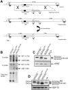
Figure 1
Figure 1
Generation and confirmation of SOCS3ΔSB/ΔSB mice. (A) The SOCS3 locus is shown at the top with the targeting construct and targeted allele below. The structure of the knock-in allele after removal of the neomycin selection cassette (PGKNEOpA) by cre-mediated excision is also shown. Exons are indicated as boxes with the coding region shaded. The 120-bp deletion in exon 2 of nucleotides 573 to 692 of the SOCS3 transcript that encodes the SOCS box is indicated by ΔSB. 5′ and 3′ probes used for Southern analysis are indicated. H indicates HindIII; RV, EcoRV. (B) Correct homologous recombination and deletion of the selection cassette was confirmed by Southern blot analyses of tail DNA digested with HindIII using 5′ and 3′ probes. DNA digested with PvuII was probed with a 130-bp SOCS box probe, confirming the absence of this sequence in SOCS3ΔSB/ΔSB samples. (C) RT-PCR analysis of cDNA prepared from RNA from livers from WT, SOCS3ΔSB/+, and SOCS3ΔSB/ΔSB mice injected with IL-6 (5 μg, intraperitoneally 60 minutes prior to the time that they were killed). Primers spanning the SOCS3 SOCS box were used. In SOCS3ΔSB/ΔSB samples, only the 200-bp transcript lacking the SOCS box was amplified. HPRT was amplified as a loading control. (D) Immunoblot of liver lysates prepared from mice injected with IL-6 (5 μg, intraperitoneally 60 minutes prior to the time that they were killed). SOCS3 was immunoprecipitated with a monoclonal antibody to SOCS3 and immunoblotted using a polyclonal SOCS3 antibody. In SOCS3ΔSB/ΔSB lysates, only the smaller (20 kDa) SOCS3 protein was detectable. NS indicates nonspecific band.
Figure 2
Figure 2
Removal of the SOCS3 SOCS box alters the regulation of the JAK/STAT pathway in vivo. (A) Activation of STAT3 in response to G-CSF. Bone marrow cells from SOCS3+/+ and SOCS3ΔSB/ΔSB or SOCS3ΔVAV/− mice were left untreated (NT) or stimulated with 50 ng/mL G-CSF for 15 minutes, washed, then lysed at intervals between 5 minutes and 120 minutes. Lysates were analyzed by immunoblot using antibodies specific for phospho-STAT3 or total STAT3. (B) Activation of ERK in response to G-CSF. Bone marrow cells from SOCS3+/+ and SOCS3ΔSB/ΔSB or SOCS3ΔVAV/− mice were left unstimulated (0 time point) or stimulated with 50 ng/mL G-CSF for intervals between 5 minutes and 60 minutes. Lysates were analyzed by immunoblot using antibodies specific for phospho-ERK or total ERK. (C) Induction of WT and SOCS box–deleted SOCS3 protein in SOCS3+/+ and SOCS3ΔSB/ΔSB bone marrow cells. Cells were left unstimulated (0 time point) or incubated in the presence of 10 ng/mL G-CSF and 10 μM MG132 for 1 to 8 hours and analyzed by immunoblot using antibodies specific for SOCS3 or HSP-70. BM indicates bone marrow.
Figure 3
Figure 3
SOCS3ΔSB/ΔSB bone marrow cells are hyperresponsive to G-CSF. (A) Bone marrow cells (25 000) from WT, SOCS3ΔSB/ΔSB, or SOCS3ΔVAV/− mice were plated in semisolid agar cultures with cytokines as indicated and colonies were counted after 7 days. Results represent the mean (± SD) from at least 3 mice of each genotype. (B) To ascertain colony size, cultures were plucked, resuspended, and counted. Results represent the mean (± SD) for at least 3 mice of each genotype. (C) To assess synergy between SCF and other cytokines, 25 000 bone marrow cells from SOCS3+/+, SOCS3ΔSB/ΔSB, or SOCS3ΔVAV/− mice were plated in semisolid agar cultures stimulated by SCF and/or additional cytokines as indicated and colonies were counted after 7 days. Results for each cytokine are represented as groups containing WT, SOCS3ΔSB/ΔSB, and SOCS3ΔVAV/− data for the single cytokine, SCF, and the combination of the 2. Results represent the mean (± SD) from at least 3 mice of each genotype. Synergy between SCF and IL-6 was not detected in cultures of SOCS3ΔVAV/− bone marrow cells. (D) Average colony size. Results represent the mean (± SD) for at least 3 mice for each genotype. Again, synergy between SCF and IL-6 was not observed in cultures of SOCS3ΔVAV/− cells. (E-F) Bone marrow cells were stimulated with G-CSF (E) or IL-3 (F) and proliferative activity was assessed after 48 hours by 3[H]-thymidine incorporation. Results represent the mean (± SD) for 2 mice per genotype; analysis of each mouse included 6 replicates (*P < .05; **P < .01).
Figure 4
Figure 4
Increased progenitor cell mobilization in SOCS3ΔSB/ΔSB mice in response to G-CSF stimulation in vivo. SOCS3+/+ and SOCS3ΔSB/ΔSB mice (4 per group) were injected intraperitoneally twice daily with either 2.5 μg/kg G-CSF or endotoxin-free saline/BSA vehicle for 5 days. Mice were killed on day 6. Results are means (± SD). (A) Peripheral blood neutrophil count. (B) Spleen weight. (C) Peripheral blood colony-forming cells. Peripheral blood (10 μL for G-CSF–treated, 30 μL for vehicle-treated) from all mice was cultured in triplicate in the presence of SCF/IL-3 for 7 days, fixed and stained, and the number of colonies was counted at × 40 magnification. Histologic examination showed increased neutrophil infiltration into the following: (D,E) Parasternal muscles. The arrow indicates neutrophils between the muscle fibers. (E,F) Lung. Note the thickening of the alveolar walls, due to increased numbers of neutrophils, in SOCS3ΔSB/ΔSB mice. (H,I) Intrathecal space. A transverse section through the spinal column at the level of the thoracic vertebrae reveals increased numbers of intrathecal granulocytes in the SOCS3ΔSB/ΔSB mice. BM indicates bone marrow; V, vertebral column; and SC, spinal cord.
Figure 5
Figure 5
Comparison of the response of SOCS3ΔSB/ΔSB and SOCS3ΔVAV/− bone marrow cells to administration of G-CSF in vivo using radiation chimeras. C57BL/6.SJL (Ly5.1) mice reconstituted with SOCS3+/+, SOCS3ΔSB/ΔSB, and SOCS3ΔVAV/− bone marrow cells (4 of each genotype per group) were injected intraperitoneally twice daily with either 2.5 μg/kg G-CSF or saline/BSA vehicle for 4 or 8 days. Mice were killed on days 0, 4, or 8. (A) Peripheral blood neutrophil count. (B) Spleen weight. (C) Peripheral blood colony-forming cells. Peripheral blood (10 μL for G-CSF–treated, 30 μL for vehicle-treated) from all mice was cultured in triplicate in the presence of SCF/IL-3 for 7 days, fixed, and stained, and the number of colonies was counted at × 40 magnification. Results are means (± SD).
Figure 6
Figure 6
SOCS3ΔSB/ΔSB mice exhibit exacerbated mBSA/IL-1–induced acute arthritis. (A-D) Acute inflammatory arthritis was induced by intra-articular injection of mBSA into the knee joint followed by 3 daily subcutaneous injections of IL-1. Mice were killed on day 7. Frontal hematoxylin and eosin–stained sections through knee joints from arthritic WT (A-B) and SOCS3ΔSB/ΔSB (C-D) mice. E indicates exudate; P, patella; PN, pannus; F, femur; and arrows, increased exudate and inflammatory cells, predominantly neutrophils, in the joint space of treated SOCS3ΔSB/ΔSB mice. (E) Joint sections were graded for 5 features of inflammatory arthritis, each on a scale of 0 (normal) to 5 (severe) by an investigator blinded to the experimental groups. Results are shown as the mean (± SD) for 5 joints per group (*P < .05; **P < .01).

Similar articles

Cited by

References

    1. Roberts AW. G-CSF: a key regulator of neutrophil production, but that's not all! Growth Factors. 2005;23:33–41. - PubMed
    1. Starr R, Willson TA, Viney EM, et al. A family of cytokine-inducible inhibitors of signalling. Nature. 1997;387:917–920. - PubMed
    1. Naka T, Narazaki M, Hirate M, et al. Structure and function of a new STAT-induced STAT inhibitor. Nature. 1997;387:924–929. - PubMed
    1. Endo TA, Masuhara M, Yokochi M, et al. A new protein containing an SH2 domain that inhibits JAK kinases. Nature. 1997;387:921–924. - PubMed
    1. Alexander WS. Suppressors of cytokine signalling (SOCS) in the immune system. Nat Rev Immunol. 2002;2:410–416. - PubMed

Publication types

MeSH terms

Substances