. 2007 May 15;67(10):4605-19.
doi: 10.1158/0008-5472.CAN-06-3619.

Fundamental differences in cell cycle deregulation in human papillomavirus-positive and human papillomavirus-negative head/neck and cervical cancers

Affiliations

Fundamental differences in cell cycle deregulation in human papillomavirus-positive and human papillomavirus-negative head/neck and cervical cancers

Dohun Pyeon et al. Cancer Res. .

Abstract

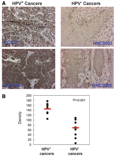
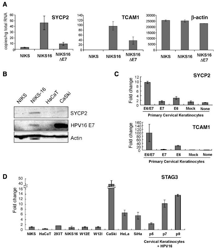
Human papillomaviruses (HPV) are associated with nearly all cervical cancers, 20% to 30% of head and neck cancers (HNC), and other cancers. Because HNCs also arise in HPV-negative patients, this type of cancer provides unique opportunities to define similarities and differences of HPV-positive versus HPV-negative cancers arising in the same tissue. Here, we describe genome-wide expression profiling of 84 HNCs, cervical cancers, and site-matched normal epithelial samples in which we used laser capture microdissection to enrich samples for tumor-derived versus normal epithelial cells. This analysis revealed that HPV(+) HNCs and cervical cancers differed in their patterns of gene expression yet shared many changes compared with HPV(-) HNCs. Some of these shared changes were predicted, but many others were not. Notably, HPV(+) HNCs and cervical cancers were found to be up-regulated in their expression of a distinct and larger subset of cell cycle genes than that observed in HPV(-) HNC. Moreover, HPV(+) cancers overexpressed testis-specific genes that are normally expressed only in meiotic cells. Many, although not all, of the hallmark differences between HPV(+) HNC and HPV(-) HNC were a direct consequence of HPV and in particular the viral E6 and E7 oncogenes. This included a novel association of HPV oncogenes with testis-specific gene expression. These findings in primary human tumors provide novel biomarkers for early detection of HPV(+) and HPV(-) cancers, and emphasize the potential value of targeting E6 and E7 function, alone or combined with radiation and/or traditional chemotherapy, in the treatment of HPV(+) cancers.


Figures

Figure 1
Figure 1
Global gene expression analysis shows similarities and differences among HPV+ HNC, HPV HNC, and cervical cancer. A, multidimensional scaling measurements between all indicated pairs of tumor and normal classes of the distances between class-averaged log 2 expression levels over all 54,675 Affymetrix probe sets. The relative distances between each class are approximated in the two-dimensional projection (top) and were tabulated (bottom). CC, cervical cancer; CN, cervical normal. B, pairwise comparisons of expression alterations from normal for three cancers are shown as scatter plots of average log 2 fold change from normal. Pearson correlations (R) measure global concordance in expression alterations between cancer pairs. Genes that show differential expression between HPV+ HNC and HPV HNC are highlighted; tracking into the HPV+ HNC versus HPV+ cervical cancer comparison, these genes are predominantly equivalently expressed between these HPV+ cancers. Dotted lines, median expression changes of red and blue genes; red and blue arrows, median shifted from HPV+ HNC/HPV HNC compared with HPV+ HNC/cervical cancer comparison. C, differential expression analysis reveals lists of genes significantly altered between the respective tissue classes. The results of three pairwise comparisons are summarized in the Venn diagram and tabulated fully in Table 3 (HPV+ versus HPV), Supplementary Table S5 (tumor versus normal), and Supplementary Table S6 (HNC versus cervical cancer).
Figure 2
Figure 2
Gene expression signatures for HPV+ versus HPV cancers and HNC versus cervical cancer cancers. A, normalized expression values for all 84 samples and 137 probe sets that are significantly differentially expressed between the HPV+ cancers and the HPV cancers. Bottom right key, high (red) and low (green) expression, corresponding to a +7.5 to −8.2 log 2 scale of fold change relative to the average of each gene across all 84 microarrays. These genes were ordered by hierarchical clustering based on similarities in their expression changes across the samples (see dendogram, left). Gene sets III and IV show significantly up- or down-regulated probe sets, respectively. HPV+ cancer samples (red text) and HPV cancer samples (blue text) are shown at the bottom of the heat map. B, like (A), but using 291 probe sets that are significantly differentially expressed between cervical cancer and HNC. Gene sets V and VII show significantly up-regulated probe sets in cervical cancer versus HNC, whereas gene set VI shows significantly down-regulated probe sets. Cervical cancer samples (red text) and HNC samples (blue text) are shown at the bottom of the heat map. *, probe set ID that does not have annotated gene name. HPV status is shown as + and − on each sample ID.
Figure 2
Figure 2
Gene expression signatures for HPV+ versus HPV cancers and HNC versus cervical cancer cancers. A, normalized expression values for all 84 samples and 137 probe sets that are significantly differentially expressed between the HPV+ cancers and the HPV cancers. Bottom right key, high (red) and low (green) expression, corresponding to a +7.5 to −8.2 log 2 scale of fold change relative to the average of each gene across all 84 microarrays. These genes were ordered by hierarchical clustering based on similarities in their expression changes across the samples (see dendogram, left). Gene sets III and IV show significantly up- or down-regulated probe sets, respectively. HPV+ cancer samples (red text) and HPV cancer samples (blue text) are shown at the bottom of the heat map. B, like (A), but using 291 probe sets that are significantly differentially expressed between cervical cancer and HNC. Gene sets V and VII show significantly up-regulated probe sets in cervical cancer versus HNC, whereas gene set VI shows significantly down-regulated probe sets. Cervical cancer samples (red text) and HNC samples (blue text) are shown at the bottom of the heat map. *, probe set ID that does not have annotated gene name. HPV status is shown as + and − on each sample ID.
Figure 3
Figure 3
Cell cycle–related genes are up-regulated in HPV+ cancers. A, highly up-regulated genes in HPV+ cancers were analyzed by GO grouping. Cell cycle–related genes were selected and plotted on the heat map. Blue bars, HPV cervical cancers. B, up- and down-regulated genes indicated in the cell cycle pathway provided by the KEGG database. Red and blue boxes, up-regulated genes in HPV+ and HPV cancers compared with corresponding normal tissue, respectively. C, a part of the cell cycle–related genes was analyzed using qRT-PCR. Fold changes of the gene expression in NIKS-16 relative to gene expression in NIKS. Columns, mean; bars, SD.
Figure 4
Figure 4
PCNA protein expression is up-regulated in HPV+ cancers. Using anti-human PCNA antibody, immunohistochemistry was done with sections of 11 HPV+ and 10 HPV cancers. Immunohistochemical images were analyzed and quantified as described previously (ref. 53; see Supplementary Methods). Representative immunohistochemical images (A) and calculated density of all samples (B). Red bars, mean values of each class. The tissue was also briefly counterstained with hematoxylin.
Figure 5
Figure 5
Testis-specific genes SYCP2 and TCAM1 are induced by HPV16. A, real-time qRT-PCR was done with total RNA extracted from NIKS cells with and without HPV16. Also, total RNA from NIKS-16 cells without HPV16 E7 protein expression was used to show that testis-specific gene induction by E7 protein was partial. B, SYCP2 induction in HPV+ cell lines were confirmed with Western blotting using anti-human SYCP2 antibody. Real-time qRT-PCR was done with total RNA extracted from primary cervical keratinocytes with either, or both, HPV16 E6 and E7 delivered by recombinant retrovirus. C, retrovirus without HPV16 gene was used as mock control. D, STAG3 mRNA expression in various cell lines was quantified using qRT-PCR and relative fold change to NIKS cells are plotted. Columns, mean; bars, SD.

References

    1. Burd EM. Human papillomavirus and cervical cancer. Clin Microbiol Rev. 2003;16:1–17. - PMC - PubMed
    1. Gillison ML, Lowy DR. A causal role for human papillomavirus in head and neck cancer. Lancet. 2004;363:1488–9. - PubMed
    1. Smith EM, Ritchie JM, Summersgill KF, et al. Human papillomavirus in oral exfoliated cells and risk of head and neck cancer. J Natl Cancer Inst. 2004;96:449–55. - PubMed
    1. Hunter KD, Parkinson EK, Harrison PR. Profiling early head and neck cancer. Nat Rev Cancer. 2005;5:127–35. - PubMed
    1. Chung CH, Parker JS, Karaca G, et al. Molecular classification of head and neck squamous cell carcinomas using patterns of gene expression. Cancer Cell. 2004;5:489–500. - PubMed

Publication types

MeSH terms