Comparative Study
. 2007 Jun 6;26(11):2670-81.
doi: 10.1038/sj.emboj.7601712. Epub 2007 May 17.

Staufen1 regulates diverse classes of mammalian transcripts

Affiliations
Comparative Study

Staufen1 regulates diverse classes of mammalian transcripts

Yoon Ki Kim et al. EMBO J. .

Abstract

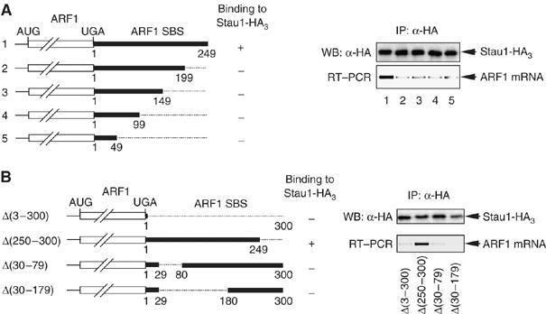
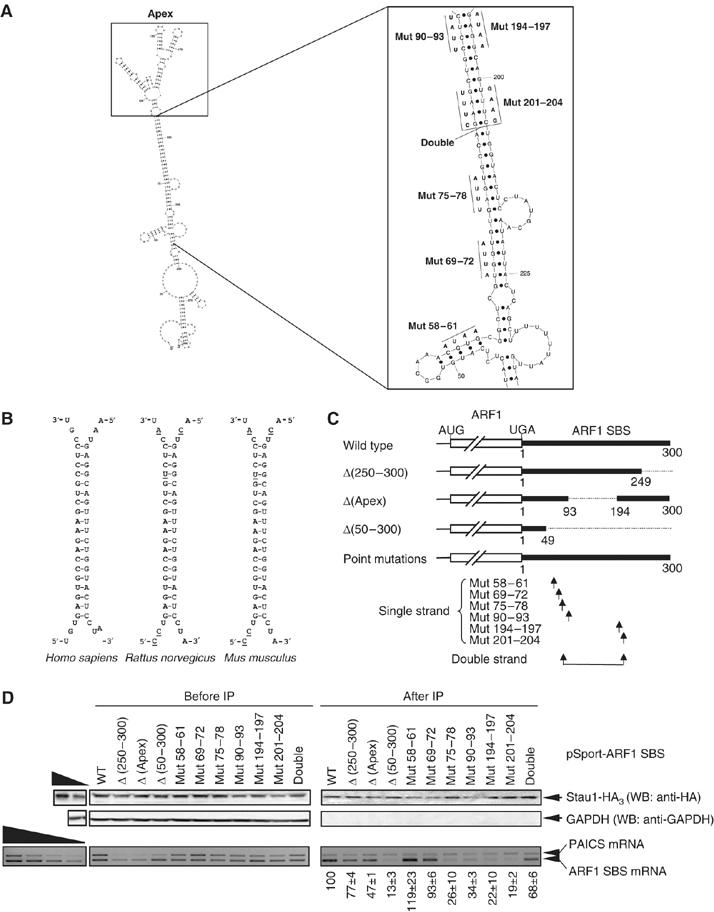
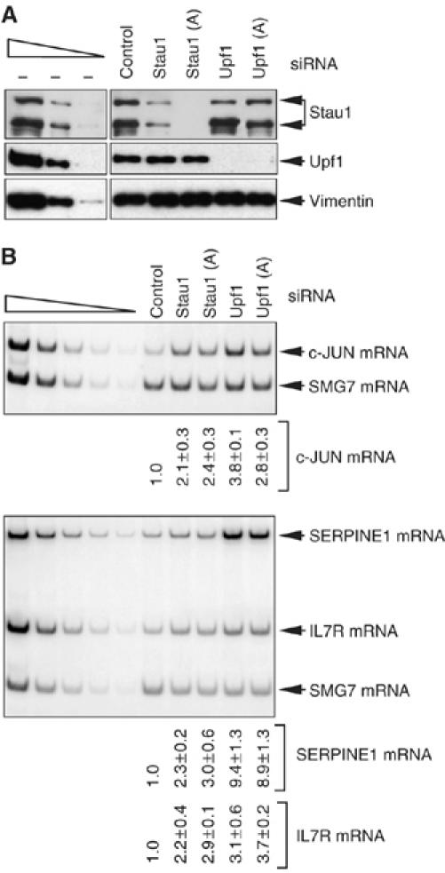
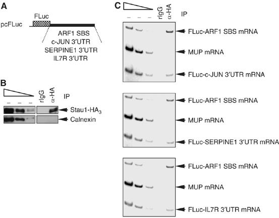
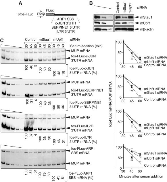
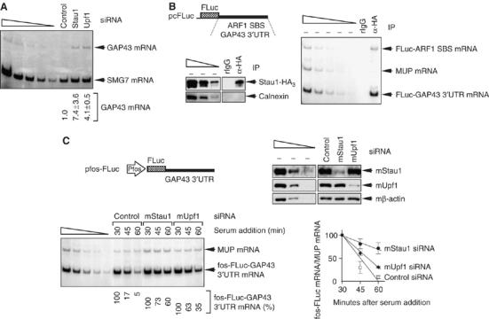
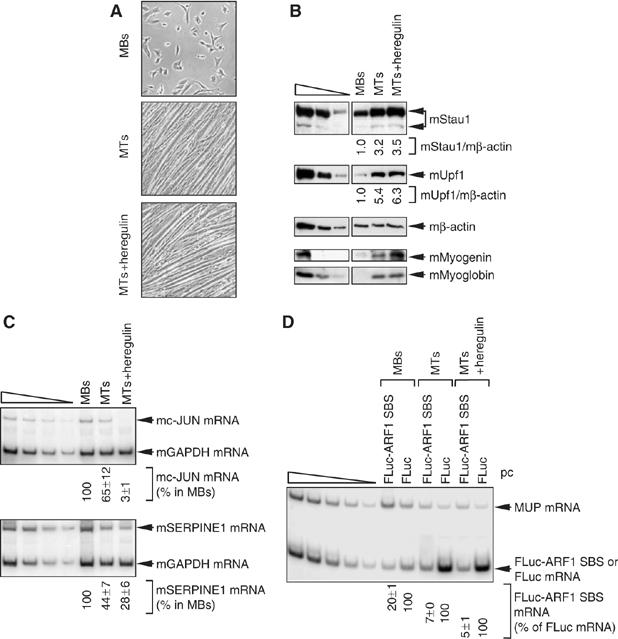
It is currently unknown how extensively the double-stranded RNA-binding protein Staufen (Stau)1 is utilized by mammalian cells to regulate gene expression. To date, Stau1 binding to the 3'-untranslated region (3'-UTR) of ADP ribosylation factor (ARF)1 mRNA has been shown to target ARF1 mRNA for Stau1-mediated mRNA decay (SMD). ARF1 SMD depends on translation and recruitment of the nonsense-mediated mRNA decay factor Upf1 to the ARF1 3'-UTR by Stau1. Here, we demonstrate that Stau1 binds to a complex structure within the ARF1 3'-UTR. We also use microarrays to show that 1.1 and 1.0% of the 11 569 HeLa-cell transcripts that were analyzed are upregulated and downregulated, respectively, at least two-fold upon Stau1 depletion in three independently performed experiments. We localize the Stau1 binding site to the 3'-UTR of four mRNAs that we define as natural SMD targets. Additionally, we provide evidence that the efficiency of SMD increases during the differentiation of C2C12 myoblasts to myotubes. We propose that Stau1 influences the expression of a wide variety of physiologic transcripts and metabolic pathways.


Figures

Figure 1
Figure 1
Deletions within the ARF1 SBS indicate that a central core region is required for Stau1 binding in vivo. (A) (Left) Schematic representations of the various 3′-end-deleted mRNAs that derive from pSport-ARF1 SBS derivatives. Numbering is relative to the first nucleotide following the termination codon, which is defined as 1. Plus (+) and minus (−) signs at the right of each representation indicate the ability or failure to bind Stau1, respectively. (Right) Human 293 cells were transfected with a derivative of the pSport-ARF1 SBS test plasmid and a plasmid expressing Stau1-HA3. After immunopurification (IP) using anti(α)-HA, protein was analyzed using Western blotting (WB) and anti-HA (upper), and RNA was analyzed using RT–PCR and ethidium bromide staining (lower). (B) (Left) as in (A, left) except for the pSport-ARF1 SBS derivatives that were analyzed. (Right) as in A, right. Results are representative of two independently performed experiments.
Figure 2
Figure 2
Stau1 binds in vivo to a complex structure within the ARF1 SBS. (A) Model for the secondary structure of the ARF1 SBS showing nucleotides 1 through 300. The plot was generated using Sfold v2.0 software (Wadsworth Bioinformatics Center). A larger view of the regions targeted by mutagenesis is shown in the inset to the right, along with the specific nucleotide changes that were made. See Supplementary Figure 1 for a full-page image. (B) The predicted 19-bp stem within the human ARF1 SBS is conserved in rat and mouse ARF1 mRNAs. Nucleotides that are not conserved with respect to the human sequence are underlined. (C) Schematic representation of the mutated mRNAs synthesized from pSport-ARF1 SBS derivatives. ‘Single strand' arrows indicate the relative positions of each of the 4-nt mutations, most of which individually disrupt the 19-bp stem, and ‘Double strand' arrows indicate the relative positions of the two 4-nt mutations made in cis that restore the stem. Δ(Apex) mRNA contains a replacement of nucleotides 94 through 193 that normally constitute what is predicted to be a complex structure containing a number of small loops and stems with a UGCA (see A for details). (D) As in Figure 1, except that pSport-PAICS was included in the transfections, wild-type (WT) ARF1 SBS was used as a positive control, and Δ(50–300) was used as a negative control. (Upper) Protein was analyzed before and after immunopurification (IP) using Western blotting (WB) and anti(α)-HA, or, as a negative control, anti-GAPDH. (Lower) RNA was analyzed using RT–PCR and ethidium bromide staining. The level of each ARF1-SBS-derived mRNA after IP was normalized to the level of PAICS mRNA after IP and divided by the ratio of the level of the same ARF1-SBS-derived mRNA relative to PAICS mRNA before IP. This value is defined as 100 for WT, and values for the mutated transcripts were calculated as a percentage of 100. Results are representative of three independently performed experiments that did not differ by the amount specified.
Figure 3
Figure 3
c-JUN, SERPINE1 and IL7R mRNAs are increased in abundance in human cells depleted of either Stau1 or Upf1. HeLa cells were transiently transfected with Stau1, Stau1(A), Upf1, Upf1(A) or, to Control for nonspecific depletion, Control siRNA. Three days later, protein and RNA were purified. (A) Western blot analysis using anti-Stau1, anti-Upf1 or, to control for variations in protein loading, anti-vimentin. The normal level of Stau1 or Upf1 is defined as the level in the presence of Control siRNA after normalization to the level of vimentin. (B) RT–PCR analysis of the level of endogenous c-JUN mRNA (upper), SERPINE1 mRNA or IL7R mRNA (lower). The level of each mRNA is normalized to the level of endogenous SMG7 mRNA, which is insensitive to Stau1 and Upf1 siRNAs (data not shown). The normalized level of each mRNA in the presence of Control siRNA is defined as 1. RT–PCR results are representative of three independently performed experiments that did not differ by the amount specified.
Figure 4
Figure 4
Stau1 binds within the 3′-UTR of c-JUN, SERPINE1 and IL7R mRNAs. Cos cells were transiently transfected with six plasmids: (i) pcFLuc-c-JUN 3′-UTR, pcFLuc-SERPINE1 3′-UTR and pcFLuc-IL7R 3′-UTR test plasmids; (ii) the Stau1-HA3 expression vector; (iii) the pcFLuc-ARF1 SBS test plasmid that serves as a positive control for Stau1-HA3 binding; and (iv) phCMV-MUP, which encodes MUP mRNA that serves as a negative control for Stau1-HA3 binding. Two days later, cells were lysed, and a fraction of each lysate was immunopurified using anti-HA or, as a control for nonspecific immunopurification (IP), rat (r)IgG. (A) Schematic representations of the pcFLuc-ARF1 SBS, pcFLuc-c-JUN 3′-UTR, pcFLuc-SERPINE1 3′-UTR and pcFLuc-IL7R 3′-UTR test plasmids. (B) Western blot analysis using anti(α)-HA or anti-Calnexin demonstrates that Stau1-HA3 but, as expected, not Calnexin was immunopurified. (C) RT–PCR analysis demonstrates that c-JUN, SERPINE1 and IL7R 3′-UTRs, like ARF1 SBS, bind Stau1-HA3, whereas MUP mRNA does not. Results are representative of three independently performed experiments.
Figure 5
Figure 5
Depleting cells of Stau1 or Upf1 increases the half-life of FLuc mRNA when the 3′-UTR consists of c-JUN, SERPINE1 or IL7R 3′-UTR sequences that bind Stau1. L cells were transfected with mouse (m)Stau1, mUpf1 or Control siRNA. Two days later, cells were re-transfected with the pfos-FLuc-ARF1 SBS, pfos-FLuc-c-JUN 3′-UTR, pfos-FLuc-SERPINE1 3′-UTR and pfos-FLuc-IL7R 3′-UTR test plasmids and the phCMV-MUP reference plasmid in the absence of serum. Serum was added to 15% after an additional 24 h. Protein was immediately purified from the cytoplasmic fraction (at 0 min) for Western blot analysis. RNA was purified from the nuclear fraction for RT–PCR analysis at the specified times. (A) Schematic representations of each test plasmid. (B) Western blotting of mStau1 or mUpf1, where the level of mβ-actin serves to control for variations in protein loading. mStau1 was depleted to at least 35% of its normal level, and mUpf1 was depleted to at least 30% of its normal level. (C) (Left) RT–PCR analysis of the SMD candidate mRNAs. For each time point, the level of each mRNA transcribed from the fos promoter is normalized to the level of MUP mRNA. Normalized levels are calculated as a percentage of the normalized level of each mRNA transcribed at 30 min in the presence of each siRNA, which is defined as 100%. (Right) These levels are plotted as a function of time after serum addition. Error bars specify the extent of variation among two or three independently performed experiments.
Figure 6
Figure 6
GAP43 mRNA is an SMD target. (A) RT–PCR demonstrating that depleting cells of Stau1 or Upf1 increases the cellular abundance of GAP43 mRNA. RNA derived from samples used in Figure 3. (B) As in Figure 4, except that FLuc-GAP43 3′-UTR and FLuc-ARF1 SBS mRNAs were analyzed. (C) As in Figure 5, except that fos-FLuc-GAP43 3′-UTR mRNA was analyzed. Results are representative of two independently performed experiments.
Figure 7
Figure 7
Evidence that the efficiency of SMD is upregulated during the differentiation of mouse C2C12 MBs to MTs. C2C12 cells at 20–30% confluency were maintained as MBs or induced to differentiate to MTs by exposure to 5% horse serum with or without 200 ng/ml of heregulin, a type of growth factor important for muscle development and function (Gramolini et al, 1999). (A) Light microscopy of C2C12 cells as MBs, MTs or MTs+heregulin. (B) Western blotting of mouse (m)Stau1, mUpf1, mMyogenin, mMyoglobin or, as a control for protein loading, mβ-actin. (C) RT–PCR analysis of the level of endogenous c-JUN mRNA (upper) or mSERPINE1 mRNA (lower), each of which is normalized to the level of endogenous mGAPDH mRNA. The normalized level of mc-JUN mRNA or mSERPINE1 mRNA in MBs is defined as 100%. (D) Using RNA from cells that had been transiently transfected with pcFLuc-ARF1 SBS or pcFLuc and phCMV-MUP, RT–PCR analysis of FLuc-ARF1 SBS or FLuc mRNA, each of which is normalized to the level of MUP mRNA. The normalized level of FLuc mRNA under each of the three conditions is defined as 100%. All RT–PCR results are representative of two independently performed experiments that did not differ by the amount specified.

References

    1. Bélanger G, Stocksley MA, Vandromme M, Schaeffer L, Furic L, DesGroseillers L, Jasmin BJ (2003) Localization of the RNA-binding proteins Staufen1 and Staufen2 at the mammalian neuromuscular junction. J Neurochem 86: 669–677 - PubMed
    1. Blais A, Tsikitis M, Acosta-Alvear D, Sharan R, Kluger Y, Dynlacht BD (2005) An initial blueprint for myogenic differentiation. Genes Dev 19: 553–569 - PMC - PubMed
    1. Brennan CM, Steitz JA (2001) HuR and mRNA stability. Cell Mol Life Sci 58: 266–277 - PMC - PubMed
    1. Chiu SY, Lejeune F, Ranganathan AC, Maquat LE (2004) The pioneer translation initiation complex is functionally distinct from but structurally overlaps with the steady-state translation initiation complex. Genes Dev 18: 745–754 - PMC - PubMed
    1. Ding Y, Chan CY, Lawrence CE (2004) Sfold web server for statistical folding and rational design of nucleic acids. Nucleic Acids Res 32: W135–W141 - PMC - PubMed

Publication types

MeSH terms