. 2007 Jun;8(6):576-82.
doi: 10.1038/sj.embor.7400982. Epub 2007 May 18.

Conserved roles of Sam50 and metaxins in VDAC biogenesis

Affiliations

Conserved roles of Sam50 and metaxins in VDAC biogenesis

Vera Kozjak-Pavlovic et al. EMBO Rep. 2007 Jun.

Abstract

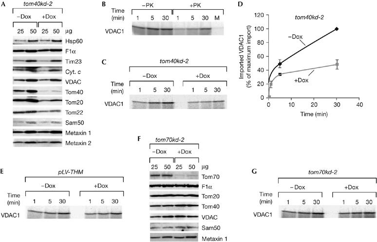
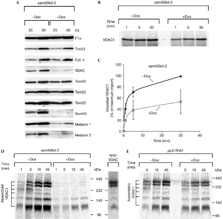
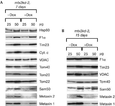
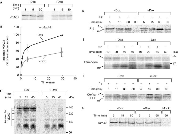
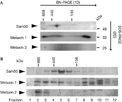
Voltage-dependent anion-selective channel (VDAC) is a beta-barrel protein in the outer mitochondrial membrane that is necessary for metabolite exchange with the cytosol and is proposed to be involved in certain forms of apoptosis. We studied the biogenesis of VDAC in human mitochondria by depleting the components of the mitochondrial import machinery by using RNA interference. Here, we show the importance of the translocase of the outer mitochondrial membrane (TOM) complex in the import of the VDAC precursor. The deletion of Sam50, the central component of the sorting and assembly machinery (SAM), led to both a strong defect in the assembly of VDAC and a reduction in the steady-state level of VDAC. Metaxin 2-depleted mitochondria had reduced levels of metaxin 1 and were deficient in import and assembly of VDAC and Tom40, but not of three matrix-targeted precursors. We also observed a reduction in the levels of metaxin 1 and metaxin 2 in Sam50-depleted mitochondria, implying a connection between these three proteins, although Sam50 and metaxins seemed to be in different complexes. We conclude that the pathway of VDAC biogenesis in human mitochondria involves the TOM complex, Sam50 and metaxins, and that it is evolutionarily conserved.


Figures

Figure 1
Figure 1
The TOM complex is necessary for VDAC import. (A) Protein levels of mitochondria isolated from tom40kd-2 cells grown for 5 days in the absence (−Dox) or presence (+Dox) of doxycycline. Mitochondria (μg of protein) were analysed by using SDS–PAGE and western blot. (B) 35S-labelled VDAC1 precursor was incubated with isolated HeLa mitochondria for the indicated time periods. The samples were left untreated (−PK) or treated (+PK) with 50 μg/ml protease K upon import and analysed by SDS–PAGE and autoradiography. Mock (M) represents a sample incubated for 30 min, containing no mitochondria. (C,D) Tom40-depleted mitochondria are defective in the import of the VDAC1 precursor. Isolated mitochondria, as in (A), were incubated with 35S-labelled VDAC1 for the indicated time periods, treated with protease K and analysed as in (B). Values on the graph represent the average of three independent import experiments. The longest time point of import into mitochondria from non-induced cells was set to 100%. (E) The VDAC1 precursor was imported, as described in (C), into mitochondria isolated from control pLV-THM cells after 7 days of induction. (F,G) Levels, of proteins in mitochondria isolated from tom70kd-1 after 7 days of induction (F), and the corresponding import of the VDAC1 precursor (G). Cyt. c, cytochrome c; Dox, doxycycline; F1α, ATPase F1 subunit α; Hsp60, heat shock protein 60; pLV-THM, single-cell clones transduced with an empty lentivirus vector; Sam, sorting and assembly machinery; SDS–PAGE, SDS–polyacrylamide gel electrophoresis; Tim, translocase of the inner mitochondrial membrane; Tom, translocase of the outer mitochondrial membrane; VDAC, voltage-dependent anion-selective channel.
Figure 2
Figure 2
Sam50 is involved in import and assembly of VDAC. (A) Protein levels in sam50kd-2 mitochondria analysed by SDS–PAGE and western blot after 7 days of knockdown induction with doxycycline. (B,C) Mitochondria isolated from induced and non-induced sam50kd-2 cells were incubated with 35S-labelled VDAC1 precursor, treated with 50 μg/ml protease K and analysed by SDS–PAGE. The average of three independent experiments is represented on the graph (C). The longest time point of the import into mitochondria from cells in which Sam50 knockdown was not induced was set to 100%. (D) VDAC1 import into sam50kd-2 mitochondria was carried out as in (B) and protein complexes were analysed by BN-PAGE, autoradiography and western blot. (E) Mitochondria were isolated from control pLV-THM cells after 7 days of doxycycline induction, and import of VDAC1 precursor was carried out as in (B) and (D). BN-PAGE, blue native-polyacrylamide gel electrophoresis; Cyt. c, cytochrome c; Dox, doxycycline; pLV-THM, single-cell clones transduced with an empty lentivirus vector; SAM, sorting and assembly machinery; SDS–PAGE, SDS–polyacrylamide gel electrophoresis; Tim, translocase of the inner mitochondrial membrane; Tom, translocase of the outer mitochondrial membrane; VDAC, voltage-dependent anion-selective channel.
Figure 3
Figure 3
Protein levels in mitochondria depleted of metaxin 2. (A,B) mtx2kd-2 cells were grown in the presence (+Dox) or absence (−Dox) of doxycycline for 7 days (A) or 15 days (B). Mitochondria were isolated and analysed by using SDS–polyacrylamide gel electrophoresis and western blot. Cyt. c, cytochrome c; Dox, doxycycline; Hsp60, heat shock protein 60; Sam, sorting and assembly machinery; Tim, translocase of the inner mitochondrial membrane; Tom, translocase of the outer mitochondrial membrane; VDAC, voltage-dependent anion-selective channel.
Figure 4
Figure 4
Metaxins are required for VDAC biogenesis, but not for the import of matrix precursors. (A,B) Knockdown of metaxins was induced with doxycycline in mtx2kd-2 cells for 7 days. Mitochondria were isolated and incubated with 35S-labelled VDAC1 precursor for the indicated time periods. After protease K treatment, mitochondria were analysed by SDS–polyacrylamide gel electrophoresis and autoradiography. (B) The graph represents an average of three independent import experiments. The longest time point of import into mitochondria from doxycyline-untreated cells was set to 100%. (C) Import was carried out as in (A), and protein complexes in mitochondria were analysed by blue native-polyacrylamide gel electrophoresis. (DF) 35S-labelled matrix precursors F1β (D), ferredoxin (E) and CoxVa-DHFR (F) were imported into metaxin 2-depleted mitochondria as in (A). (G) 35S-labelled precursor of outer-membrane β-barrel protein Tom40 was imported in metaxin 2-depleted mitochondria as in (A) for the indicated time periods. Mock represents a sample containing no mitochondria. CoxVa-DHFR, cytochrome oxidase subunit Va presequence fused to full-length human dihydrofolate reductase; Dox, doxycyline; Δψ, membrane potential; m, mature protein; p, precursor; Tom40, major subunit of the translocase of the outer mitochondrial membrane 40; VDAC, voltage-dependent anion-selective channel; *, nonspecific translation product.
Figure 5
Figure 5
Analysis of Sam50 and metaxin complexes by two-dimensional electrophoresis and gel filtration. (A) Isolated mitochondria (100 μg of protein) were solubilized in 1% digitonin buffer and subjected to blue native-polyacrylamide gel electrophoresis (BN-PAGE) in the first dimension (1D) and SDS–PAGE in the second dimension (2D), followed by western blot with antibodies against Sam50, metaxin 1 and metaxin 2. (B) Isolated mitochondria (400 μg of protein) were solubilized in 1% digitonin buffer and subjected to gel filtration chromatography. Proteins were precipitated by trichloroacetic acid and analysed by SDS–PAGE and western blot. SAM, sorting and assembly machinery; SDS–PAGE, SDS–polyacrylamide gel electrophoresis.

References

    1. Abdul KM, Terada K, Yano M, Ryan MT, Streimann I, Hoogenraad NJ, Mori M (2000) Functional analysis of human metaxin in mitochondrial protein import in cultured cells and its relationship with the Tom complex. Biochem Biophys Res Commun 276: 1028–1034 - PubMed
    1. Armstrong LC, Komiya T, Bergman BE, Mihara K, Bornstein P (1997) Metaxin is a component of a preprotein import complex in the outer membrane of the mammalian mitochondrion. J Biol Chem 272: 6510–6518 - PubMed
    1. Armstrong LC, Saenz AJ, Bornstein P (1999) Metaxin 1 interacts with metaxin 2, a novel related protein associated with the mammalian mitochondrial outer membrane. J Cell Biochem 74: 11–22 - PubMed
    1. Blachly-Dyson E, Baldini A, Litt M, McCabe ERB, Forte M (1994) Human genes encoding the voltage-dependent anion channel (VDAC) of the outer mitochondrial membrane: mapping and identification of two new isoforms. Genomics 20: 62–67 - PubMed
    1. Cheng EHY, Sheiko TV, Fisher JK, Craigen WJ, Korsmeyer SJ (2003) VDAC2 inhibits BAK activation and mitochondrial apoptosis. Science 301: 513–517 - PubMed

Publication types