. 2007 Jun;117(6):1647-57.
doi: 10.1172/JCI30168. Epub 2007 May 17.

Prostate cell differentiation status determines transient receptor potential melastatin member 8 channel subcellular localization and function

Affiliations

Prostate cell differentiation status determines transient receptor potential melastatin member 8 channel subcellular localization and function

Gabriel Bidaux et al. J Clin Invest. 2007 Jun.

Abstract

In recent years, the transient receptor potential melastatin member 8 (TRPM8) channel has emerged as a promising prognostic marker and putative therapeutic target in prostate cancer (PCa). However, the mechanisms of prostate-specific regulation and functional evolution of TRPM8 during PCa progression remain unclear. Here we show, for the first time to our knowledge, that only secretory mature differentiated human prostate primary epithelial (PrPE) luminal cells expressed functional plasma membrane TRPM8 ((PM)TRPM8) channels. Moreover, PCa epithelial cells obtained from in situ PCa were characterized by a significantly stronger (PM)TRPM8-mediated current than that in normal cells. This (PM)TRPM8 activity was abolished in dedifferentiated PrPE cells that had lost their luminal secretory phenotype. However, we found that in contrast to (PM)TRPM8, endoplasmic reticulum TRPM8 ((ER)TRPM8) retained its function as an ER Ca(2+) release channel, independent of cell differentiation. We hypothesize that the constitutive activity of (ER)TRPM8 may result from the expression of a truncated TRPM8 splice variant. Our study provides insight into the role of TRPM8 in PCa progression and suggests that TRPM8 is a potentially attractive target for therapeutic intervention: specific inhibition of either (ER)TRPM8 or (PM)TRPM8 may be useful, depending on the stage and androgen sensitivity of the targeted PCa.


Figures

Figure 1
Figure 1. TRPM8 channel expression and activity in human prostate cells.
(A) Immunoblot showing detection of 128-kDa protein in representative human NP, BPH, and PCa samples. PC-3 cells were used for negative control; detection of recombinant (rec) TRPM8-His fusion protein was used for positive control. Calnexin was used to control the amount of proteins. (B) Confocal examination of immunohistochemical sections reporting specific expression of the TRPM8 protein (green) in CK18-positive (red) cells in PCa. Arrows denote TRPM8 expression on the luminal side membrane of apical epithelial cells. Boxed area in top left panel is shown at a higher magnification in the other panels. (C) Representative confocal image of a PrPE cell from human BPH showing colocalization of TRPM8 (green) with CK18 (red) 6 days after tissue dissociation. Note that a thin green signal was localized on PM. Top right panel shows the cell viewed with transmitted light. (D) PM localization of TRPM8 (green) in PrPE cells was confirmed by its colocalization with membrane marker CD10 (red). (E) Representative time courses of menthol-activated iTRPM8 in PrPE cells transfected with 50 nM of either siTRPM8 or scramble siRNA (siCon). Inset shows the representative current/voltage relationships of the menthol-activated membrane currents. Scale bars: 10 μm.
Figure 2
Figure 2. iTRPM8 is functional in PrPE cells and presents increased activity in PCa.
(AC) Representative time courses of icilin-activated iTRPM8 in PrPE (A), PrPCa (B), and PC-3 cells (C). Insets show the representative current/voltage relationships of the icilin-activated iTRPM8. Inset in A shows an agarose gel indicating the enhancement of TRPM8 expression in PCa. M, protein ladder. (D) Cumulative data (mean ± SEM) of maximum currents measured at 100 mV. (EG) Typical traces of the estimated passive Ca2+ leak induced by 10 μM icilin in digitotin-permeabilized PrPE (E), PrPCa (F), and PC-3 cells (G). IM, ionomycin. (H) Cumulative data (mean ± SEM) of icilin-evoked ER Ca2+ release. *P < 0.05; **P < 0.01; ***P < 0.001.
Figure 3
Figure 3. TRPM8 localization and activity in PrPE cells depends on the phenotype of the differentiated epithelial cells.
(A and B) Confocal images showing immunolocalization of TRPM8 in either PrPE-6d (A) or PrPE-20d cells (B). Higher magnifications of boxed areas are presented in the insets (original magnification, ×600). Scale bars: 10 μm. (C) Representative time courses of menthol-activated (100 μM) iTRPM8 in PrPE-6d and PrPE-20d cells at 36°C. Currents were recorded from voltage ramps at 100 mV. Inset shows the representative current/voltage relationships of iTRPM8. (D) Typical [Ca2+]c in response to menthol (100 μM) in PrPE-6d and PrPE-20d cells. CCE, capacitative Ca2+ entry.
Figure 4
Figure 4. TRPM8 expression is stimulated by AR in PCa cells.
(A) Agarose gel showing amplification of the 2 TRPM8 amplicons (TRPM8F12/R15), AR and PSA in PC-3 transfected with either empty vector (Con) or AR for 3 or 5 days (d3 and d5, respectively). GAPDH was used as an internal reporter. (B) Quantification of PCR experiment in A. (C) Immunoblotting showing detection of TRPM8 proteins and AR in PC-3 cells transfected with empty vector, with AR for 3 or 5 days, or with TRPM8 encoding vector. Actin was used to control protein loading. **P < 0.01.
Figure 5
Figure 5. TRPM8 activity correlated with AR expression levels in PC3 cells.
(A) Typical traces of the estimated ER Ca2+ release induced by 250 μM menthol in digitotin-permeabilized control PC-3 cells, PC3 cells transfected with AR for 3 or 5 days, and TRPM8-transfected PC3 cells. (B) Cumulative data (mean ± SEM) for percent release from internal Ca2+ stores measured at 375 s. (C and D) Representative time courses of menthol-activated (250 μM) iTRPM8 in control PC3 cells and in PC3 cells transfected with AR for 3 or 5 days (C) as well as in TRPM8-transfected PC3 cells (D). Insets show the representative current/voltage relationships of the baseline and menthol-activated membrane currents. (E) Cumulative data (mean ± SEM) of maximum currents measured at 100 mV. **P < 0.01.
Figure 6
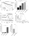
Figure 6. The trpm8 gene encodes for classical TRPM8 channel and a putative truncated TRPM8 splice variant.
(A) The trpm8 gene localized on chromosome 2 in position 37.1. Alignments of several TRPM8 mRNAs and proposed structures of TRPM8 genomic DNA and mRNA to scale. (B) Genomic map of TRPM8 (not to scale; numbered boxes denote exons) with its associated protein structure (boxes 1–6 represent putative transmembrane domains). The 3 pairs of PCR primers and siTRPM8-1 and -2 are aligned with their matching exons. (C) Real-time analysis of exons 8 to 9 amplicon (F8/R9) and exons 21 to 22 amplicon (F21/R22). Data are presented as a ratio of F8/R9 to F21/R22. HEK-TRPM8 cells represent the control condition with only 1 TRPM8 mRNA variant. (D) Ratio of TRPM8 F8/R9 to F21/R22 expression normalized to hypoxanthine-guanine phosphoribosyl transferase (HPRT) expression after quantification of PCR products obtained from single cells. Each population is represented by 10 prostate apical epithelial cells. ***P < 0.001.
Figure 7
Figure 7. Specific siRNA-mediated ablation of either classical TRPM8 or total TRPM8 mRNAs has different effects on menthol-evoked Ca2+ release.
(A and D) Real-time quantification of TRPM8 F8/R9 and F21/R22 amplicon normalized to hypoxanthine-guanine phosphoribosyl transferase expression in PC-3 cells (A) or HEK-TRPM8 cells (D) transfected with 100 nM of control siRNA, siTRPM8-1, or siTRPM8-2. Each experiment was performed 6 times. (B, C, and F) Typical traces of the estimated ER Ca2+ release induced by 250 μM menthol in digitotin-permeabilized PC-3 (B), PrPE-20d (C), and HEK-TRPM8 cells (F). Insets show cumulative data (mean ± SEM) for percentage of menthol-evoked Ca2+ release from internal Ca2+ stores. Each condition included at least 40 cells from 3 independent experiments. (E) Cumulative data (mean ± SEM) of cold- and menthol-activated iTRPM8 in HEK-TRPM8 cells with control siRNA, siTRPM8-1, or siTRPM8-2 (currents recorded from voltage ramps at 100 mV). *P < 0.05; **P < 0.01; ***P < 0.001.
Figure 8
Figure 8. Schematic diagram summarizing the principal findings of this study and showing a simplified representation of differential TRPM8 localization and function depending on the AR activity, differentiation, and oncogenic status of human prostate epithelial cells.
(A) General pattern of nondifferentiated TA/I cells and dedifferentiated metastatic cells. In these cells AR level was low, and only the ERTRPM8 isoform was expressed in the ER membrane. ERTRPM8 functioned as a Ca2+-release ER channel, which, by depleting ER stores, activated SOC localized on the PM. Under these conditions, TRPM8 agonists did not induce classical PMTRPM8-mediated current. (B) Normal, fully differentiated prostate cell with an apical secretory phenotype. In these cells, with high AR levels, the AR-dependent classical TRPM8 channel was expressed on both ER and PM. Under these conditions, TRPM8 agonists may induce not only SOCE, but also substantial PMTRPM8-mediated current. (C) Differentiated apical secretory in situ cancer cells. In these cells, with enhanced AR activity, the AR-dependent classical TRPM8 channel was overexpressed and TRPM8 agonists induced high levels of PMTRPM8-mediated current. iTRPM8 traces are schematic.

References

    1. Reya T., Morrison S.J., Clarke M.F., Weissman I.L. Stem cells, cancer, and cancer stem cells. Nature. 2001;414:105–111. - PubMed
    1. Schalken J.A., van Leenders G. Cellular and molecular biology of the prostate: stem cell biology. Urology. 2003;62:11–20. - PubMed
    1. Hendriksen P.J., et al. Evolution of the androgen receptor pathway during progression of prostate cancer. Cancer Res. 2006;66:5012–5020. - PubMed
    1. Legrand G., et al. Ca2+ pools and cell growth. Evidence for sarcoendoplasmic Ca2+-ATPases 2B involvement in human prostate cancer cell growth control. J. Biol. Chem. 2001;276:47608–47614. - PubMed
    1. Thebault S., et al. Differential role of transient receptor potential channels in Ca2+ entry and proliferation of prostate cancer epithelial cells. Cancer Res. 2006;66:2038–2047. - PubMed

Publication types

MeSH terms