. 2007 Jun;117(6):1527-37.
doi: 10.1172/JCI28338. Epub 2007 May 17.

PSGL-1-mediated activation of EphB4 increases the proangiogenic potential of endothelial progenitor cells

Affiliations

PSGL-1-mediated activation of EphB4 increases the proangiogenic potential of endothelial progenitor cells

Philippe Foubert et al. J Clin Invest. 2007 Jun.

Abstract

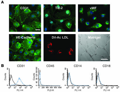
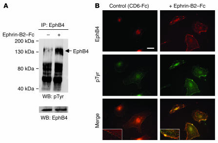
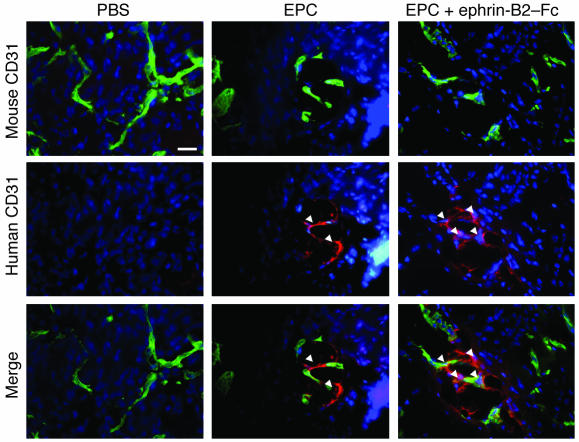
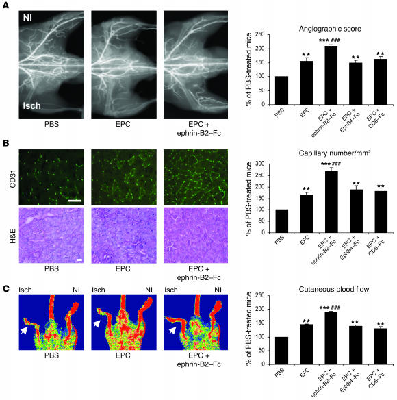
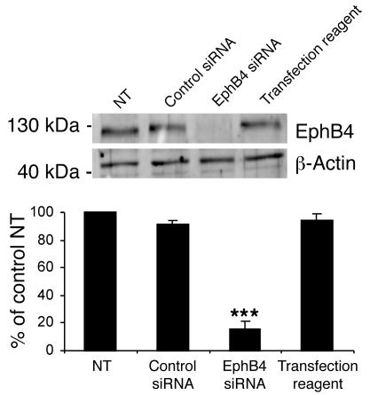
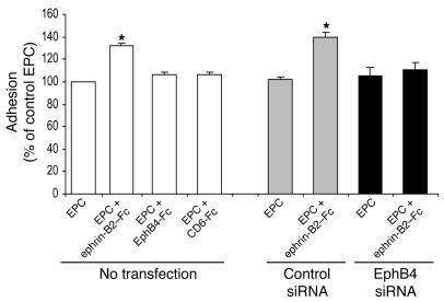
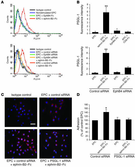
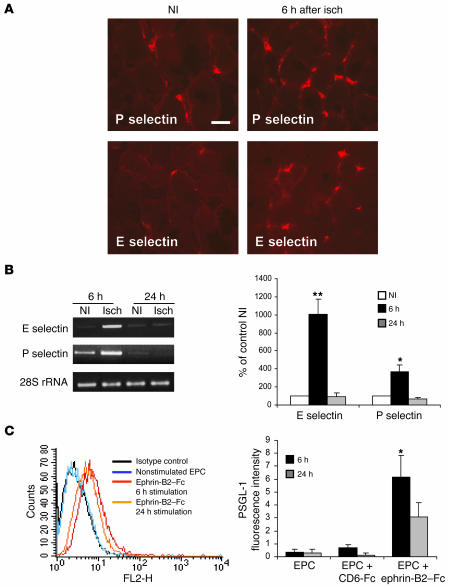
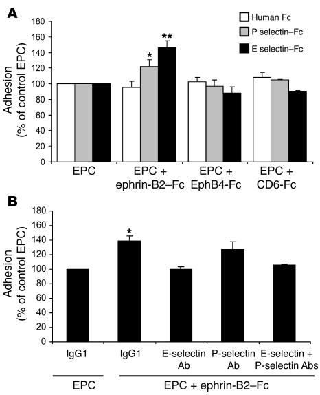
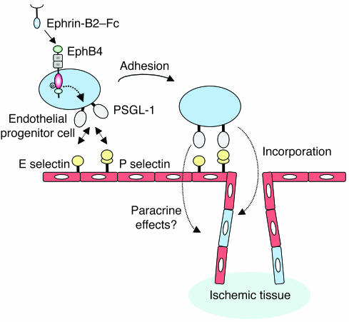
Endothelial progenitor cell (EPC) transplantation has beneficial effects for therapeutic neovascularization; however, only a small proportion of injected cells home to the lesion and incorporate into the neocapillaries. Consequently, this type of cell therapy requires substantial improvement to be of clinical value. Erythropoietin-producing human hepatocellular carcinoma (Eph) receptors and their ephrin ligands are key regulators of vascular development. We postulated that activation of the EphB4/ephrin-B2 system may enhance EPC proangiogenic potential. In this report, we demonstrate in a nude mouse model of hind limb ischemia that EphB4 activation with an ephrin-B2-Fc chimeric protein increases the angiogenic potential of human EPCs. This effect was abolished by EphB4 siRNA, confirming that it is mediated by EphB4. EphB4 activation enhanced P selectin glycoprotein ligand-1 (PSGL-1) expression and EPC adhesion. Inhibition of PSGL-1 by siRNA reversed the proangiogenic and adhesive effects of EphB4 activation. Moreover, neutralizing antibodies to E selectin and P selectin blocked ephrin-B2-Fc-stimulated EPC adhesion properties. Thus, activation of EphB4 enhances EPC proangiogenic capacity through induction of PSGL-1 expression and adhesion to E selectin and P selectin. Therefore, activation of EphB4 is an innovative and potentially valuable therapeutic strategy for improving the recruitment of EPCs to sites of neovascularization and thereby the efficiency of cell-based proangiogenic therapy.


Figures

Figure 1
Figure 1. Phenotypic and functional characterization of EPCs.
(A) EPCs were fixed in 90% cold acetone and incubated with the appropriate primary antibody, then with a FITC-coupled secondary antibody. EPCs were characterized according to the presence or absence of endothelial-specific markers including CD31, Tie-2, vWF, vascular endothelial–cadherin (VE-cadherin), and uptake of Dil-acetylated LDL (Dil-Ac LDL) (scale bar: 10 μm) and by their capacity to induce tube formation on Matrigel (scale bar: 100 μm). (B) Flow cytometric analysis of surface antigens on EPCs. EPCs were positive for CD31 but not for monocytic markers CD45, CD14, and CD18 (blue histogram). Isotypic control is represented by the black histogram.
Figure 2
Figure 2. EphB4 activation by ephrin-B2–Fc.
(A) EPCs were stimulated with 3 μg/ml ephrin-B2–Fc for 30 minutes at 37°C. Cell lysates were prepared, subjected to immunoprecipitation with an anti-human EphB4 antibody, and resolved by SDS-PAGE, and proteins were transferred to nitrocellulose membranes as described in Methods. Membranes were then blotted with the 4G10 anti-phosphotyrosine antibody. To check for equal protein loading, membranes were stripped and reprobed with an anti-EphB4 antibody. pTyr, phosphotyrosine; WB, Western blot. (B) To confirm EphB4 activation, ephrin-B2–Fc–stimulated EPCs were subjected to double immunostaining with EphB4 and anti-phosphotyrosine antibodies. Note the membrane colocalization of the EphB4 and the 4G10 signals corresponding to clusters (yellow color) in ephrin-B2–stimulated cells. Inset: magnification showing clustering and internalization of the phosphorylated EphB4 when cells were stimulated with ephrin-B2–Fc (right) but not with control CD6-Fc (left). Scale bar: 10 μm.
Figure 3
Figure 3. Transplanted EPCs home to the ischemic muscle.
Representative photomicrographs of incorporated EPCs identified by double-fluorescence labeling in ischemic muscles. Transplanted human EPCs were stained using a biotinylated anti-human CD31 antibody (red fluorescence) in histological sections retrieved from ischemic muscles 4 days after injection. Mouse vasculature was identified by CD31 staining (green fluorescence) in the same tissue sections. Nuclei were stained with DAPI (blue labeling). Arrows indicate labeled EPCs. Scale bar: 50 μm.
Figure 4
Figure 4. Ephrin-B2–Fc increases EPC proangiogenic potential in hind limb ischemia.
(A) Representative photomicrographs and quantitative analysis of microangiography. (B) H&E staining and capillary density. CD31-positive capillaries appear in green. Scale bar: 100 μm. (C) Foot perfusion in mice injected with PBS, nonstimulated EPCs (EPCs), and stimulated EPCs (EPCs + ephrin-B2–Fc, EPC + EphB4-Fc, or EPC + CD6-Fc). Values are expressed as means ± SEM; n = 10 per group. **P < 0.01, ***P < 0.001 versus PBS-injected mice; ##P < 0.01, ###P < 0.001 versus nonstimulated EPC-injected mice. NI, nonischemic; Isch, ischemic.
Figure 5
Figure 5. EphB4 knockdown in EPCs.
Western blot quantification of EphB4 protein demonstrating the efficiency of EphB4 siRNA in transfected cells. The membrane was reprobed for β-actin to check for equal loading. NT, nontransfected cells; control siRNA, luciferase siRNA; transfection reagent, Dharmafect2. n = 3. ***P < 0.001 versus nontransfected cells.
Figure 6
Figure 6. Ephrin-B2–Fc induces EPC adhesion to IL-1β prestimulated HUVEC.
EPCs were stimulated for 6 hours with 3 μg/ml of either EphB4-Fc, ephrin-B2–Fc, or CD6-Fc or left unstimulated and then allowed to attach to IL-1β preactivated HUVEC monolayer. EPCs transfected with EphB4 siRNA were stimulated with 3 μg/ml ephrin-B2–Fc or left unstimulated and then allowed to attach in a similar manner. Cells transfected with luciferase siRNA were used as control (siRNA control cells). Data are expressed as means ± SEM. n = 3. *P < 0.05 versus nonstimulated EPCs (EPCs). EPCs represented in this figure were nonstimulated.
Figure 7
Figure 7. EphB4 activation mediates EPC adhesion through PSGL-1.
(A and B) PSGL-1 expression in EPCs. EPCs were stimulated with 3 μg/ml of either ephrin-B2–Fc, EphB4-Fc, or CD6-Fc and then processed for FACS analysis of PSGL-1 expression. (A) FACS profiles of nontransfected (top panel) and EphB4 siRNA–transfected (bottom panel) EPCs. (B) Expression of PSGL-1 in nontransfected (top panel) and EphB4 siRNA–transfected (bottom panel) EPCs. n = 3. **P < 0.01 versus nonstimulated EPCs. (C) Effect of PSGL-1 siRNA on PSGL-1 protein expression. Control siRNA– and PSGL-1 siRNA–transfected EPCs were stimulated with 3 μg/ml ephrin-B2–Fc or left unstimulated and then processed for immunocytochemistry with an anti–PSGL-1 antibody as described in Methods. PSGL-1–positive staining appears in red. Scale bar: 20 μm. (D) Effect of PSGL-1 siRNA on EPC adhesion to IL-1β prestimulated HUVEC monolayer. Adhesion was quantified by measuring OD at 570 nm. n = 3. *P < 0.05 versus nonstimulated EPCs transfected with control siRNA.
Figure 8
Figure 8. Time-dependent expression of selectins and PSGL-1.
(A) Representative photomicrographs of ischemic (6 hours) and nonischemic gastrocnemius muscle sections stained with rat anti-mouse E selectin or P selectin. Positive staining (red) is localized in blood vessels between the muscle fibers. Scale bar: 40 μm. (B) RT-PCR analysis of E selectin and P selectin mRNA expression in ischemic muscles 6 hours or 24 hours after the onset of ischemia and in nonischemic muscles at the same time intervals. Data were normalized for loading with 28S RNA. n = 3. *P < 0.05; **P < 0.01 versus nonischemic muscle. (C) PSGL-1 expression on EPCs following ephrin-B2–Fc stimulation. EPCs were stimulated with ephrin-B2–Fc for 6 hours or 24 hours and then processed for FACS analysis of PSGL-1 expression. FACS profiles are shown in the left panel, and quantification of PSGL-1 expression in the right panel. n = 3. *P < 0.05 versus EPCs.
Figure 9
Figure 9. E selectin and P selectin mediate ephrin-B2–Fc–induced EPC adhesion.
(A) Ephrin-B2–Fc treatment increased EPC adhesion to immobilized E selectin and P selectin fusion proteins. EPCs were stimulated with 3 μg/ml of either ephrin-B2–Fc, EphB4-Fc, or CD6-Fc. Then the cells were allowed to adhere to immobilized recombinant E selectin–Fc (black bars), P selectin–Fc (gray bars), or to human IgG Fcγ fragments (white bars). Adhesion was quantified by measuring OD at 570 nm. Results are expressed as percentages of control nonstimulated EPCs (EPC). EPCs represented in this figure were nonstimulated. n = 3. *P < 0.05; **P < 0.01 versus nonstimulated EPCs. (B) Blocking antibodies directed against E selectin and P selectin neutralized ephrin-B2–Fc–induced EPC adhesion to IL-1β–prestimulated HUVEC monolayers. n = 3. *P < 0.05 versus EPCs treated with control IgG1.
Figure 10
Figure 10. Schematic model showing how EphB4 activation increases adhesion potential of EPCs.
E selectin and P selectin were overexpressed by the ischemic endothelium. EphB4 activation enhances PSGL-1 expression at the surface of EPCs. This allows attachment of the circulating ephrin-B2–Fc–stimulated EPCs via interaction between PSGL-1 and E selectin and P selectin. The attached cells can then migrate to the ischemic tissue where they can integrate into the nascent vessels and/or participate in a paracrine fashion in the neoangiogenic process.

References

    1. Kalka C., et al. Transplantation of ex vivo expanded endothelial progenitor cells for therapeutic neovascularization. Proc. Natl. Acad. Sci. U. S. A. 2000;97:3422–3427. - PMC - PubMed
    1. Kawamoto A., et al. Therapeutic potential of ex vivo expanded endothelial progenitor cells for myocardial ischemia. Circulation. 2001;103:634–637. - PubMed
    1. Asahara T., et al. Bone marrow origin of endothelial progenitor cells responsible for postnatal vasculogenesis in physiological and pathological neovascularization. Circ. Res. 1999;85:221–228. - PubMed
    1. Murohara T., et al. Transplanted cord blood-derived endothelial precursor cells augment postnatal neovascularization. J. Clin. Invest. 2000;105:1527–1536. - PMC - PubMed
    1. Hristov M., Weber C. Endothelial progenitor cells: characterization, pathophysiology, and possible clinical relevance. J. Cell. Mol. Med. 2004;8:498–508. - PMC - PubMed

Publication types

MeSH terms

LinkOut - more resources