. 2007 Jun;117(6):1627-36.
doi: 10.1172/JCI30765. Epub 2007 May 17.

Blocking aggrecanase cleavage in the aggrecan interglobular domain abrogates cartilage erosion and promotes cartilage repair

Affiliations

Blocking aggrecanase cleavage in the aggrecan interglobular domain abrogates cartilage erosion and promotes cartilage repair

Christopher B Little et al. J Clin Invest. 2007 Jun.

Erratum in

  • J Clin Invest. 2008 Nov;118(11):3812

Abstract

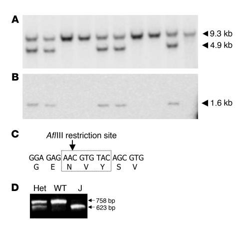
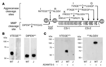
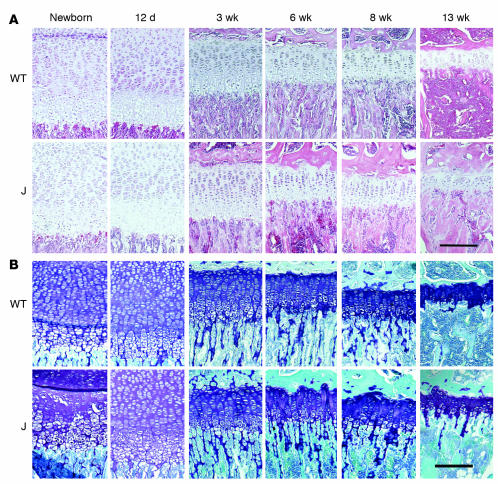
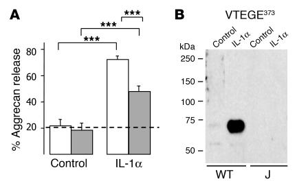
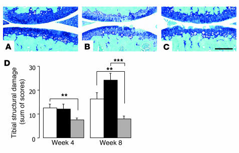
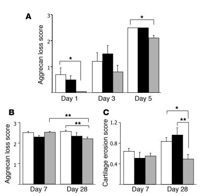
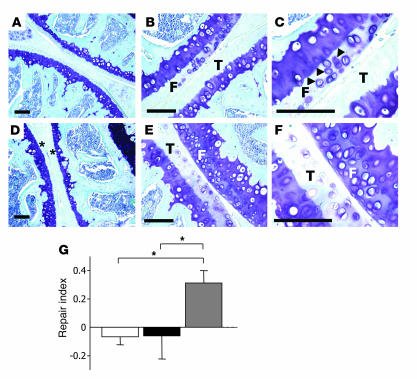
Aggrecan loss from cartilage in arthritis is mediated by aggrecanases. Aggrecanases cleave aggrecan preferentially in the chondroitin sulfate-2 (CS-2) domain and secondarily at the E(373) downward arrow(374)A bond in the interglobular domain (IGD). However, IGD cleavage may be more deleterious for cartilage biomechanics because it releases the entire CS-containing portion of aggrecan. Recent studies identifying aggrecanase-2 (ADAMTS-5) as the predominant aggrecanase in mouse cartilage have not distinguished aggrecanolysis in the IGD from aggrecanolysis in the CS-2 domain. We generated aggrecan knockin mice with a mutation that rendered only the IGD resistant to aggrecanases in order to assess the contribution of this specific cleavage to cartilage pathology. The knockin mice were viable and fertile. Aggrecanase cleavage in the aggrecan IGD was not detected in knockin mouse cartilage in situ nor following digestion with ADAMTS-5 or treatment of cartilage explant cultures with IL-1 alpha. Blocking cleavage in the IGD not only diminished aggrecan loss and cartilage erosion in surgically induced osteoarthritis and a model of inflammatory arthritis, but appeared to stimulate cartilage repair following acute inflammation. We conclude that blocking aggrecanolysis in the aggrecan IGD alone protects against cartilage erosion and may potentiate cartilage repair.


Figures

Figure 1
Figure 1. Mutation detection by Southern blotting and PCR.
(A) Southern blotting of genomic DNA detected 9.3- and 4.9-kb BamHI fragments in wild-type and Jaffa alleles, respectively. (B) After stripping, a 1.6-kb fragment was detected in Jaffa DNA on the same filter probed with a neo cDNA probe. (C) The mutation that changed amino acids 374ALG to 374NVY introduced a unique AflIII restriction site in exon 7. (D) A 758-bp PCR fragment amplified from wild-type DNA was resistant to AflIII digestion. AflIII digestion of the PCR fragment amplified from Jaffa (J) and heterozygote (Het) DNA yielded 623-bp and 135-bp fragments, although the 135-bp fragment was rarely visible on the gels.
Figure 2
Figure 2. G1 fragments in Jaffa and wild-type cartilage extracts.
(A) The domain structure of the aggrecan core protein, with 2 N-terminal globular domains, G1 and G2, and a C-terminal G3 domain. The extended region between G2 and G3 is heavily substituted with CS chains (wavy lines). Aggrecanase and MMP cleavage site sequences in the mouse IGD and CS-2 domains are shown, and amino acids that are different in human aggrecan are shown above. Numbering corresponds with the mouse sequence. (B) Cartilage extracts from wild-type and Jaffa mice were analyzed for VTEGE373 or DIPEN341 neoepitopes. (C) Dialyzed extracts of equal cartilage wet weight from wild-type and Jaffa mice were digested with or without recombinant human ADAMTS-5 and analyzed for the VTEGE373 and 374ALGSV neoepitopes. Stripping and reprobing the membranes with monoclonal antibody 2B6 confirmed sample loading in each lane (data not shown).
Figure 3
Figure 3. Jaffa has normal growth plate morphology and aggrecan content.
Paraffin sections of wild-type and Jaffa proximal tibial growth plates from birth to 13 weeks of age were stained with H&E (A) or toluidine blue (B). There was no difference in growth plate architecture between genotypes at any age. Scale bars: 200 μm.
Figure 4
Figure 4. Aggrecan degradation in vitro.
(A) Aggrecan release from wild-type (white bars) and Jaffa (gray bars) cartilage cultured with or without 10 ng/ml IL-1α (n = 8–10 per treatment). Results are mean ± SEM. The dotted line shows the level of baseline release. ***P < 0.001. (B) Cartilage extracts from wild-type and Jaffa mice with or without IL-1α treatment were analyzed by Western blot. Membranes were immunolocalized for VTEGE373 neoepitope.
Figure 5
Figure 5. DMM model in wild-type and aggrecan knockin mice.
(AC) Representative toluidine blue/fast green–stained sagittal sections of medial femorotibial joints from wild-type mice demonstrating mild (A), moderate (B), and severe lesions (C). Scale bar: 200 μm. (D) Sum of scores for tibial cartilage structural damage (mean ± SEM) in the medial tibial plateau at 4 or 8 weeks after surgery in wild-type (n = 12 [4 wk], 11 [8 wk]; open bars), Chloe (n = 13 [4 wk], 11 [8 wk]; black bars), and Jaffa (n = 11 [4 wk], 8 [8 wk]; gray bars) joints after DMM induction. **P < 0.01; ***P < 0.001.
Figure 6
Figure 6. AIA in wild-type and aggrecan knockin mice.
Mean ± SEM of AIA scores for (A) aggrecan loss at days 1, 3, and 5 (n = 5 per group); (B) aggrecan loss at days 7 (n = 35 [wild-type], 15 [Chloe], 32 [Jaffa]) and 28 (n = 49 [wild-type], 19 [Chloe], 21 [Jaffa]); and (C) cartilage erosion at days 7 and 28 (n as in B) in wild-type (white bars), Chloe (black bars), and Jaffa (gray bars) mice. *P < 0.05; **P < 0.01.
Figure 7
Figure 7. Cartilage repair in Jaffa mice.
(AF) Representative toluidine blue–stained sections of AIA joints from wild-type (AC) or Jaffa (DF) cartilage at day 28. A and D show no evidence of repair at day 28. Note the complete loss of toluidine blue in the articular cartilage (asterisks). Repair in wild-type (B and C) and Jaffa mice (E and F) at day 28 was seen predominantly in the femur. F, femoral surface; T, tibial surface. Arrowheads in C point to pericellular staining. Images in C and F are higher magnifications of the same sections shown in B and E. Scale bars: 100 μm. (G) Repair index, calculated as improvement in cartilage aggrecan loss score between days 7 and 28, in wild-type (n = 49; white bar), Chloe (n = 19; black bar), and Jaffa (n = 21; gray bar) femorotibial sections. Results are mean ± SEM. *P < 0.05.

References

    1. Sandy J.D., Neame P.J., Boynton R.E., Flannery C.R. Catabolism of aggrecan in cartilage explants. Identification of a major cleavage site within the interglobular domain. J. Biol. Chem. 1991;266:8683–8685. - PubMed
    1. Loulakis P., Shrikhande A., Davis G., Maniglia C.A. N-terminal sequence of proteoglycan fragments isolated from medium of interleukin-1-treated articular-cartilage cultures. Putative site(s) of enzymic cleavage. Biochem. J. 1992;284:589–593. - PMC - PubMed
    1. Ilic M.Z., Handley C.J., Robinson H.C., Mok M.T. Mechanism of catabolism of aggrecan by articular cartilage. Arch. Biochem. Biophys. 1992;294:115–122. - PubMed
    1. Sandy J.D., Flannery C.R., Neame P.J., Lohmander L.S. The structure of aggrecan fragments in human synovial fluid. Evidence for the involvement in osteoarthritis of a novel proteinase which cleaves the Glu 373-Ala 374 bond of the interglobular domain. J. Clin. Invest. 1992;89:1512–1516. - PMC - PubMed
    1. Lohmander L.S., Neame P.J., Sandy J.D. The structure of aggrecan fragments in human synovial fluid. Evidence that aggrecanase mediates cartilage degradation in inflammatory joint disease, joint injury, and osteoarthritis. Arthritis Rheum. 1993;36:1214–1222. - PubMed

Publication types

MeSH terms