. 2007 Jun;117(6):1708-17.
doi: 10.1172/JCI30630. Epub 2007 May 17.

Disruption of erythroid K-Cl cotransporters alters erythrocyte volume and partially rescues erythrocyte dehydration in SAD mice

Affiliations

Disruption of erythroid K-Cl cotransporters alters erythrocyte volume and partially rescues erythrocyte dehydration in SAD mice

Marco B Rust et al. J Clin Invest. 2007 Jun.

Abstract

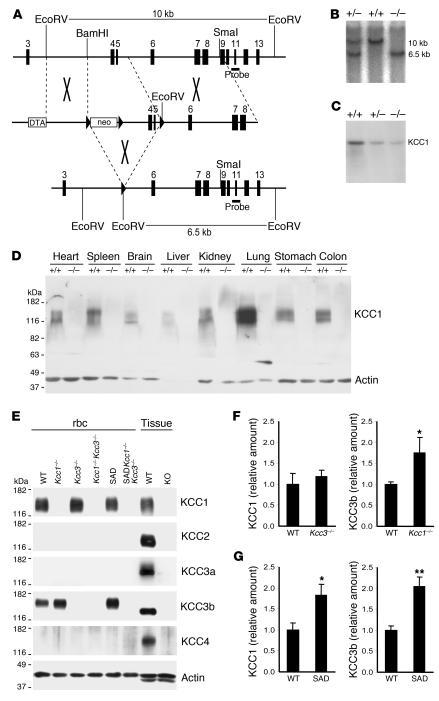
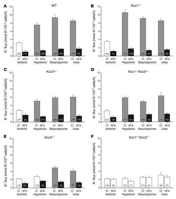
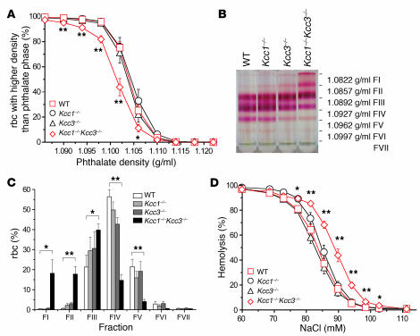
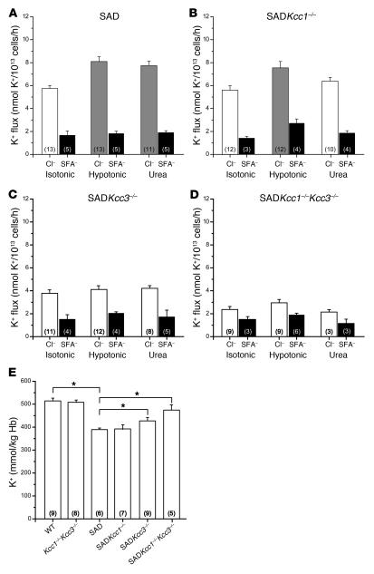
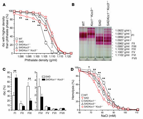
K-Cl cotransport activity in rbc is a major determinant of rbc volume and density. Pathologic activation of erythroid K-Cl cotransport activity in sickle cell disease contributes to rbc dehydration and cell sickling. To address the roles of individual K-Cl cotransporter isoforms in rbc volume homeostasis, we disrupted the Kcc1 and Kcc3 genes in mice. As rbc K-Cl cotransport activity was undiminished in Kcc1(-/-) mice, decreased in Kcc3(-/-) mice, and almost completely abolished in mice lacking both isoforms, we conclude that K-Cl cotransport activity of mouse rbc is mediated largely by KCC3. Whereas rbc of either Kcc1(-/-) or Kcc3(-/-) mice were of normal density, rbc of Kcc1(-/-)Kcc3(-/-) mice exhibited defective volume regulation, including increased mean corpuscular volume, decreased density, and increased susceptibility to osmotic lysis. K-Cl cotransport activity was increased in rbc of SAD mice, which are transgenic for a hypersickling human hemoglobin S variant. Kcc1(-/-)Kcc3(-/-) SAD rbc lacked nearly all K-Cl cotransport activity and exhibited normalized values of mean corpuscular volume, corpuscular hemoglobin concentration mean, and K(+) content. Although disruption of K-Cl cotransport rescued the dehydration phenotype of most SAD rbc, the proportion of the densest red blood cell population remained unaffected.


Figures

Figure 1
Figure 1. Generation of Kcc1–/– mice.
(A) Partial genomic organization of Slc12a4 (top) and the targeting construct (middle). Exons are shown as vertical bars, loxP sites as arrowheads. Transient Cre expression resulted in excision of exons 4 and 5 (bottom), producing a frameshift and premature stop. DTA, diphtheria toxin A cassette. (B) An additional EcoRV site was exploited for Southern blot analysis and resulted in the approximately 6.5-kb KO compared with the approximately 10-kb Kcc1+/+ fragment with the probe shown in A. (C) Northern blot analysis of Kcc1+/+, Kcc1+/–, and Kcc1–/– liver tissue with a full-length KCC1 probe revealed some residual aberrant transcript. (D) A membrane protein immunoblot with a KCC1 antibody confirmed the absence of KCC1 in tissues of Kcc1–/– mice. (E) rbc membrane protein immunoblot demonstrated KCC3b expression in WT rbc and absence of KCC3a, KCC2, and KCC4 in erythrocytes of WT, Kcc1–/–Kcc3–/–, and SADKcc1–/–Kcc3–/– mice. Actin served as a loading control. Kidney served as a positive control for KCC1 and KCC3b, brain for KCC2 and KCC3a, and lung for KCC4. (F and G) Expression levels of KCC1 and KCC3 proteins in rbc ghosts of various genotypes. (F) KCC3b protein level was upregulated in ghosts lacking KCC1, but KCC1 levels were unchanged in KCC3-KO ghosts. (G) Levels of both KCC1 and KCC3b proteins were increased in ghosts from SAD mice. Protein levels were determined by Western blot analysis (see Supplemental Figure 1) and normalized to actin. Bars represent arithmetic means from 6 mice normalized to WT. Error bars represent SEM. *P < 0.05, **P < 0.005 compared with WT.
Figure 2
Figure 2. K-Cl cotransport activity in rbc of WT and KO mice.
K-Cl cotransport was determined by measuring K+ efflux in the presence or absence of Cl (replaced with SFA) in isotonic or hypotonic conditions and in the presence of staurosporine (1 μM) or urea (500 mM) added to the isotonic solution. Gray bars indicate a significant difference between stimulated and isotonic K+ fluxes in the presence of Cl. Black bars indicate a significant difference between K+ fluxes in a single condition in the presence and absence of Cl. WT K-Cl cotransport activity (A) was unaltered in rbc of Kcc1–/– mice (B). (C, D, and E) Disruption of Kcc3 reduced K-Cl cotransport activity in all conditions examined, with an apparent gene-dosage effect. (F) In rbc of Kcc1–/–Kcc3–/–mice, equivalent K+ fluxes in the presence and absence of Cl indicated complete loss of K-Cl cotransport activity. Numbers in parentheses refer to the number of independent samples in each category.
Figure 3
Figure 3. Hematologic characterization of Kcc-KO mice.
(A) rbc densities determined by centrifugation through phthalate mixtures of defined density. (B) rbc densities determined by centrifugation through discontinuous Stractan gradients. FI, fraction I. (C) Distinct rbc bands were collected and the rbc numbers quantified to evaluate relative number of rbc per fraction. Kcc1–/–and Kcc3–/–rbc densities were indistinguishable from WT, but Kcc1–/–Kcc3–/–rbc densities were reduced. (D) Osmotic lysis curve shows significantly increased osmotic sensitivity only for rbc lacking both KCCs. *P ≤ 0.05; **P ≤ 0.005.
Figure 4
Figure 4. rbc K-Cl cotransport activity and K+ content of SAD mice of various Kcc genotypes.
(AD) K+ efflux from rbc measured as described in Methods in the presence of Cl or upon its substitution by SFA, measured under isotonic or hypotonic conditions and in the presence of 500 mM urea added to the isotonic solutions. Gray bars indicate a significant difference between isotonic and stimulated K+ fluxes. Black bars indicate a significant difference between K+ fluxes in a single condition in the presence and absence of Cl. (A) The elevated isotonic K-Cl cotransport activity of SAD rbc (compare with WT rbc in Figure 2A) was further stimulated by hypotonicity and by urea. Absence of KCC1 had minimal effect on K-Cl cotransport activity (B), whereas absence of KCC3 reduced K-Cl cotransport activity (C), and absence of both KCC1 and KCC3 nearly abolished cotransport activity (D). (E) Disruption of KCC1 and KCC3 did not alter rbc K+ content in the absence of the SAD transgene but rescued nearly completely the reduced K+ content of SAD rbc. *P ≤ 0.05.
Figure 5
Figure 5. Hematological characterization of SAD mice of various Kcc genotypes.
(A) Phthalate density profile of SAD mice of various Kcc genotypes. (B) rbc density distribution revealed by discontinuous Stractan gradient centrifugation. (C) Disruption of erythroid K-Cl cotransport in SADKcc1–/–Kcc3–/–mice shifted the density distribution profile toward lower densities only in the low-density fractions, but not in high-density fractions. (D) Osmotic fragility curve. The osmotic resistance phenotype of SAD rbc was partially rescued by the absence of both KCC3 and KCC1. *P ≤ 0.05; **P ≤ 0.005.

References

    1. Lauf P.K., Theg B.E. A chloride dependent K+ flux induced by N-ethylmaleimide in genetically low K+ sheep and goat erythrocytes. . Biochem. Biophys. Res. Commun. 1980;92:1422–1428. - PubMed
    1. Dunham P.B., Stewart G.W., Ellory J.C. Chloride-activated passive potassium transport in human erythrocytes. Proc. Natl. Acad. Sci. U. S. A. 1980;77:1711–1715. - PMC - PubMed
    1. Kregenow F.M. The response of duck erythrocytes to nonhemolytic hypotonic media. Evidence for a volume-controlling mechanism. J. Gen. Physiol. 1971;58:372–395. - PMC - PubMed
    1. Gamba G. Molecular physiology and pathophysiology of electroneutral cation-chloride cotransporters. Physiol. Rev. 2005;85:423–493. - PubMed
    1. Howard H.C., et al. The K-Cl cotransporter KCC3 is mutant in a severe peripheral neuropathy associated with agenesis of the corpus callosum. Nat. Genet. 2002;32:384–392. - PubMed

Publication types

MeSH terms