. 2007 Jun 12;46(23):6774-83.
doi: 10.1021/bi700391b. Epub 2007 May 19.

Structure of the Na,K-ATPase regulatory protein FXYD1 in micelles

Affiliations

Structure of the Na,K-ATPase regulatory protein FXYD1 in micelles

Peter Teriete et al. Biochemistry. .

Abstract

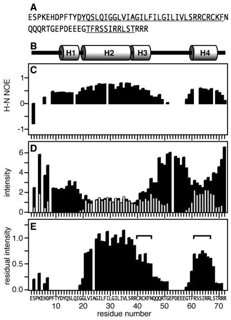
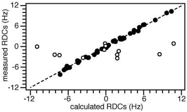
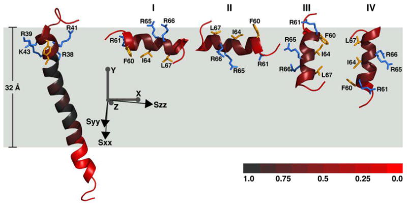
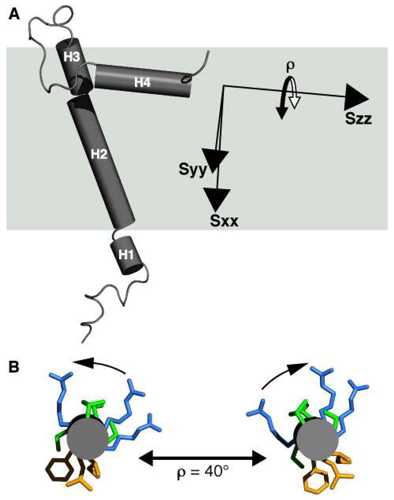
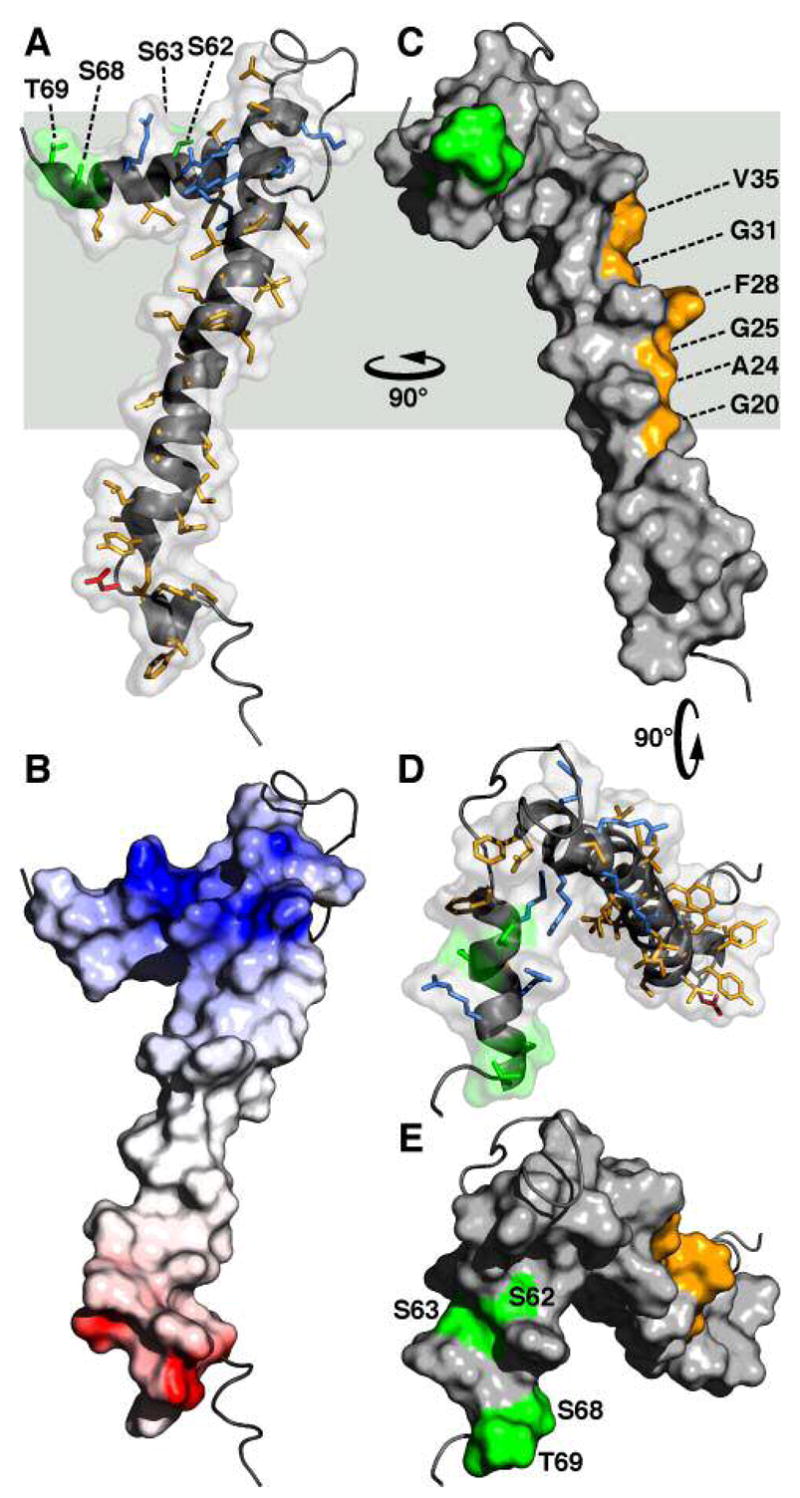
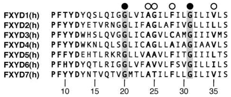
FXYD1 is a major regulatory subunit of the Na,K-ATPase and the principal substrate of hormone-regulated phosphorylation by c-AMP dependent protein kinases A and C in heart and skeletal muscle sarcolemma. It is a member of an evolutionarily conserved family of membrane proteins that regulate the function of the enzyme complex in a tissue-specific and physiological-state-specific manner. Here, we present the three-dimensional structure of FXYD1 determined in micelles by NMR spectroscopy. Structure determination was made possible by measuring residual dipolar couplings in weakly oriented micelle samples of the protein. This allowed us to obtain the relative orientations of the helical segments and information about the protein dynamics. The structural analysis was further facilitated by the inclusion of distance restraints, obtained from paramagnetic spin label relaxation enhancements, and by refinement with a micelle depth restraint, derived from paramagnetic Mn line broadening effects. The structure of FXYD1 provides the foundation for understanding its intra-membrane association with the Na,K-ATPase alpha subunit and suggests a mechanism whereby the phosphorylation of conserved Ser residues, by protein kinases A and C, could induce a conformational change in the cytoplasmic domain of the protein to modulate its interaction with the alpha subunit.


Figures

Figure 1
Figure 1
SDS-PAGE of purified FXYD1 obtained with or without the reducing agent β-mercaptoethanol. (A) molecular weight markers. (B) FXYD1 without β-mercaptoethanol. (C) FXYD1 with β-mercaptoethanol.
Figure 2
Figure 2
Effect of Mn-induced PRE on the resonance intensities. (A) Amino acid sequence of human FXYD1 with helical regions underlined. Residue numbering begins at 1 after the signal sequence (NCBI protein accession: NP_068702). (B) Protein secondary structure. (C) 1H/15N heteronuclear NOEs. (D) Normalized 1H/15N HSQC peak intensities obtained without (I−Mn, black bars) or with (I+Mn, gray bars) 1.6 mM MnCl2. Positions that are left blank correspond to prolines (P3, P8, P53) or overlapped resonances (E5, A24). (E) Residual normalized peak intensity (I−Mn/I+Mn). The horizontal square brackets mark residues in H3 and H4 with similar protection from aqueous Mn.
Figure 3
Figure 3
Correlation between HN RDCs measured experimentally and HN RDCs back-calculated from the refined structure of FXYD1. The RDCs from the flexible linker and termini (white circles) correlate poorly with those from the helical region of the protein (black circles; RMSD = 0.31; R factor = 0.07).
Figure 4
Figure 4
Molecular fragments of FXYD1 oriented in the frame of the alignment tensor (Sxx, Syy, Szz; black axes) and of the membrane (X, Y, Z; gray axes). Fragment A (shown at left) can be linked with one of four possible orientations of fragment B (I to IV). The red color gradation reflects the degree of protection from Mn-induced PRE, with full protection in the center of the membrane (gray), and no protection outside the membrane (red). The total Mn protection profile corresponds to a length of 32 Å along the membrane Y axis. Side-chains from basic residues in helix H3 (R37, R39, R41, K43), and helix H4 (R61, R65, R66), are shown in blue. Side-chains from apolar residues in H3 (C40, C42, F44), and H4 (F60, I64, L67) are shown in yellow.
Figure 5
Figure 5
Tube representation of the FXYD1 structure in the frame of the membrane (gray box), and of the alignment tensor (Sxx, Syy, Szz). (A) Helix H4 is nearly parallel to Szz. This is consistent with helix reorientation around Szz, resulting both in a GDO value similar to that of helices H1–H3, as well as greater dynamics observed from the heteronuclear NOEs and resonance intensities. (B) Helix H4 viewed from the C-terminus. A maximum helix reorientation of ρ≈ 40°, around Szz, maintains the amphiphilic polarity of hydrophobic and hydrophilic side-chains at the membrane surface.
Figure 6
Figure 6
Molecular backbone and surface representations of FXYD1. (A, D) In the helical regions, basic side-chains are shown in blue, acidic side-chains are red, and apolar side-chains are yellow. The three Ser residues (S62, S63, S68) and Thr69 in the cytoplasmic helix are in green. (B) The surface is color coded with regions of electrostatic potential <−8kBT in red, and regions of electrostatic potential >+8kBT in blue, where kB is the Boltzmann constant and T is the temperature. (C) The structure is viewed 90° around the membrane Y axis from (A, B). Residues in the transmembrane helix (G20, A24, G25, F28, G31, V35), predicted to interact with the Na,K-ATPase α subunit are shown in yellow. (D, E) The structures are viewed down the membrane surface from the cytoplasm, 90° around the membrane X axis from (C). The structure has been deposited in the databank (PDB accession code: 2JOL).
Figure 7
Figure 7
Amino acid sequences of the transmembrane domains from the human FXYD proteins. The fully conserved Gly residues, at positions 20 and 31, are highlighted in gray, and marked by black circles. Other key residues, at positions 24, 25, 28 and 35, are marked by white circles. Residue numbering is according to FXYD1.

References

    1. Sweadner KJ, Rael E. The FXYD gene family of small ion transport regulators or channels: cDNA sequence, protein signature sequence, and expression. Genomics. 2000;68:41–56. - PubMed
    1. Crambert G, Geering K. FXYD proteins: new tissue-specific regulators of the ubiquitous Na, K-ATPase. Sci STKE 2003. 2003:RE1. - PubMed
    1. Cornelius F, Mahmmoud YA. Functional modulation of the sodium pump: the regulatory proteins “Fixit”. News Physiol Sci. 2003;18:119–124. - PubMed
    1. Garty H, Karlish SJ. Role of fxyd proteins in ion transport. Annu Rev Physiol. 2006;68:431–459. - PubMed
    1. Crambert G, Fuzesi M, Garty H, Karlish S, Geering K. Phospholemman (FXYD1) associates with Na, K-ATPase and regulates its transport properties. Proc Natl Acad Sci U S A. 2002;99:11476–11481. - PMC - PubMed

Publication types

LinkOut - more resources