. 2007 Jun;64(6):1515-28.
doi: 10.1111/j.1365-2958.2007.05748.x. Epub 2007 May 18.

Identification and characterization of the immunity repressor (ImmR) that controls the mobile genetic element ICEBs1 of Bacillus subtilis

Affiliations

Identification and characterization of the immunity repressor (ImmR) that controls the mobile genetic element ICEBs1 of Bacillus subtilis

Jennifer M Auchtung et al. Mol Microbiol. 2007 Jun.

Abstract

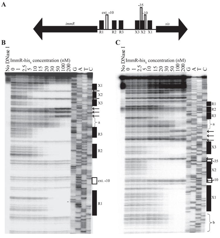
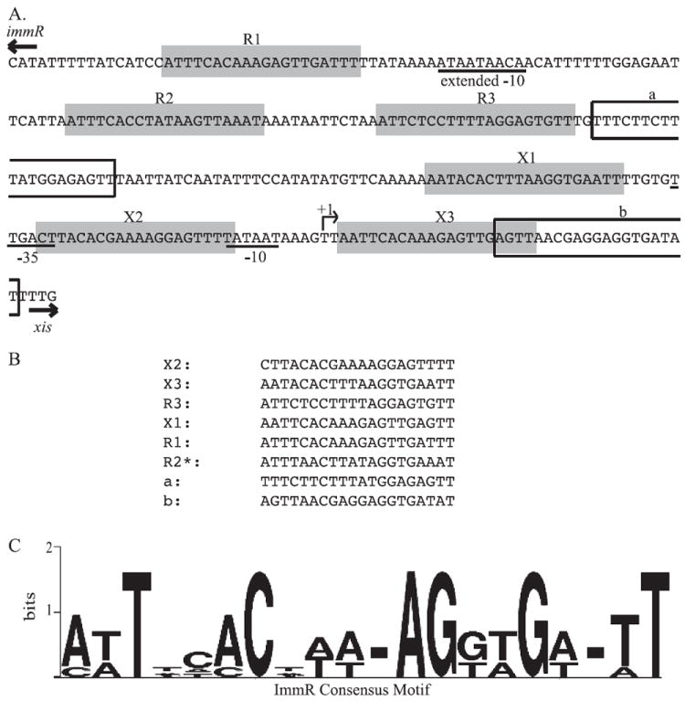
ICEBs1 is a mobile genetic element found in the chromosome of Bacillus subtilis. Excision and transfer of ICEBs1 is regulated by the global DNA damage response and intercellular peptide signalling. We identified and characterized a repressor, ImmR (formerly YdcN), encoded by ICEBs1. ImmR represses transcription of genes required for excision and transfer, and both activates and represses its own transcription. ImmR regulates transcription within ICEBs1 by binding to several sites in the region of DNA that contains promoters for both immR and xis (encoding excisionase). In addition, we found that ImmR confers immunity from acquisition of additional copies of ICEBs1. ImmR-mediated regulation serves to keep a single copy of ICEBs1 stably maintained in the absence of induction, allows a rapid response to inducing signals, and helps limit acquisition of additional copies of ICEBs1.


Figures

Fig. 1
Fig. 1. Organization of open reading frames in ICEBs1
The 24 open reading frames of ICEBs1 are indicated by thick black arrows, oriented in the direction of transcription, with the name of each gene indicated below the arrow. ydcO-ydcT and yddA-yddK are indicated by the terminal unique letter directly under each arrow and the appropriate initial three-letter designation indicated underneath each underlined section of arrows. ydc and ydd indicate genes with unknown function. The positions of the promoters for xis, immR, rapI (Jarmer et al., 2001; Auchtung et al., 2005) and phrI (McQuade et al., 2001) are indicated by vertical lines with small arrows pointed in the direction of transcription. The 60 bp direct repeats marking the ends of the element are indicated by white boxes.
Fig. 2
Fig. 2. Characterization of the xis promoter and its activation by RapI overexpression and the global DNA damage response
A. Schematic of xis, immR, and the shared intergenic region. The thick arrows indicate the locations of the xis and immR genes, and show the orientation of transcription of each. The white arrowhead indicates the position of the primer used for the primer extension assay in (B). The 5′ end of the xis transcript identified in (B) is indicated by a vertical line and arrow pointing to the right. The black box indicates the region upstream of xis fused to lacZ and used to monitor xis expression in (C) and (D). B. The 5′ end of the xis transcripts was determined by primer extension assays. G, A, T and C indicate the lanes containing dideoxynucleotide sequencing reactions with the indicated nucleotide. RNA was isolated from untreated wild-type cells (JH642, lane 1); from wild-type cells 1 h after treatment with mitomycin C (lane 2); and from Pspank(hy)-rapI cells (JMA28) 30 min after treatment with IPTG (lane 3). Results of reverse transcription reactions with the primer indicated in (A) are shown; similar results were seen when reverse transcription reactions were carried out with a primer more proximal to +1 (data not shown). The sequences complementary to the consensus −10 and −35 regions are indicated on the left of the gel. The arrow indicates the nucleotide complementary to the end of the major transcript. C and D. Cells containing a Pxis–lacZΩ343 fusion were grown in minimal medium and samples for β-galactosidase assays were collected at the times indicated. Cells were treated with 1 mM IPTG (C) or 1 μg ml−1 MMC (D) in mid-exponential phase (OD600 = 0.4–0.6). β-Galactosidase-specific activities were calculated relative to the cell densities (OD600) of the cultures. β-Galactosidase-specific activities are plotted relative to the time (in minutes) before and after addition of IPTG or MMC. In these graphs, β-galactosidase specific activity in wild-type cells appears to be at or near background (zero) levels. However, there is a low level of activity above background (Fig. 3A and B). C. xis expression in cells (KLG126) containing Pxis–lacZΩ343 and Pspank(hy)-rapI (●) and cells (KLG125) containing Pxis–lacZΩ343 and Pspank(hy) empty vector (○). D. xis expression in Pxis–lacZΩ343 cells (JMA201) (□) and Pxis–lacZΩ343 cells treated with MMC (■).
Fig. 3
Fig. 3. ImmR regulates excision through transcription of xis
A and B. Pxis–lacZΩ343 expression was monitored throughout exponential growth in minimal medium as described in Fig. 2. IPTG (0.025 or 1 mM, as indicated) was present throughout growth. A. Pxis–lacZΩ343 expression in wild-type (ICEBs1+) cells (JMA201, ▽, wt); ICEBs10 (JMA264, □, ICE0); ICEBs10 Pspank-immRΩ26 (JMA362) cells grown in the presence of 0.025 mM [○, ICE0/Pspank-immR(0.025)] or 1 mM IPTG [●, ICE0/Pspank-immR(1)]; and ICEBs10 PimmR-immRΩ267 (JMA421, ▲, ICE0/PimmR-immR) cells. B. Pxis–lacZΩ343 expression in wild-type cells (JMA201, ▽, wt; same data as in A); ΔimmR cells (JMA214, ◊, ΔimmR); and ΔimmR Pspank-immRΩ26 cells in the presence of 1 mM IPTG (JMA541, ◆, ΔimmR/Pspank-immR). C. Schematic representation of the excision assay performed in (D). Upon excision of ICEBs1 from the chromosome, two products are formed, an ICEBs1 circular intermediate and a repaired chromosomal junction. These products can be detected through PCR using primers b + c and a + d (respectively), which are represented by small arrows in the diagram. The sequences of these primers [oJMA93 (a), oJMA95 (b), oJMA97 (c) and oJMA100 (d)] were described previously (Auchtung et al., 2005). D. Excision was monitored in wild-type (JMA201, lane 1), ΔimmR (JMAM214, lane 2) and ΔimmR Pspank-immRΩ26 (JMA541, grown in the presence of 1 mM IPTG, lane 3) cells. DNA was extracted from cells in exponential phase and 100 ng was used as template to amplify the indicated products described in (C). Quantitative PCR performed on DNA extracted from a population of ΔimmR cells revealed that excision occurred in ~97% of cells whereas excision occurred in ~0.003% of wild-type cells.
Fig. 4
Fig. 4. ImmR represses and activates its own transcription
A. Schematic of xis, immR, and the intergenic region. xis, immR, and their direction of transcription are indicated by the large black arrows. The location of the putative extended −10 recognition sequence for EσA is indicated by a vertical black rectangle (ext. −10). The sequences present in the immR constructs used in (B) and (C) are indicated by white boxes underneath the diagram. B. Functional analysis of the immR promoter. Constructs containing the entire immR open reading frame and segments of its upstream sequence (diagrammed in A) were tested for their ability to reconstitute ImmR function by repressing transcription of Pxis–lacZΩ343 in ICEBs10 cells. β-Galactosidase-specific activity in cells grown to mid-late exponential phase (OD600 ~ 1) in minimal medium was determined. Pxis–lacZΩ343 containing cells assayed were wild type (JMA201, wt); ICEBs10 (JMA264, ICE0); ICEBs10 Pspank-immRΩ26 (JMA362, ICE0/immRΩ26); ICEBs10 PimmR-immRΩ141 (JMA266, ICE0/immRΩ141); and ICEBs10 PimmR-immRΩ267 (JMA421, ICE0/immRΩ267). C. Expression of a PimmR–lacZ fusion was monitored throughout exponential growth in minimal medium in otherwise wild-type cells (JMA309, □), ΔimmR (JMA310, Δ), ICEBs10 (JMA576, ◆) and ΔimmR Pspank-immRΩ26 cells (JMA638) grown in the in the presence of 0.025 (○) or 1 mM IPTG (●). IPTG, when used, was present throughout growth. β-Galactosidase-specific activities are plotted relative to the OD600 of the cultures.
Fig. 5
Fig. 5. ImmR binds to the xis and immR intergenic region
A. Detailed schematic of xis, immR, and the intergenic region. xis, immR, and the directions of transcription are indicated by the big black arrows. The location of the putative extended −10 recognition sequence for sigma-A-containing RNA polymerase in the immR promoter, and the −10 and −35 recognition sequences for sigma-A-containing RNA polymerase in the xis promoter are indicated by vertical white rectangles. The positions of the six regions protected by ImmR (sites X1, X2, X3, R1, R2 and R3) are indicated by shorter vertical black boxes. B and C. Binding of ImmR to the xis-immR intergenic region was monitored through DNase I protection assays. Increasing concentrations (1–200 nM) of purified ImmR-his6 protein were incubated with radiolabelled DNA from the xis-immR intergenic region. DNase I was added to each reaction to digest DNA not protected by ImmR. Reactions were analysed by electrophoresis along with dideoxynucleotide sequencing reactions of the xis-immR intergenic region. The concentrations of ImmR used in each reaction are indicated above each lane of the gel. G, A, T and C indicate the dideoxynucleotide used in each sequencing reaction. Positions of the extended −10 recognition sequence for EσA in the immR promoter, and the −10 and −35 recognition sequences for EσA in the xis promoter, the six protected regions described in (A), and the two additional sites of ImmR protection (a and b) are indicated to the right of each gel image. The arrows indicate the positions of DNase I hypersensitive sites. In (B), the 5′ end of DNA at the immR end (the top strand) was labelled; in (C), the 5′ end of DNA near xis (the bottom strand) was labelled.
Fig. 6
Fig. 6. Identification of a conserved ImmR binding motif
A. The sequence of a single strand of the xis-immR intergenic region is shown. The positions of the immR and xis start codons are indicated by arrows above the appropriate sequences. The positions of the PimmR extended −10 and the Pxis −35 and −10 recognition sequences for RNA polymerase containing sigma-A are indicated by the underlined nucleotides. The positions of R1, R2 and R3, and X1, X2 and X3 are indicated by grey boxes. R1, R3, and X1, X2 and X3 are all on the same strand of DNA whereas R2 is on the complementary strand. The positions of the a and b sites are indicated by white boxes. B. An alignment of the nucleotide sequences of all eight sites protected by ImmR listed in order of ImmR affinities observed in the DNase I protection experiments (Fig. 5). C. A representation of the consensus motif for the ImmR-binding sequence was generated using Weblogo (Crooks et al., 2004). The size of each nucleotide corresponds to the frequency with which that nucleotide was observed in that position; dashes at a position indicate lack of consensus.

References

    1. Achtman M, Kennedy N, Skurray R. Cell–cell interactions in conjugating Escherichia coli: role of traT protein in surface exclusion. Proc Natl Acad Sci USA. 1977;74:5104–5108. - PMC - PubMed
    1. Anthony KG, Klimke WA, Manchak J, Frost LS. Comparison of proteins involved in pilus synthesis and mating pair stabilization from the related plasmids F and R100-1: insights into the mechanism of conjugation. J Bacteriol. 1999;181:5149–5159. - PMC - PubMed
    1. Auchtung JM, Lee CA, Monson RE, Lehman AP, Grossman AD. Regulation of a Bacillus subtilis mobile genetic element by intercellular signaling and the global DNA damage response. Proc Natl Acad Sci USA. 2005;102:12554–12559. - PMC - PubMed
    1. Bailey TL, Elkan C. Fitting a mixture model by expectation maximization to discover motifs in biopolymers. Proc Int Conf Intell Syst Mol Biol. 1994;2:28–36. - PubMed
    1. Bailey TL, Gribskov M. Combining evidence using p-values: application to sequence homology searches. Bioinformatics. 1998;14:48–54. - PubMed

Publication types

MeSH terms

LinkOut - more resources