. 2007 Jul;47(15):2055-66.
doi: 10.1016/j.visres.2007.04.005. Epub 2007 May 21.

Novel RDH12 mutations associated with Leber congenital amaurosis and cone-rod dystrophy: biochemical and clinical evaluations

Affiliations

Novel RDH12 mutations associated with Leber congenital amaurosis and cone-rod dystrophy: biochemical and clinical evaluations

Wenyu Sun et al. Vision Res. 2007 Jul.

Abstract

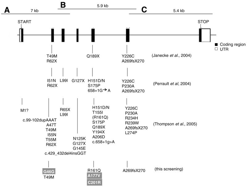
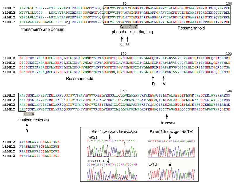
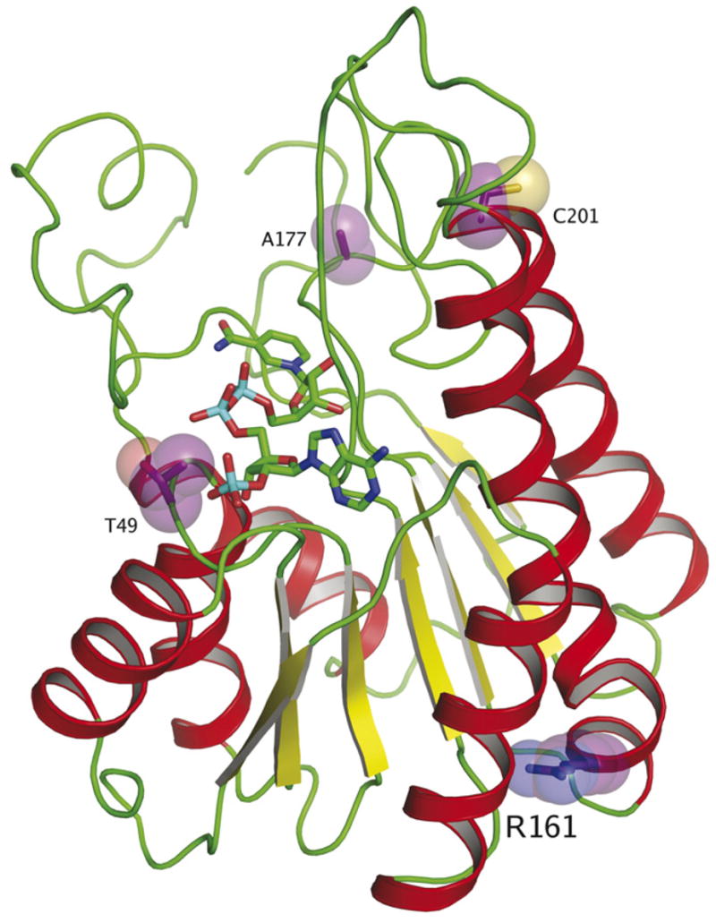
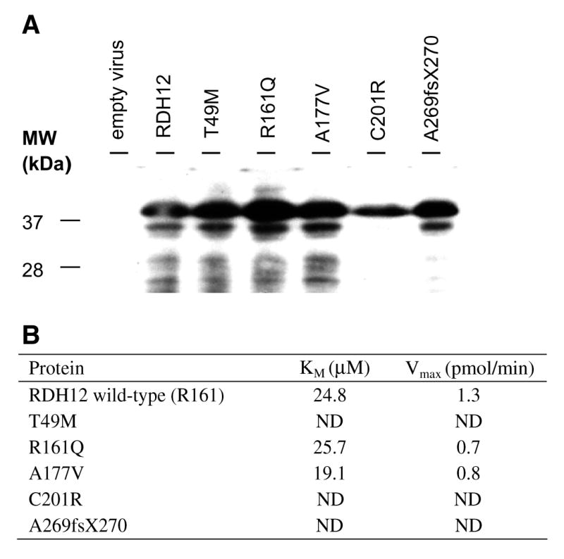
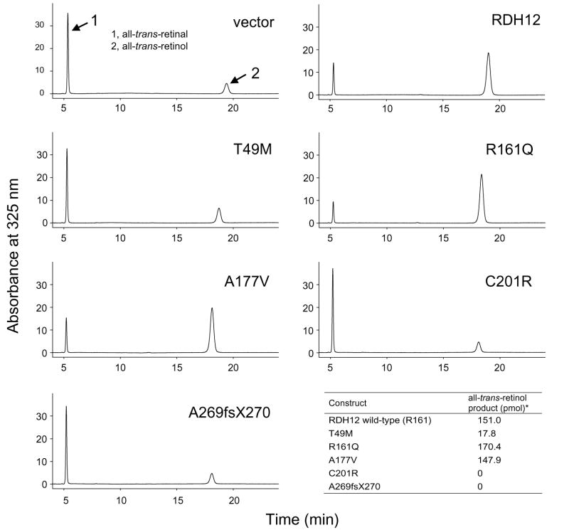
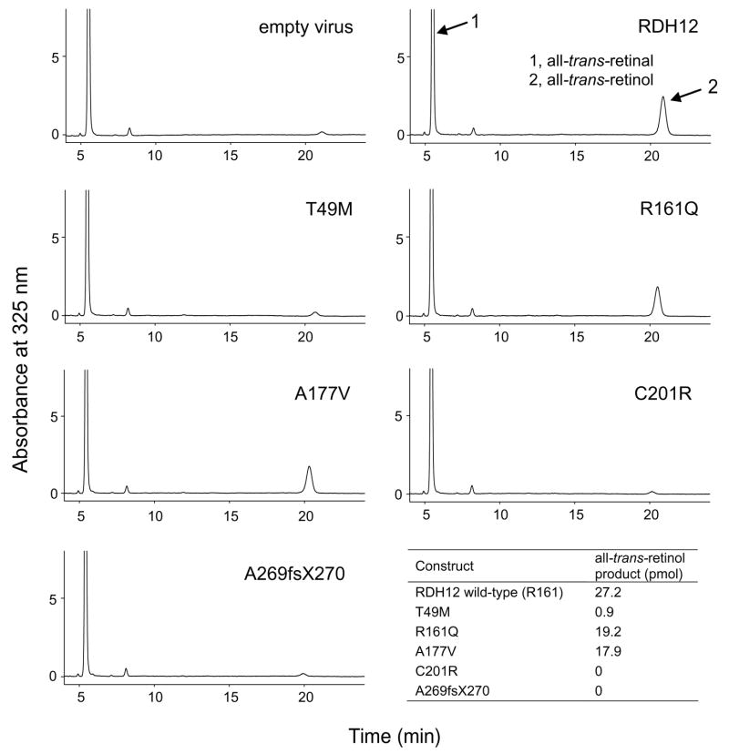
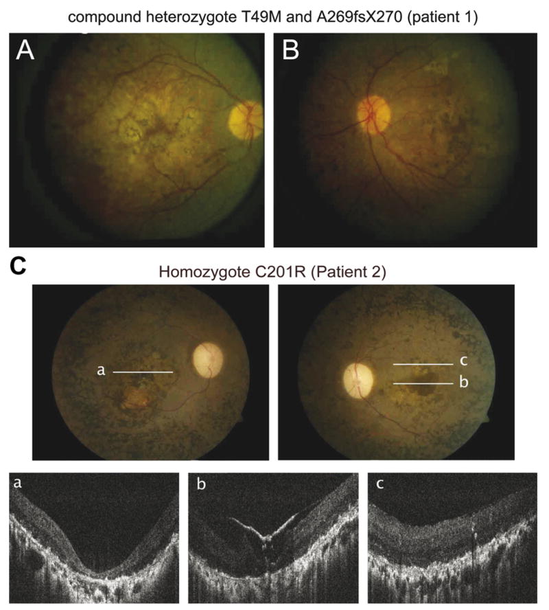
The purpose of this study was to determine the role of the retinol dehydrogenase 12 (RDH12) gene in patients affected with Leber congenital amaurosis (LCA), autosomal recessive retinitis pigmentosa (arRP) and autosomal dominant/recessive cone-rod dystrophies (CORD). Changes in the promoter region, coding regions and exon/intron junctions of the RDH12 gene were evaluated using direct DNA sequencing of patients affected with LCA (n=36 cases), RP (n=62) and CORD (n=21). The allele frequency of changes observed was assessed in a multiethnic control population (n=159 individuals). Detailed biochemical and structural modeling analysis of the observed mutations were performed to assess their biological role in the inactivation of Rdh12. A comprehensive clinical assessment of retinal structure and function in LCA patients carrying mutations in the RDH12 gene was completed. Of the six changes identified, three were novel including a homozygous C201R change in a patient affected with LCA, a heterozygous A177V change in patients affected with CORD and a heterozygous G46G change in a patient affected with LCA. A novel compound heterozygote T49M/A269fsX270 mutation was also found in a patient with LCA, and both homozygous and heterozygous R161Q changes were seen in 26 patients affected with LCA, CORD or RP. These R161Q, G46G and the A177V sequence changes were shown to be polymorphic. We found that Rdh12 mutant proteins associated with LCA were inactive or displayed only residual activity when expressed in COS-7 and Sf9 cells, whereas those mutants that were considered polymorphisms were fully active. Thus, impairment of retinal structure and function for patients carrying these mutations correlated with the biochemical properties of the mutants.


Figures

Fig. 1
Fig. 1
Intron/exon gene structure of human RDH12 and all identified genetic variants. A, B, and C correspond to three PCR fragments that overlap each other to cover the entire RDH12 gene. For comparison, genetic variants identified previously by other groups and variants identified in this study are listed below the structure. New genetic variants found in this study are denoted with a gray background.
Fig. 2
Fig. 2
Homology of Rdh12 among different species. Rdh12 from human, bovine, mouse and Drosophila are aligned together here to show the sequence homology. The putative transmembrane domain is located at the N-terminus. Regions corresponding to the Rossmann fold, phosphate-binding site and catalytic site are also labeled. Polymorphisms/mutations identified in this study are also indicated. The chromatogram figures show compound heterozygote of 146C → T/806delCCCTG (patient 1) and homozygote of 601T → C (patient 2).
Fig. 3
Fig. 3
Homology model of Rdh12 showing sites of mutations/polymorphisms identified in this study. Residues 49, 161, 177, and 201 are shown. The T49M mutation can interfere with NADP binding, whereas change A177V may interfere with substrate binding. That the R161Q mutation does not appear to exhibit any changes in enzymatic activity is unsurprising, because the location is far from the active site and is likely not involved in catalysis. C201R, which lies close to the catalytic residues, can disrupt the formation of the active site, leading to a loss of enzymatic activity. The homology model was built using β-ketoacyl-[ACP]-reductase from Escherichia coli as a backbone model (PDB ID:1Q7B), using the Geno3D server (Combet et al., 2002) and O (Jones et al., 1991) for generation of an initial homology model and manual model building respectively. Figure was created with PYMOL (DeLano, W.L. The PyMOL Molecular Graphics System (2002) DeLano Scientific, San Carlos, CA, USA. http://www.pymol.org).
Fig. 4
Fig. 4
(A) Expression of Rdh12 and its mutants. Lanes were loaded with extracts from Sf9 cells infected with virus only, virus containing RDH12, T49M, R161Q, A177V, C201R, and A269fsX270 from left to right, respectively. Immunoblotting was carried out according to standard protocols using Immobilon-P to adsorb proteins (polyvinylidene difluoride; Millipore Corp.) (Ohguro et al., 1993). Mouse polyclonal antibodies against bacterially expressed full-length mouse Rdh12 were raised in BALB/c mice as described previously (Maeda et al., 2006). Secondary antibody is HRP conjugated anti-mouse IgG (Promega Corporation, Madison, WI) and signal was detected using SuperSignal West Pico Chemiluminescent Substrate (Pierce Biotechnology, Rockford, IL). (B) Calculated Km and Vmax values of Rdh12 constructs expressed in Sf9 cells. ND, not determined.
Fig. 5
Fig. 5
HPLC analysis of the enzymatic activity of Rdh12 variants in COS-7 cells. Enzyme activity was detected by monitoring all-trans-retinol production via normal phase HPLC.
Fig. 6
Fig. 6
HPLC analysis of the enzymatic activity of Rdh12 variants in Sf9 cells. The reaction was carried out as described for the COS-7 cells.
Fig. 7
Fig. 7
Fundus photography. A (right eye) and B (left eye) are fundus photography of patient 1 (21 years), with compound mutations (T49M/A269fsX270) and a LCA phenotype shows diffuse retinopathy with pigment clumping and bone spicule pigmentation, cystoid macular edema, attenuated blood vessels with mild optic atrophy in both eyes. C is fundus photography and corresponding FD-OCT images of patient 2 (32 years) who is homozygous for the C201R mutation and has a LCA phenotype. Lines are symbolizing area of FD-OCT scan acquisition. Both eyes show a macular pseudo-coloboma with dense pigmentation, optic atrophy, arteriole and venous attenuation and peripheral granular pigmentation. Foveal and extrafoveal FD-OCT images a–c demonstrate re-organization of retinal lamination and loss of photoreceptors and epiretinal membrane in the left eye.

Similar articles

Cited by

References

    1. Acland GM, Aguirre GD, Bennett J, Aleman TS, Cideciyan AV, Bennicelli J, et al. Long-term restoration of rod and cone vision by single dose rAAV-mediated gene transfer to the retina in a canine model of childhood blindness. Molecular Therapeutics. 2005;12(6):1072–1082. - PMC - PubMed
    1. Acland GM, Aguirre GD, Ray J, Zhang Q, Aleman TS, Cideciyan AV, et al. Gene therapy restores vision in a canine model of childhood blindness. Nature Genetics. 2001;28(1):92–95. - PubMed
    1. Aleman TS, Jacobson SG, Chico JD, Scott ML, Cheung AY, Windsor EA, et al. Impairment of the transient pupillary light reflex in Rpe65(−/−) mice and humans with leber congenital amaurosis. Investigative Ophthalmology and Visual Science. 2004;45(4):1259–1271. - PubMed
    1. Batten ML, Imanishi Y, Tu DC, Doan T, Zhu L, Pang J, et al. Pharmacological and rAAV gene therapy rescue of visual functions in a blind mouse model of Leber congenital amaurosis. PLoS Medicine. 2005;2(11):e333. - PMC - PubMed
    1. Belyaeva OV, Korkina OV, Stetsenko AV, Kim T, Nelson PS, Kedishvili NY. Biochemical properties of purified human retinol dehydrogenase 12 (RDH12): Catalytic efficiency toward retinoids and C9 aldehydes and effects of cellular retinol-binding protein type I (CRBPI) and cellular retinaldehyde-binding protein (CRALBP) on the oxidation and reduction of retinoids. Biochemistry. 2005;44(18):7035–7047. - PMC - PubMed

Publication types

Substances