

Volumetric cerebral characteristics of children exposed to opiates and other substances in utero
- PMID: 17513131
- PMCID: PMC2039875
- DOI: 10.1016/j.neuroimage.2007.03.070
Volumetric cerebral characteristics of children exposed to opiates and other substances in utero
Erratum in
- Neuroimage. 2008 Jul 15;41(4):1514-6
Abstract
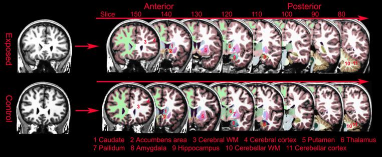
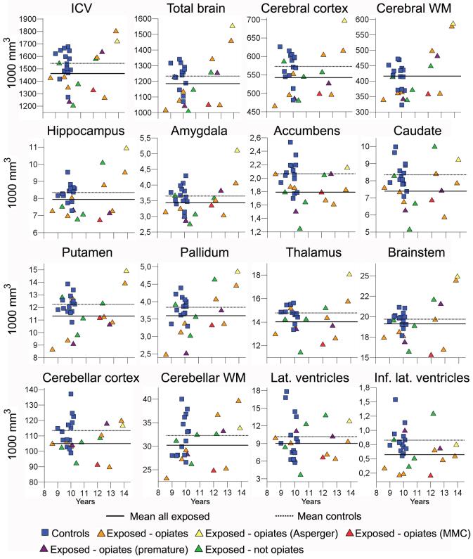
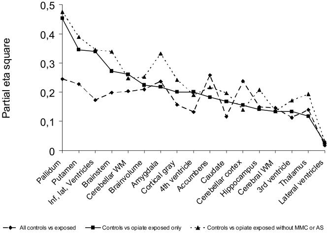
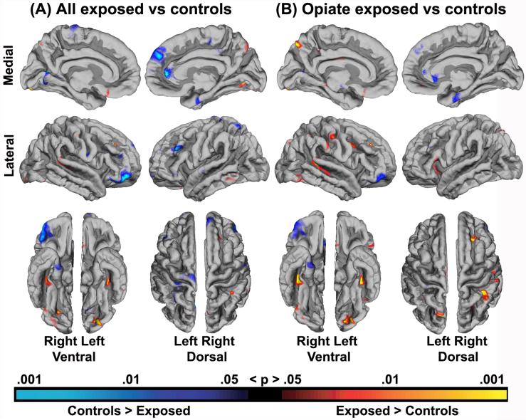
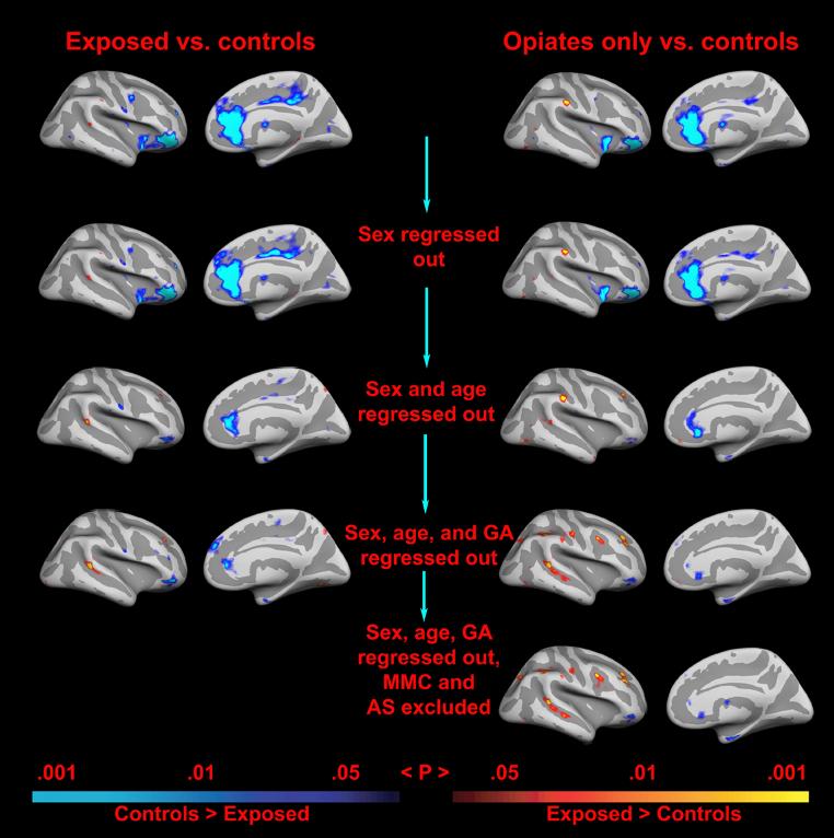
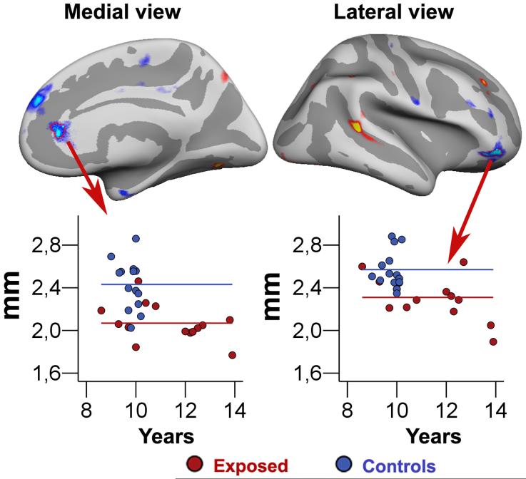
Morphometric cerebral characteristics were studied in children with prenatal poly-substance exposure (n=14) compared to controls (n=14) without such exposure. Ten of the substance-exposed children were born to mothers who used opiates (heroin) throughout the pregnancy. Groups were compared across 16 brain measures: cortical gray matter, cerebral white matter, hippocampus, amygdala, thalamus, accumbens area, caudate, putamen, pallidum, brainstem, cerebellar cortex, cerebellar white matter, lateral ventricles, inferior lateral ventricles, and the 3rd and 4th ventricles. In addition, continuous measurement of thickness across the entire cortical mantle was performed. Volumetric characteristics were correlated with ability and questionnaire assessments 2 years prior to scan. Compared to controls, the substance-exposed children had smaller intracranial and brain volumes, including smaller cerebral cortex, amygdala, accumbens area, putamen, pallidum, brainstem, cerebellar cortex, cerebellar white matter, and inferior lateral ventricles, and thinner cortex of the right anterior cingulate and lateral orbitofrontal cortex. Pallidum and putamen appeared especially reduced in the subgroup exposed to opiates. Only volumes of the right anterior cingulate, the right lateral orbitofrontal cortex and the accumbens area, showed some association with ability and questionnaire measures. The sample studied is rare and hence small, so conclusions cannot be drawn with certainty. Morphometric group differences were observed, but associations with previous behavioral assessment were generally weak. Some of the volumetric differences, particularly thinner cortex in part of the right lateral orbitofrontal cortex, may be moderately involved in cognitive and behavioral difficulties more frequently experienced by opiate and poly-substance-exposed children.
Figures
References
-
- Achenbach T,M. Manual for the Child Behavior Checklist/4-18 and 1991 Profile. University of Vermont Department of Psychiatry; Burlington: 1991.
-
- Alexander G,E, Delong M,R, Strick P,L. Parallel organization of functionally segregated circuits linking basal ganglia and cortex. Annu Rev Neurosci. 1986;9:357–381. - PubMed
-
- Bhat R, Chari G, Rao R. Effects of prenatal cocaine, morphine, or both on postnatal opioid (μ) receptor development. Life Sci. 2006;78:1478–1482. - PubMed
-
- Bush G, Valera E,M, Seidman L,E. Functional neuroimaging of attention-deficit/hyperactivity disorder: a review and suggested future directions. Biol Psychiatry. 2005;57:1273–1284. - PubMed
-
- Canivez GL, Watkins MW. Long term stability of the Wechsler Intelligence Scale for Children-Third Edition among Demographic Subgroups: Gender, Race/Ethnicity, and Age. Journal of Psychoeducational Assessment. 1999;17:300–313.
Publication types
MeSH terms
Substances
Grants and funding
- P41-RR14075/RR/NCRR NIH HHS/United States
- R01-RR13609/RR/NCRR NIH HHS/United States
- BIRN002/PHS HHS/United States
- R01 EB001550/EB/NIBIB NIH HHS/United States
- U54 EB005149/EB/NIBIB NIH HHS/United States
- K02 DA000354/DA/NIDA NIH HHS/United States
- P41 RR014075/RR/NCRR NIH HHS/United States
- R01 NS052585/NS/NINDS NIH HHS/United States
- R01-RR16594/RR/NCRR NIH HHS/United States
- R01 RR016594/RR/NCRR NIH HHS/United States
- R01-NS39581/NS/NINDS NIH HHS/United States
- R01 EB006758/EB/NIBIB NIH HHS/United States
- U24 RR021382/RR/NCRR NIH HHS/United States
- R01 DA017905/DA/NIDA NIH HHS/United States
LinkOut - more resources
Full Text Sources
Medical
