. 2007 Aug 15;93(4):1116-23.
doi: 10.1529/biophysj.107.106708. Epub 2007 May 18.

Stochastic entry of enveloped viruses: fusion versus endocytosis

Affiliations

Stochastic entry of enveloped viruses: fusion versus endocytosis

Tom Chou. Biophys J. .

Abstract

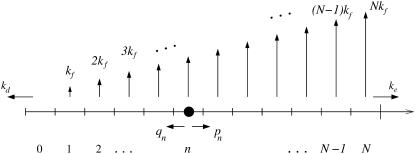
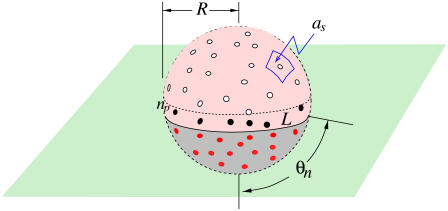
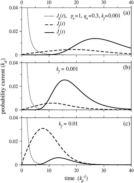
Infection by membrane-enveloped viruses requires the binding of receptors on the target cell membrane to glycoproteins, or "spikes," on the viral membrane. The initial entry mechanism is usually classified as fusogenic or endocytotic. However, binding of viral spikes to cell surface receptors not only initiates the viral adhesion and the wrapping process necessary for internalization, but can simultaneously initiate direct fusion with the cell membrane. Both fusion and internalization have been observed to be viable pathways for many viruses. We develop a stochastic model for viral entry that incorporates a competition between receptor-mediated fusion and endocytosis. The relative probabilities of fusion and endocytosis of a virus particle initially nonspecifically adsorbed on the host cell membrane are computed as functions of receptor concentration, binding strength, and number of spikes. We find different parameter regimes where the entry pathway probabilities can be analytically expressed. Experimental tests of our mechanistic hypotheses are proposed and discussed.


Figures

FIGURE 1
FIGURE 1
A schematic of viral entry pathways. (a) A nonspecifically adsorbed virus particle can desorb with rate kd, or it can (b) recruit and specifically bind a receptor. The receptor can immediately initiate membrane fusion with rate kf as shown in panel c, or, it can recruit additional receptor molecules, inducing wrapping of the virus particle. From partially wrapped states (d), the virus can at any stage undergo membrane fusion (e), or, it can completely wrap and internalize the virus particle (f and g).
FIGURE 2
FIGURE 2
The stochastic process representing the competition between membrane fusion and endocytosis. The states n correspond to the number of receptor-spike complexes formed, while N is the total number of spikes on the virus membrane. Each receptor-spike complex can initiate membrane fusion with rate kf. As more receptors are bound, the total rate of fusion increases linearly. The irreversible pinch-off and endocytosis rate is denoted ke.
FIGURE 3
FIGURE 3
Schematic of a partially wrapped virus particle. The unbound spikes above the contact region are represented by open circles, while the receptor-bound spikes in the contact region are represented by the red dots. The number np of spikes or spike-receptor complexes near the contact perimeter used to compute pn or qn via Eq. 2 are shown as black dots.
FIGURE 4
FIGURE 4
The currents through each pathway for N = 20 spikes. The thin dashed black curve is the current for desorption, while the thick dashed and solid curves are the currents for fusion and endocytosis, respectively. Time is measured in units of formula image and all rates are normalized with respect to kd. The parameters used in all plots are p0 = 1, q1 = 0.3, and ke = 0.3. (a) The currents for constant pn = 1, qn = 0.3, and fusion rate kf = 0.001. (b) The same parameters except that Eq. 2 is used for the rates pn, qn. (c) The currents with pn, qn as in panel b, except that the fusion rate of each spike-receptor complex is increased to kf = 0.01.
FIGURE 5
FIGURE 5
Numerical solutions of entry probabilities Qi (all rates normalized by kd). (a) The entry probabilities as a function of p0. Endocytosis arises only for larger p0 > q1, after the fusion probability becomes significant. Parameters used are N = 20, kf = 0.003, and ke = 0.3 (all rates are normalized by kd). (b) The probabilities of dissociation (thin dashed), fusion (thick dashed), and endocytosis (thick solid) as functions of the individual receptor-spike fusion rate kf. Here, q1 = 0.3. The thin dotted curve corresponds to a faster receptor detachment rate (q1 = 2), which prevents endocytosis.
FIGURE 6
FIGURE 6
The pathway dependence on receptor association/dissociation rates and the number N of virus spikes. The number of spikes controls which regime of Eqs. 4 or 5 is valid. Large N enhances fusion almost entirely at the expense of endocytosis.

References

    1. Barocchi, M. A., V. Masignani, and R. Rappuoli. 2005. Cell entry machines: a common theme in nature? Nat. Rev. Microbiol. 3:349–358. - PMC - PubMed
    1. Lakadamyali, M., M. J. Rust, H. P. Babcock, and X. Zhuang. 2003. Visualizing infection of individual influenza viruses. Proc. Natl. Acad. Sci. USA. 100:9280–9285. - PMC - PubMed
    1. Markosyan, R. M., F. S. Cohen, and G. B. Melikyan. 2005. Time-resolved imaging of HIV-1 Env-mediated lipid and content mixing between a single virion and cell membrane. Mol. Biol. Cell. 16:5502–5513. - PMC - PubMed
    1. Cross, K. J., L. M. Burleigh, and D. A. Steinhauer. 2001. Mechanisms of cell entry by influenza virus. Expert Rev. Mol. Med. Aug. 6:1–18. - PubMed
    1. Shekel, J. J., and D. C. H. Wiley. 2000. Receptor binding and membrane fusion in virus entry: the influenza hemagglutinin. Annu. Rev. Biochem. 69:531–569. - PubMed

Publication types

LinkOut - more resources