. 2007 May;19(5):1549-64.
doi: 10.1105/tpc.106.047621. Epub 2007 May 18.

Components of the Arabidopsis mRNA decapping complex are required for early seedling development

Affiliations

Components of the Arabidopsis mRNA decapping complex are required for early seedling development

David C Goeres et al. Plant Cell. 2007 May.

Abstract

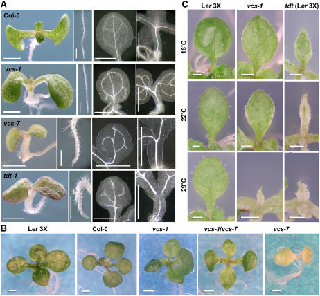
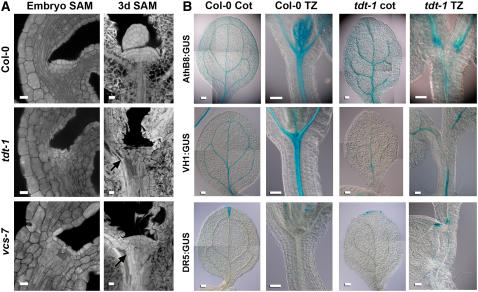
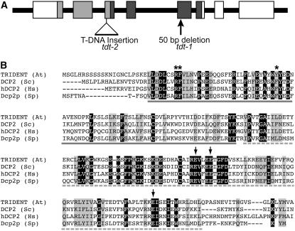
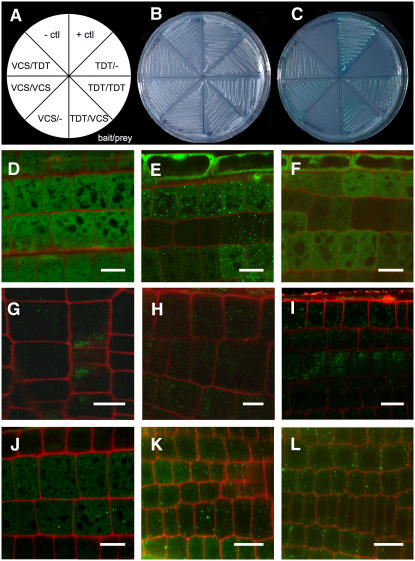
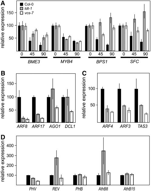
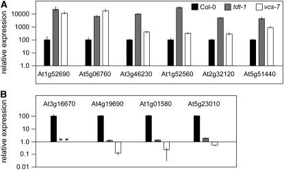
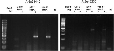
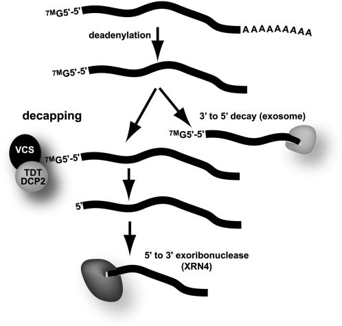
To understand the mechanisms controlling vein patterning in Arabidopsis thaliana, we analyzed two phenotypically similar mutants, varicose (vcs) and trident (tdt). We had previously identified VCS, and recently, human VCS was shown to function in mRNA decapping. Here, we report that TDT encodes the mRNA-decapping enzyme. VCS and TDT function together in small cytoplasmic foci that appear to be processing bodies. To understand the developmental requirements for mRNA decapping, we characterized the vcs and tdt phenotypes. These mutants were small and chlorotic, with severe defects in shoot apical meristem formation and cotyledon vein patterning. Many capped mRNAs accumulated in tdt and vcs mutants, but surprisingly, some mRNAs were specifically depleted. In addition, loss of decapping arrested the decay of some mRNAs, while others showed either modest or no decay defects, suggesting that mRNAs may show specificity for particular decay pathways (3' to 5' and 5' to 3'). Furthermore, the severe block to postembryonic development in vcs and tdt and the accompanying accumulation of embryonic mRNAs indicate that decapping is important for the embryo-to-seedling developmental transition.


Figures

Figure 1.
Figure 1.
Phenotypes of vcs and tdt Seedlings. (A) Phenotypes of seedlings grown for 6 d at 22°C. Panels show seedling, root, cotyledon vein pattern, and hypocotyl vasculature (left to right). (B) The vcs-1/vcs-7 transheterozygote shows a suppressed phenotype similar to that of vcs-1 homozygotes. These seedlings were grown for 15 d at 16°C. (C) Leaf development of vcs-1, and tdt-1 in the Ler genetic background, depends on growth temperature. Bars = 1 mm.
Figure 2.
Figure 2.
SAM and Vascular Defects in tdt and vcs-7. (A) Confocal analysis of SAM organization in mature embryos and 3-d-old seedlings. Arrows indicate ectopic differentiating tracheary elements. Bars = 10 μm. (B) Reporter gene expression in cotyledon and hypocotyl transition zones (TZ) of the wild type (Col-0) and tdt-1. AthB8:GUS confers expression in provascular, procambial, and vascular cells; VH1:GUS confers strong expression in provascular, procambial, and phloem cells; DR5:GUS confers expression in cells responding to auxin. Bars = 100 μm.
Figure 3.
Figure 3.
TDT Encodes the mRNA-Decapping Enzyme. (A) Diagram of the TDT gene. Boxes correspond to exons, and the 5′ end is to the left. Light gray boxes correspond to the DCP2 domain, and dark gray boxes correspond to the NUDIX domain. Locations of the T-DNA insertion and the lesion identified in tdt-1 are indicated. (B) Alignment of DCP2 and NUDIX domains of TDT with those of Saccharomyces cerevisiae (Sc), Schizosaccharomyces pombe (Sp), and Homo sapiens (Hs). Arrows indicate conserved E residues that are required for catalytic activity (all are conserved in TDT), and asterisks indicate residues required for interaction with DCP1 (She et al., 2006). Solid gray underlining indicates the DCP2 domain, and the dashed underline indicates the NUDIX domain.
Figure 4.
Figure 4.
VCS and TDT Interact, and TDT Localizes to P-Body–Like Cytoplasmic Foci. (A) to (C) Yeast two-hybrid analysis of VCS and TDT interaction. (A) Map of yeast strains shown in (B) and (C). (B) trp-leu- medium shows growth of each transformed yeast line. (C) trp-leu-his-ade-x-α-gal+ medium shows interaction in the positive control, combinations that include both VCS and TDT, and VCS–VCS and TDT–TDT self-interactions. (D) to (L) Confocal micrographs of root elongation zones of 4-d-old seedlings. Bars = 10 μm. (D) 35S:GFP in wild-type roots shows signal distributed throughout the cells. (E) 35S:GFP:TDT in wild-type roots shows GFP signal that is largely localized to small spots dispersed through the cytoplasm. (F) 35S:GFP:TDT in wild-type roots treated with cycloheximide no longer shows localized spots of GFP signal; instead, the GFP signal resembles that of 35S:GFP. (G) to (I) 35S:GFP:TDT in roots of vcs-7 mutants. (G) Untreated vcs-7 seedlings show weak and diffuse signal from 35S:GFP:TDT. (H) DMSO-treated controls also show weak and diffuse staining. (I) vcs-7 mutants treated for 1 h with the proteasome inhibitor MG132 show slightly brighter, but still diffuse, signal from the 35S:GFP:TDT transgene. (J) to (L) 35S:GFP:TDT in wild-type roots shows similar localization and intensity in untreated (J), DMSO-treated (K), and MG132-treated (L) seedlings.
Figure 5.
Figure 5.
vcs and tdt Mutants Show Defects in RNA Decay and Aberrant Patterns of RNA Accumulation. In these graphs, data for the wild type are represented by black bars, data for tdt-1 by gray bars, and data for vcs-7 by white bars. Four-day-old seedlings are represented in (A), and 3-d-old seedlings are represented in (B) to (D). Error bars indicate sd; n = 4. (A) RNA decay monitored by analyzing the relative expression of four mRNAs following treatment with the transcription inhibitor cordycepin (0, 45, or 90 min after the addition of cordycepin). (B) Relative expression levels of select miRNA target RNAs in 3-d-old seedlings grown at 22°C. In tdt-1 and vcs-7, these miRNA target RNAs show levels that are either similar to, or lower than, that of the wild type. (C) Analysis of two trans-acting silencing RNA targets (ARF3 and ARF4) and the TAS3 RNA. (D) Relative expression levels of the HD-ZIPIII RNAs in tdt-1 and vcs-7 reveal no consistent pattern of mRNA overaccumulation in tdt-1 and vcs-7 mutants.
Figure 6.
Figure 6.
RNA Accumulation and Depletion in tdt-1 and vcs-7 Mutants. Real-time RT-PCR analysis of relative expression of genes with altered expression detected in the microarray. Data for the wild type are represented by black bars, data for tdt-1 by gray bars, and data for vcs-7 by white bars. Asterisks indicate not detectable levels; error bars indicate sd. (A) The relative levels of six RNAs that microarray analysis indicated increased in tdt-1 show strong increases; the levels are also elevated in vcs-7. (B) The relative levels of four RNAs that microarray analysis indicated as underaccumulated in tdt-1 show strong differences; the levels are also depleted in vcs-7.
Figure 7.
Figure 7.
tdt-1 and vcs-7 Accumulate Capped mRNAs. Capped RNAs were assayed by ligation to a 5′ RNA adapter either before (−) or after (+) treatment with TAP to remove the cap. We detected an abundant product only in the mutants, and only after TAP treatment, confirming that these abundant mRNAs were capped. ctl indicates a reaction without template addition, and (G) indicates genomic DNA supplied as the template.
Figure 8.
Figure 8.
Model for mRNA Bulk Decay Pathways. Two key features of an mRNA in the cytoplasm are the 5′-5′ 7-methylguanosine (7MG) cap and the 3′ poly(A) tail. Bulk mRNA decay is typically initiated by deadenylation. The deadenylated mRNA can then undergo further decay from its 3′ end by the activity of the exosome. Alternatively, the deadenylated mRNA can have its 5′ cap removed (decapped), followed by further decay from its 5′ end by XRN4 activity.

Similar articles

Cited by

References

    1. Adenot, X., Elmayan, T., Lauressergues, D., Boutet, S., Bouché, N., Gasciolli, V., and Vaucheret, H. (2006). DRB4-dependent TAS3 trans-acting siRNAs control leaf morphology through AGO7. Curr. Biol. 16 927–932. - PubMed
    1. Alonso, J.M., et al. (2003). Genome-wide insertional mutagenesis of Arabidopsis thaliana. Science 301 653–657. - PubMed
    1. Badis, G., Saveanu, C., Fromont-Racine, M., and Jacquier, A. (2004). Targeted mRNA degradation by deadenylation-independent decapping. Mol. Cell 15 5–15. - PubMed
    1. Baima, S., Nobili, F., Sessa, G., Lucchetti, S., Ruberti, I., and Morelli, G. (1995). The expression of the Athb-8 homeobox gene is restricted to provascular cells in Arabidopsis thaliana. Development 121 4171–4182. - PubMed
    1. Barton, M.K., and Poethig, R.S. (1993). Formation of the shoot apical meristem in Arabidopsis thaliana: An analysis of development in the wild type and in the shoot meristemless mutant. Development 119 823–831.

Publication types

MeSH terms

Associated data