. 2007 Oct 1;110(7):2511-9.
doi: 10.1182/blood-2007-01-066092. Epub 2007 May 18.

The Src family kinase Hck regulates mast cell activation by suppressing an inhibitory Src family kinase Lyn

Affiliations

The Src family kinase Hck regulates mast cell activation by suppressing an inhibitory Src family kinase Lyn

Hong Hong et al. Blood. .

Erratum in

  • Blood. 2008 Mar 15;111(6):3299

Abstract

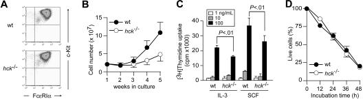
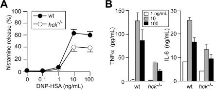
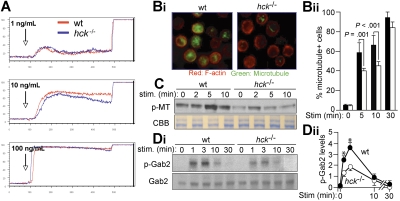
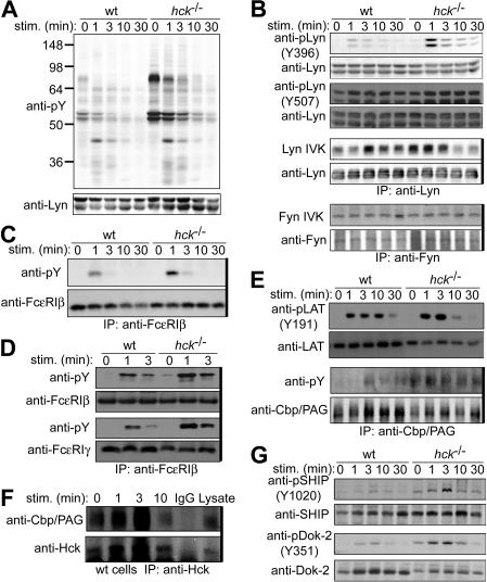
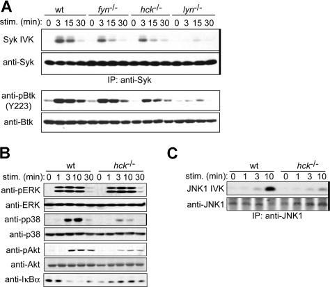
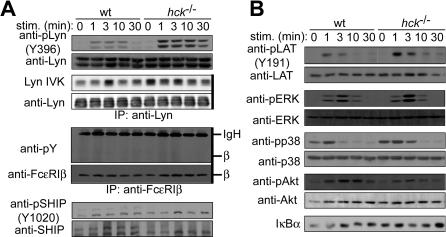
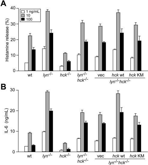
IgE/antigen-dependent mast cell activation plays a central role in immediate hypersensitivity and other allergic reactions. The Src family tyrosine kinase (SFK) Lyn is activated by the cross-linking of high-affinity IgE receptors (FcepsilonRI). Activated Lyn phosphorylates the FcepsilonRI subunits, beta and gamma, leading to subsequent activation of various signaling pathways. Lyn also plays a negative regulatory function by activating negative regulatory molecules. Another SFK, Fyn, also contributes to mast cell degranulation by inducing Gab2-dependent microtubule formation. Here we show that a third SFK, Hck, plays a critical role in mast cell activation. Degranulation and cytokine production are reduced in FcepsilonRI-stimulated hck(-/-) mast cells. The reduced degranulation can be accounted for by defects in Gab2 phosphorylation and microtubule formation. Importantly, Lyn activity is elevated in hck(-/-) cells, leading to increased phosphorylation of several negative regulators. However, positive regulatory events, such as activation of Syk, Btk, JNK, p38, Akt, and NF-kappaB, are substantially reduced in hck(-/-) mast cells. Analysis of lyn(-/-)hck(-/-), lyn(-/-)FcepsilonRIbeta(-/-), and hck(-/-)FcepsilonRIbeta(-/-) cells shows that Hck exerts these functions via both Lyn-dependent and Lyn-independent mechanisms. Thus, this study has revealed a hierarchical regulation among SFK members to fine-tune mast cell activation.


Figures

Figure 1
Figure 1
Hck deficiency results in reduced mast cell proliferation. (A) Flow cytometric analysis of FcϵRI and c-Kit expression on the surface of WT and hck−/− BMMCs. (B) Growth curves of bone marrow cells cultured in IL-3–containing medium. (C) Proliferation of WT and hck−/− BMMCs in response to the indicated concentrations of IL-3 or SCF were measured by thymidine uptake. Error bars represent standard deviation (SD) unless otherwise mentioned. (D) Growth factor-deprivation–induced apoptosis in WT and hck−/− BMMCs. Percentages of annexin V/7AAD live cells are plotted as a function of incubation time. Representative results from at least 3 independent experiments are shown.
Figure 2
Figure 2
Hck deficiency results in reduced histamine release and cytokine production when mast cells are stimulated with high concentrations of antigen. IgE-sensitized WT and hck−/− BMMCs were stimulated with the indicated concentrations of antigen for 45 minutes (A) or 20 hours (B). Histamine, tumor necrosis factor-α, and IL-6 secreted into culture media were measured. Representative results from 3 experiments are shown. Error bars represent SD.
Figure 3
Figure 3
Hck deficiency results in impaired microtubule formation associated with reduced Gab2 phosphorylation. (A) IgE-sensitized WT and hck−/− BMMCs were stimulated with the indicated concentrations of antigen at the indicated points and with 2.5 μg/mL ionomycin 400 seconds later. Ca2+ flux was measured by flow cytometry. Representative results from 3 experiments are shown. (B) IgE-sensitized cells were stimulated with 100 ng/mL DNP23-HSA for 5, 10, and 30 minutes. Immunofluorescence analysis for F-actin (stained by rhodamine-phalloidin) and microtubules (stained by anti-α-tubulin) was performed. Images shown are taken from cells stimulated for 10 minutes (Bi). The percentage of microtubule+ cells is shown in panel Bii. See “Microscopy” for image acquisition information. (C) IgE-sensitized cells were stimulated with 100 ng/mL DNP23-HSA for the indicated periods (minutes). Polymeric tubulin (p-MT) in Triton-insoluble fractions was measured as described in Document S1 (top). An SDS-PAGE gel containing Triton-soluble proteins was stained with Coomassie Brilliant Blue to show that comparable amounts of lysates were used for this assay. (Di) Immunoblot analysis of phospho-Gab2 (Tyr-452) in IgE/antigen-stimulated BMMCs (top panel). The same blot was reprobed with anti-Gab2 (bottom panel). Densitometric analysis was performed (Dii). Values shown in panel Dii represent means from at least 3 independent experiments at each time point. Error bars represent SEM. *Statistically significant differences between WT and hck−/− cells (P < .05 by Student t test).
Figure 4
Figure 4
Hck deficiency leads to increased Lyn activity and increased phosphorylation of Lyn phosphorylation targets. IgE-sensitized WT and hck−/− cells were stimulated with 100 ng/mL DNP23-HSA for the indicated periods. Cell lysates were either directly analyzed by SDS-PAGE and immunoblotting with the indicated antibodies (A,B,E,G) or first immunoprecipitated (indicated by thick vertical lines on the right of gels) with anti-FcϵRIβ mAb (C) or anti-Cbp/PAG (E,F), and followed by immunoblotting with antiphosphotyrosine mAb (C,E) or anti-Hck antibody (F). (B) Immunoprecipitated SFKs were subjected to in vitro kinase assays. (D) Cell lysates were fractionated into lipid raft and soluble compartments by sucrose density gradient ultracentrifugation. Lipid raft compartments were immunoprecipitated with anti-FcϵRIβ mAb, and followed by immunoblotting with antiphosphotyrosine mAb. Immunoprecipitated antigens were detected by reprobing the blots. Representative results from 2 experiments are shown, except for Lyn and Fyn kinase assays (B), which represent 3 experiments, and phosphotyrosine probing (A), which represent at least 4 experiments.
Figure 5
Figure 5
Hck deficiency results in reduced activities of positive regulatory molecules. IgE-sensitized WT and hck−/− cells were stimulated with 100 ng/mL DNP23-HSA for the indicated periods. (A) Syk was immunoprecipitated (indicated by thick vertical line on the right of gel) from cleared cell lysates and immune complexes subjected to in vitro kinase assays using GST-HS1 as a substrate. Portion of the autoradiogram including GST-HS1 phosphorylation is shown. Cell lysates were directly analyzed by SDS-PAGE and immunoblotting with anti-Syk or anti-phospho-Btk (Tyr223). The pBtk blot was reprobed with anti-Btk antibody. (B) Cell lysates were directly analyzed by SDS-PAGE and immunoblotting with the indicated antibodies. The same blots were reprobed with antibodies that detect antigens irrespective of their phosphorylation status. (C) Immunoprecipitated JNK1 (indicated by thick vertical line on the right of gel) was subjected to in vitro kinase assays. Representative results from 2 experiments are shown.
Figure 6
Figure 6
Stimulation with a low concentration of antigen does not induce phosphorylation of FcϵRIβ or increase SHIP phosphorylation in hck−/− cells despite increased Lyn activity. IgE-sensitized WT and hck−/− cells were stimulated with 1 ng/mL DNP23-HSA for the indicated periods. Cell lysates were directly analyzed by SDS-PAGE and immunoblotting with the indicated phospho-specific antibodies. The same blots were reprobed with antibodies that detect antigens irrespective of their phosphorylation status. (A, third and fourth rows) Immunoprecipitated Lyn was subjected to autophosphorylation assays. Comparable immunoprecipitations were confirmed by immunoblotting. (A, middle) Immunoprecipitated FcϵRIβ was analyzed by immunoblotting with anti-phosphotyrosine mAb and then reprobed with anti-FcϵRIβ mAb. Immunoprecipitations are indicated by thick vertical lines on the right of gels. Representative results from 2 experiments are shown.
Figure 7
Figure 7
Hck/Lyn doubly deficient mast cells exhibit an intermediate activation phenotype between Hck- or Lyn-deficient cells, and positive and negative regulation via FcϵRI β subunit is exerted by Lyn-mediated phosphorylation of the canonical and noncanonical tyrosine residues. IgE-sensitized mast cells of the indicated genotypes and FcϵRIβ-transduced cells were stimulated with 1 (□), 10 (░), or 100 (■) ng/mL DNP23-HSA for 45 minutes (A) or 20 hours (B). Histamine and IL-6 secreted into culture media were measured. ND indicates not detected. Representative results from 2 independent transduction experiments are shown. Error bars represent SD.

Similar articles

Cited by

References

    1. Kinet JP. The high-affinity IgE receptor (Fc epsilon RI): from physiology to pathology. Annu Rev Immunol. 1999;17:931–972. - PubMed
    1. Turner H, Kinet JP. Signalling through the high-affinity IgE receptor Fc epsilonRI. Nature. 1999;402:B24–30. - PubMed
    1. Eiseman E, Bolen JB. Engagement of the high-affinity IgE receptor activates src protein-related tyrosine kinases. Nature. 1992;355:78–80. - PubMed
    1. Jouvin MH, Adamczewski M, Numerof R, Letourneur O, Valle A, Kinet JP. Differential control of the tyrosine kinases Lyn and Syk by the two signaling chains of the high affinity immunoglobulin E receptor. J Biol Chem. 1994;269:5918–5925. - PubMed
    1. Kawakami T, Galli SJ. Regulation of mast-cell and basophil function and survival by IgE. Nat Rev Immunol. 2002;2:773–786. - PubMed

Publication types

MeSH terms