Comparative Study
. 2007 Jun 1;178(11):7072-80.
doi: 10.4049/jimmunol.178.11.7072.

C57BL/6 mice genetically deficient in IL-12/IL-23 and IFN-gamma are susceptible to experimental autoimmune myasthenia gravis, suggesting a pathogenic role of non-Th1 cells

Affiliations
Comparative Study

C57BL/6 mice genetically deficient in IL-12/IL-23 and IFN-gamma are susceptible to experimental autoimmune myasthenia gravis, suggesting a pathogenic role of non-Th1 cells

Wei Wang et al. J Immunol. .

Erratum in

  • J Immunol. 2007 Nov 15;179(10):7184. Caspi, Rachel [corrected to Caspi, Rachel R]

Abstract

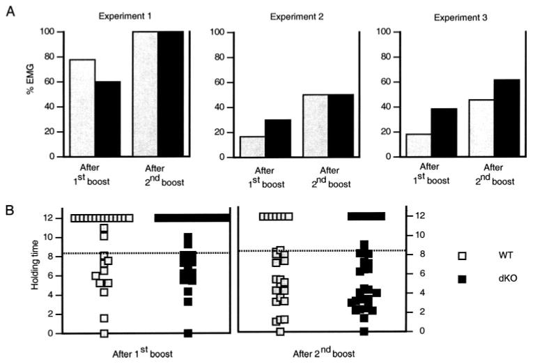
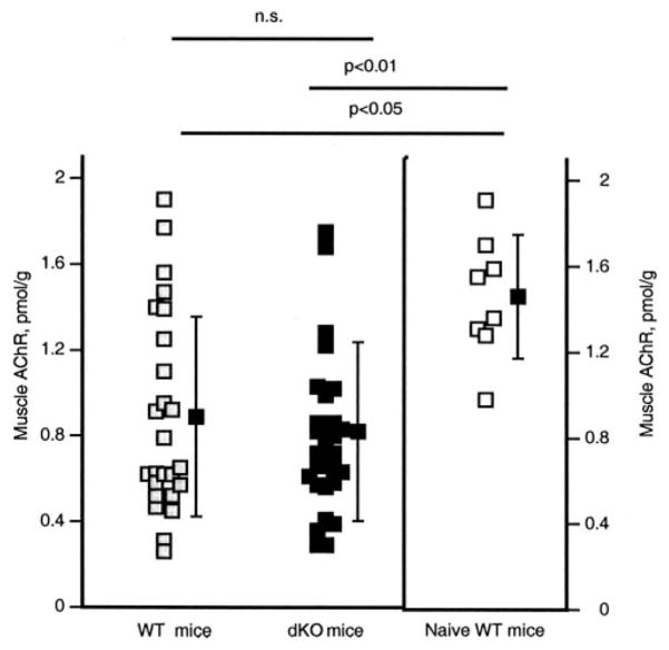
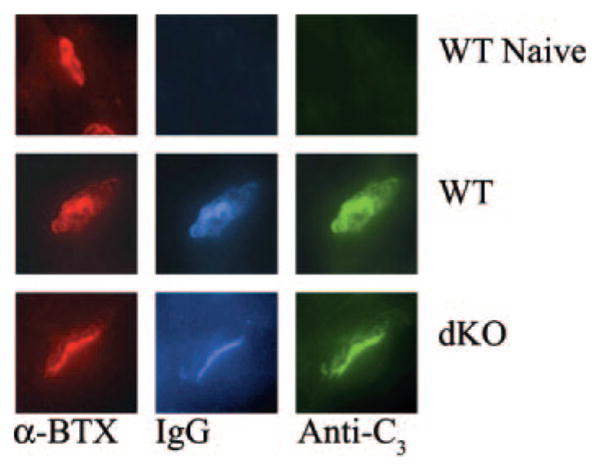
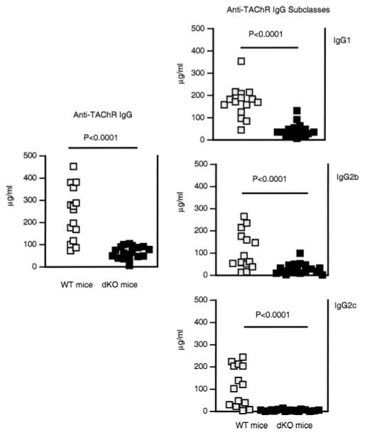
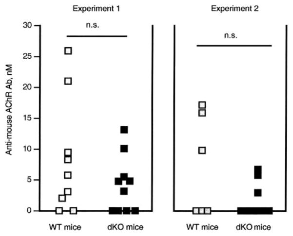
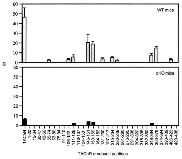
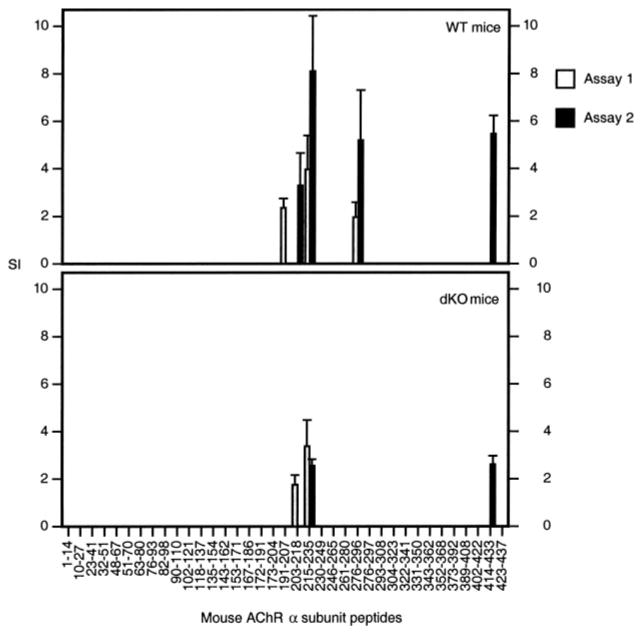
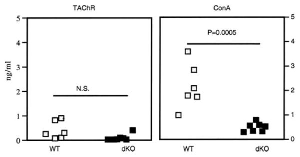
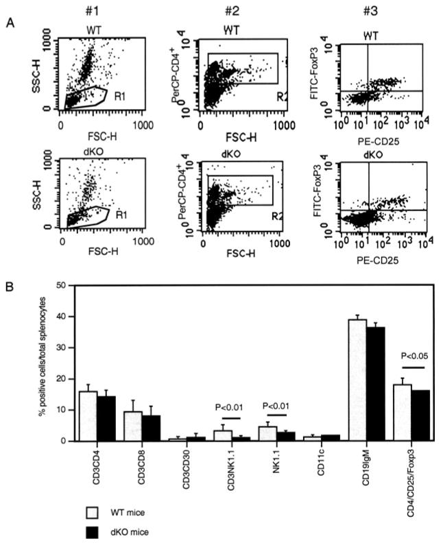
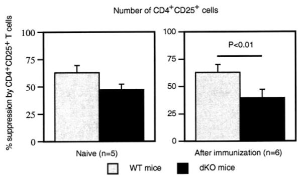
Immunization with Torpedo acetylcholine receptor (TAChR) induces experimental autoimmune myasthenia gravis (EAMG) in C57BL/6 (B6) mice. EAMG development needs IL-12, which drives differentiation of Th1 cells. The role of IFN-gamma, an important Th1 effector, is not clear and that of IL-17, a proinflammatory cytokine produced by Th17 cells, is unknown. In this study, we examined the effect of simultaneous absence of IL-12 and IFN-gamma on EAMG susceptibility, using null mutant B6 mice for the genes of both the IL-12/IL-23 p40 subunit and IFN-gamma (dKO mice). Wild-type (WT) B6 mice served as control for EAMG induction. All mice were immunized with TAChR in Freund's adjuvant. dKO mice developed weaker anti-TAChR CD4(+)T cells and Ab responses than WT mice. Yet, they developed EAMG symptoms, anti-mouse acetylcholine receptor (AChR) Ab, and CD4(+) T cell responses against mouse AChR sequences similar to those of WT mice. dKO and WT mice had similarly reduced AChR content in their muscles, and IgG and complement at the neuromuscular junction. Naive dKO mice had significantly fewer NK, NKT, and CD4(+)CD25(+)Foxp3(+) T regulatory (Treg) cells than naive WT mice. Treg cells from TAChR-immunized dKO mice had significantly less suppressive activity in vitro than Treg cells from TAChR-immunized WT mice. In contrast, TAChR-specific CD4(+) T cells from TAChR-immunized dKO and WT mice secreted comparable amounts of IL-17 after stimulation in vitro with TAChR. The susceptibility of dKO mice to EAMG may be due to reduced Treg function, in the presence of a normal function of pathogenic Th17 cells.


Conflict of interest statement

Disclosures

The authors have no financial conflict of interest.

Figures

FIGURE 1
FIGURE 1
EAMG symptoms in TAChR-immunized dKO and WT mice. A, Frequency of EAMG weakness among the WT and dKO mice used in three independent experiments evaluated about 2 wk after the first and second TAChR/IFA boost. B, Holding times of all individual mice of experiments 1, 2, and 3. The dotted line indicates the holding time of 8.3 min, above which the mouse is considered healthy. EAMG symptoms were always comparable in dKO and WT mice. See text for experimental details.
FIGURE 2
FIGURE 2
Muscle AChR content in TAChR-immunized dKO, WT mice, and naive WT mice. Concentration (picomoles per gram) of AChR in the muscle of individual dKO and WT mice sacrificed 12 wk after the first immunization and of 8 naive WT mice. The filled symbol next to each group represents the group’s average concentration ± SD. TAChR-immunized dKO and WT mice had comparable AChR muscle concentrations that were significantly lower than those of naive WT mice. See text for experimental details.
FIGURE 3
FIGURE 3
Presence of IgG bound to complement at the NMJ of TAChR-immunized dKO, WT, and of naive WT mice. Mice were sacrificed 12 wk after the first TAChR immunization; 20 –28 sections per mouse were analyzed for the presence of AChR (red fluorescence), C3 (blue fluorescence), and IgG (green fluorescence) at the NMJ. Shown is one representative section. Both TAChR-immunized dKO and WT mice had IgG and C3 bound to the AChR at the NMJ. Naive WT mice only stained for the AChR and were negative for both IgG and C3. See text for experimental details.
FIGURE 4
FIGURE 4
Anti-TAChR IgG and IgG subclasses in the sera of TAChR-immunized dKO and WT mice. Concentrations (micrograms per milliliter) of anti-TAChR IgG, IgG1, IgG2b, and IgG2c in the individual sera of dKO and WT mice of experiments 1 and 2 were tested by ELISA. Sera were collected 12 wk after the first TAChR immunization. dKO mice had significantly less anti-TAChR IgG and IgG subclasses than WT mice. See text for experimental details.
FIGURE 5
FIGURE 5
Anti-mouse AChR Ab in the sera of TAChR-immunized dKO and WT mice. Concentrations (nanomolar) of anti-mouse AChR Ab in the individual sera of dKO and WT mice of experiments 1 and 2. Sera were collected 12 wk after the first TAChR immunization. WT mice had serum anti-mouse AChR Ab more frequently than dKO, but the difference between the concentrations was not significant. See text for experimental details.
FIGURE 6
FIGURE 6
Proliferative response of CD8+-depleted splenocytes obtained from TAChR-immunized dKO and WT mice against peptides spanning the α subunit of the TAChR. CD8+-depleted splenocytes obtained from dKO and WT mice 12 wk after the first TAChR immunization were cultured in the presence of peptides spanning the α subunit of TAChR or with TAChR. The columns of the plots represent the SI ± SD calculated with the cpm of quadruplicated cultures (one of three consistent proliferation assays). The background cpm of cells incubated with an unrelated peptide as stimulant were 342.5 + 81.5 for WT mice and 980.17 + 202.28 for dKO mice. CD8+-depleted splenocytes of WT mice responded to a higher number of peptides and more vigorously than dKO mice. Although with different intensity, both dKO and WT mice responded to peptides sequences previously identified as immunodominant epitopes in B6 mice. See text for experimental details.
FIGURE 7
FIGURE 7
Proliferative response of CD8+-depleted splenocytes obtained from TAChR-immunized dKO and WT mice against peptides spanning the α subunit of the mouse AChR. The columns of the plots represent the SI ± SD of two proliferation assays conducted with CD8+ depleted splenocytes obtained from dKO and WT mice 12 wk after the first TAChR immunization. Assay 1 is represented by light gray columns and assay 2 by dark gray columns. Both dKO and WT mice responded to few peptides and their response was of the same order of magnitude. Both WT and dKO mice recognized peptides within the sequence α191–235 and α414 – 433. WT mice, but not dKO mice, recognized also peptide α276 –295. See text for experimental details.
FIGURE 8
FIGURE 8
IL-17 concentration in the supernatant of purified CD4+ splenocytes obtained from TAChR-immunized WT and dKO mice stimulated by TAChR or Con A. CD4+ T cells from TAChR-immunized WT mice (n = 6) and TAChR-immunized dKO mice (n = 7) were isolated and incubated with Ag and APC for 48 h, then the supernatants were harvested and tested by ELISA. Each dot represents the results obtained from each individual mouse. The amount of specific IL-17 production induced by TAChR stimulation is comparable in the two strains of mice. The nonspecific production stimulated by Con A is significantly higher in WT mice than in dKO mice. See text for experimental details.
FIGURE 9
FIGURE 9
Phenotype of splenocytes obtained from naive dKO and WT mice. A, The results of a representative flow cytometry analysis done with whole spleen cells stained with PerCP-CD4, FITC-FoxP3, and PE-CD25. In plot #1, R1 is the gate defining the population of interest, in plot #2 R2 is the gate defining the CD4+ cells. The right upper quadrant of plot #3 shows the cells positive for the three markers used. The average of all of the triple staining we performed is represented in B (CD4/CD25/Foxp3). B, The phenotype of splenocytes obtained from several individual mice (three to eight mice). dKO mice had significantly less NK, NKT, and CD4+CD25+Foxp3+ T cells than WT mice. See text for experimental details.
FIGURE 10
FIGURE 10
Suppressive activity of CD4+CD25+ Treg cells obtained from TAChR-immunized dKO and WT mice. The columns represent the average ± SD of the percentage of suppression obtained in five to six independent experiments as indicated. CD4+CD25+ Treg cells from TAChR-immunized dKO mice inhibited the anti-CD3-induced proliferation of CD4+CD25 T cells significantly less (p = 0.02) than CD4+CD25+ Treg cells from TAChR-immunized WT mice. The suppressive ability of CD4+CD25+ Treg cells from naive dKO and WT mice was comparable. See text for experimental details.

References

    1. Kolls JK, Linden A. Interleukin-17 family members and inflammation. Immunity. 2004;21:467– 476. - PubMed
    1. Komiyama Y, Nakae S, Matsuki T, Nambu A, Ishigame H, Kakuta S, Sudo K, Iwakura Y. IL-17 plays an important role in the development of experimental autoimmune encephalomyelitis. J Immunol. 2006;177:566 –573. - PubMed
    1. Veldhoen M, Hocking RJ, Atkins CJ, Locksley RM, Stockinger B. TGFβ in the context of an inflammatory cytokine milieu supports de novo differentiation of IL-17-producing T cells. Immunity. 2006;24:179 –189. - PubMed
    1. Sakaguchi S. Naturally arising Foxp3-expressing CD25+CD4+ regulatory T cells in immunological tolerance to self and non-self. Nat Immunol. 2005;6:345–352. - PubMed
    1. Conti-Fine BM, Protti M, Bellone M, Howard JF. Myasthenia Gravis: The Immunobiology of an Autoimmune Disease. R. G. Landes; Austin, TX: 1997.

Publication types

MeSH terms