. 2007 Jun;75(6):2679-88.
doi: 10.1128/IAI.00076-07.

Quantitative profile of the uropathogenic Escherichia coli outer membrane proteome during growth in human urine

Affiliations

Quantitative profile of the uropathogenic Escherichia coli outer membrane proteome during growth in human urine

Christopher J Alteri et al. Infect Immun. 2007 Jun.

Erratum in

  • Infect Immun. 2009 Mar;77(3):1272

Abstract

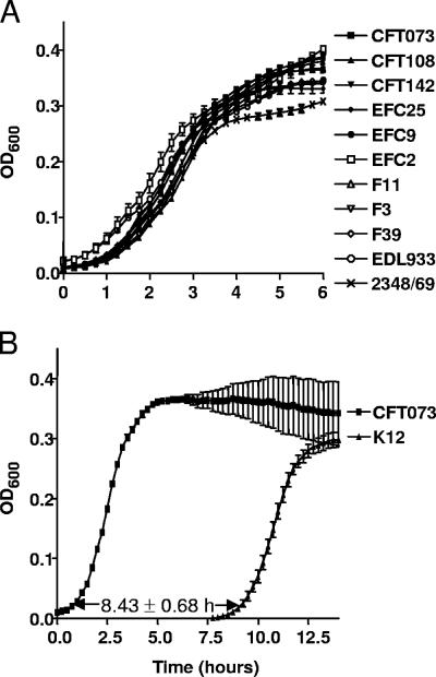
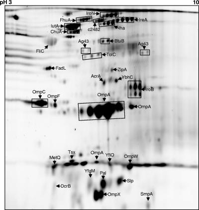
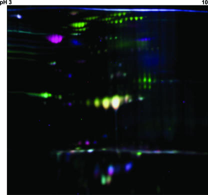
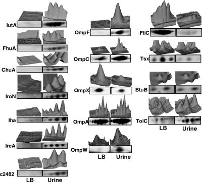
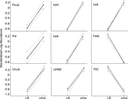
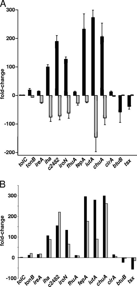
Outer membrane proteins (OMPs) of microbial pathogens are critical components that mediate direct interactions between microbes and their surrounding environment. Consequently, the study of OMPs is integral to furthering the understanding of host-pathogen interactions and to identifying key targets for development of improved antimicrobial agents and vaccines. In this study, we used two-dimensional polyacrylamide gel electrophoresis (2D-PAGE) and tandem mass spectrometry to characterize the uropathogenic Escherichia coli (UPEC) outer membrane subproteome; 30 individual OMPs present on the bacterial surface during growth in human urine were identified. Fluorescence difference gel electrophoresis was used to identify quantitative changes in levels of UPEC strain CFT073 OMPs during growth in urine; six known receptors for iron compounds were induced in this environment, i.e., ChuA, IutA, FhuA, IroN, IreA, and Iha. A seventh putative iron compound receptor, encoded by CFT073 open reading frame (ORF) c2482, was also identified and found to be induced in urine. Further, the induction of these seven iron receptors in human urine and during defined iron limitation was verified by using quantitative real-time PCR (qPCR). An eighth iron receptor, fepA, displayed similar induction levels under these conditions as measured by qPCR but was not identified by 2D-PAGE. Addition of 10 microM FeCl(2) to human urine repressed the transcription of all eight iron receptor genes. A number of fecal-commensal, intestinal pathogenic, and uropathogenic E. coli strains all displayed similar growth rates in human urine, showing that the ability to grow in urine per se is not a urovirulence trait. Thus, human urine is an iron-limiting environment and UPEC enriches its outer membrane with iron receptors to contend with this iron limitation.


Figures

FIG. 1.
FIG. 1.
Growth of uropathogenic, diarrheagenic, and fecal-commensal E. coli isolates cultured in human urine. (A) Growth profile for pyelonephritis, fecal-commensal, cystitis, enterohemorrhagic, and enteropathogenic strains during static culture at 37°C in sterilized urine pooled from 10 male and female donors. (B) Comparison of growth characteristics of E. coli CFT073 (UPEC) and MG1655 (K-12) in pooled human urine. OD600, optical density at 600 nm.
FIG. 2.
FIG. 2.
Outer membrane subproteome for UPEC during growth in human urine. 2D gel of carbonate-insoluble membrane proteins prepared from CFT073 following culture in urine. Outer membrane fraction (150 μg protein) was isoelectrically focused on a pH 3 to 10 IPG strip, followed by 10% SDS-PAGE. The image shows the proteins identified by MS/MS (arrows). Boxed areas indicate proteins where multiple isoforms produced the same identification. The CFT073 annotated gene numbers, theoretical masses, and isoelectric points of the identified proteins are listed in Table 2.
FIG. 3.
FIG. 3.
2D-DIGE of UPEC outer membrane proteins during growth in urine. Outer membrane proteins (50 μg) from E. coli CFT073 cultured in urine were labeled with Cy3 (green), those from CFT073 grown in LB medium were labeled with Cy5 (red), and the pooled internal standard representing equal amounts of urine and LB medium outer membrane proteins was labeled with Cy2 (blue). The labeled proteins, 150 μg, were pooled and applied to a pH 3 to 10 IPG strip and second-dimension 10% SDS-PAGE. Green spots indicate protein features induced in urine, red spots represent proteins induced in LB medium, and blue spots show proteins that are similarly expressed in both urine and LB medium.
FIG. 4.
FIG. 4.
Normalized spot volumes from multivariable 2D-DIGE scan of urine (Cy3) and LB medium (Cy5) UPEC outer membrane proteins. (A) Spot volumes for known and predicted CFT073 TonB-dependent outer membrane receptors for iron, (B) outer membrane porins, and (C) proteins showing decreased expression in urine or similar expression patterns in LB medium and urine. The 3D peaks represent the normalized pixel intensities for identical spot boundaries between the Cy3 and Cy5 scans. The boxed regions below the peaks are the corresponding protein isoforms present in the DIGE scan.
FIG. 5.
FIG. 5.
2D-DIGE expression patterns for UPEC outer membrane proteins differentially expressed in urine and LB medium. The standardized log10 abundance from triplicate 2D-DIGE experiments was generated by using the internal standard included in every gel. The Cy2 internal standard signals were used to normalize the ratios of LB to urine across multiple gels. The average abundance (solid lines) and individual quantitative changes (dashed lines) between normalized Cy3 (urine) and Cy5 (LB medium) signals are shown.
FIG. 6.
FIG. 6.
qPCR for UPEC iron receptor genes during growth in urine, during growth in iron-replete urine, and during defined iron limitation. (A) qPCR results for CFT073 grown in human urine in the presence (gray bars) and absence (black bars) of 10 μM FeCl2 and (B) cultured in LB medium containing 10 mM deferoxamine mesylate, an iron-chelating agent. Data were normalized to gapA (glyceraldehyde 3-phosphate dehydrogenase) expression levels, and changes were determined by using LB medium expression levels as the calibrator. For iron-replete urine qPCR, urine without supplementation with ferrous chloride was used as the calibrator to generate n-fold change values. In panel B, the black bars represent urine transcript n-fold changes and the gray bars represent LB-deferoxamine mesylate medium transcript levels compared to LB medium transcript levels.

References

    1. Alban, A., S. O. David, L. Bjorkesten, C. Andersson, E. Sloge, S. Lewis, and I. Currie. 2003. A novel experimental design for comparative two-dimensional gel analysis: two-dimensional difference gel electrophoresis incorporating a pooled internal standard. Proteomics 336-44. - PubMed
    1. Anderson, J. D., F. Eftekhar, M. Y. Aird, and J. Hammond. 1979. Role of bacterial growth rates in the epidemiology and pathogenesis of urinary infections in women. J. Clin. Microbiol. 10766-771. - PMC - PubMed
    1. Asscher, A. W., M. Sussman, W. E. Waters, R. H. Davis, and S. Chick. 1966. Urine as a medium for bacterial growth. Lancet ii1037-1041. - PubMed
    1. Busler, V. J., V. J. Torres, M. S. McClain, O. Tirado, D. B. Friedman, and T. L. Cover. 2006. Protein-protein interactions among Helicobacter pylori Cag proteins. J. Bacteriol. 1884787-4800. - PMC - PubMed
    1. Connell, I., W. Agace, P. Klemm, M. Schembri, S. Marild, and C. Svanborg. 1996. Type 1 fimbrial expression enhances Escherichia coli virulence for the urinary tract. Proc. Natl. Acad. Sci. USA 939827-9832. - PMC - PubMed

Publication types

MeSH terms