. 2007 Jul 27;282(30):21998-2010.
doi: 10.1074/jbc.M701737200. Epub 2007 May 18.

MMP25 (MT6-MMP) is highly expressed in human colon cancer, promotes tumor growth, and exhibits unique biochemical properties

Affiliations

MMP25 (MT6-MMP) is highly expressed in human colon cancer, promotes tumor growth, and exhibits unique biochemical properties

Qing Sun et al. J Biol Chem. .

Abstract

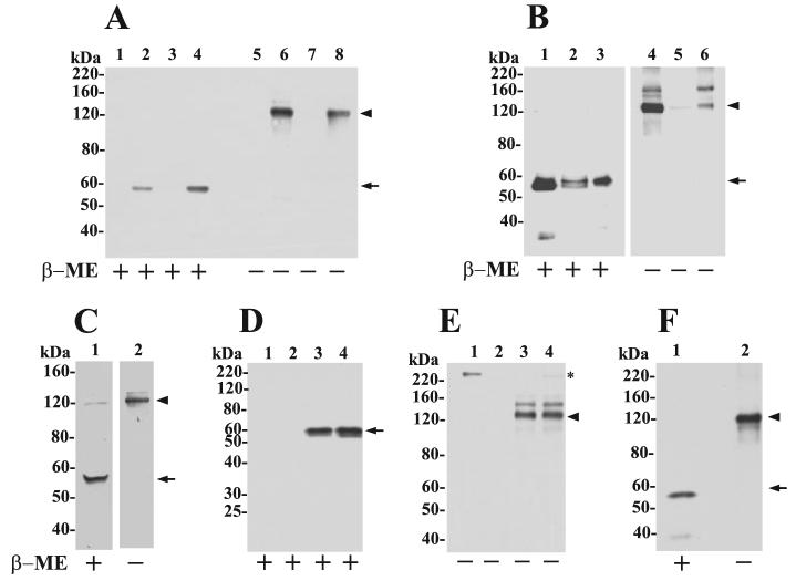
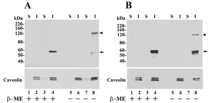
MMP25 (MT6-MMP) is one of the two glycosylphosphatidylinositol-anchored matrix metalloproteinases (MMPs) that have been suggested to play a role in pericellular proteolysis. However, its role in cancer is unknown, and its biochemical properties are not well established. Here we found a marked increase in MT6-MMP expression within in situ dysplasia and invasive cancer in 61 samples of human colon cancer. Expression of MT6-MMP in HCT-116 human colon cancer cells promoted tumori-genesis in nude mice. Histologically, the MT6-MMP-expressing tumors demonstrated an infiltrative leading edge in contrast to a rounded leading edge in vector control tumors. Biochemical and biosynthesis analyses revealed that MT6-MMP displayed on the cell surface exists as a major form of 120 kDa that likely represents enzyme homodimers linked by disulfide bonds. Upon reduction, a single 57-kDa active MT6-MMP was detected. Interestingly, neither membrane-anchored nor phosphatidylinositol-specific phospholipase C-released MT6-MMPs were found to be associated with tissue inhibitor of metalloproteinases (TIMPs) and did not activate pro-gelatinases (pro-MMP-2 and pro-MMP-9) even in the presence of exogenous TIMP-2 or TIMP-1. A catalytic domain of MT6-MMP was inhibited preferentially by TIMP-1 (K(i) = 0.2 nm) over TIMP-2 (K(i) = 2.0 nm), because of a slower association rate. These results show that MT6-MMP may play a role in colon cancer and exhibit unique biochemical and structural properties that may regulate proteolytic function at the cell surface.


Figures

FIGURE 1
FIGURE 1. Invasive human colorectal carcinomas express high levels of MT6-MMP.
Hematoxylin and eosin stains (A–E) and MT6-MMP immunostaining of adjacent sections (A′–E′) of specimens from five patients. A and A′, in situ dysplasia (area above arrows) and benign epithelium (area below arrows) are present. The immunostaining shows that the benign epithelium is negative, although faint staining can be detected in the preinvasive neoplasia (dysplasia, area above arrows). B and B′, moderately differentiated invasive colorectal carcinoma (arrowhead) is present infiltrating beneath benign epithelium (arrow). The immunostaining shows that the invasive carcinoma is strongly positive for MT6-MMP, whereas the overlying benign epithelium is negative. C and C′, moderately differentiated gland-forming invasive colorectal carcinoma. D and D′, poorly differentiated invasive colorectal carcinoma infiltrating as single cells and small nests. The invasive cells are stained positively for MT6-MMP. E and E′, lymphatic invasion in moderately differentiated invasive colorectal carcinoma. All scale bars = 100 μm. F, semi-quantitative analysis of mean MT6-MMP staining in duplicate paired samples from 61 patients (two normal, two invasive cancers from each patient). The lines connect each pair. MT6-MMP expression was increased in 50 of 61 pairs (p < 0.0001). Mean intensity for the entire study set is shown in red (control, 0.7 ± 0.08, S.E.; carcinoma, 1.7 ± 0.06 S.E.).
FIGURE 2
FIGURE 2. Expression of MT6-MMP in colon cancer cell lines and in stable transfectants.
A, RT-PCR analysis of MT6-MMP mRNA level in human colon cancer cell lines and in human PMNs. Expression of GAPDH is shown as a loading control. Arrow shows the PCR product of MT-MMP cDNA. B and C, immunoblot analyses of recombinant MT6-MMP protein expression in transfected clones of HT-29 (B) and HCT-116 (C) colon cancer cell lines. Cells were lysed with lysis buffer, and 20 μg/lane of lysate were resolved by reducing 10% SDS-PAGE followed by immunoblot analyses with anti-MT6-MMP hinge pAb Ab39031. A human PMN lysate (40 μg) was run as a control. D, immunoblot analyses of BS-C-1 cells co-infected to express MT6-MMP (lanes 2, 4, and 6) or infected with control virus (lanes 1, 3, and 5). Lysates (20 μg/lane) were resolved by reducing 10% SDS-PAGE followed by immunoblot analyses with pAb RP2-MMP25 (directed to the C terminus) (lanes 1 and 2), pAb RP4-MMP25 (directed to the prodomain) (lanes 3 and 4), and pAb107 (directed to the catalytic domain) (lanes 5 and 6). Note the lack of recognition of the 57-kDa species by the pro-domain antibody. E, PI-PLC release of MT6-MMP from HT-29 and HCT-116 stable clones. Cells were treated with (+) or without (−) PI-PLC as described under “Experimental Procedures.” The supernatant was collected, concentrated, and subjected to reducing 10% SDS-PAGE followed by immunoblot analysis with pAb Ab39031 to the hinge region of MT6-MMP. Arrow in E indicates the soluble MT-MMP released by PI-PLC.
FIGURE 3
FIGURE 3. MT6-MMP promotes the tumorigenicity and local invasion of human HCT-116 colon cancer cells.
A, MT6-HCT clone M2 (■) and EV-HCT clone E2 (□); or B, pooled clones MT6-HCT (■) and EV-HCT (□) were subcutaneously inoculated (3 × 10⁶ cells/mouse) into NCr female nude mice (n = 8). Tumors were measured at the indicated days, and the tumor volumes were calculated as described under “Experimental Procedures.” Points represent mean of tumor volume, and bars indicate S.E. *, p < 0.002; **, p < 0.02; ***, p < 0.05. C and D, histological sections of EV-HCT (C) and MT6-HCT (D)tumors stained by hematoxylin and eosin.Note the smooth border between tumor and surrounding stroma in EV-HCT tumors (C) versus the ragged infiltrative border with the surrounding desmoplastic stroma in MT6-HCT tumors (D). Bar = 100 μm. T, tumor; S, stroma.
FIGURE 4
FIGURE 4. Cell surface MT6-MMP is displayed as an ∼120-kDa species.
A, immunoblot analyses of lysates of pooled clones of EV-HT (lanes 1 and 5), MT6-HT (lanes 2 and 6), EV-HCT (lanes 3 and 7), and MT6-HCT (lanes 4 and 8). Cells were lysed in cold lysis buffer supplemented with 20 mm NEM and mixed with Laemmli SDS-sample buffer with (+) or without (−) β-ME. The lysates (10 μg/lane) were resolved by 7.5% SDS-PAGE followed by immunoblot analysis with mAb1142. B, crude plasma membrane fraction isolated from MT6-HCT cells was treated with PI-PLC (5 units per 5 mg of total protein in 1 ml of TBS) and centrifuged, and the supernatant (lanes 3 and 6,20 μg/lane) and pellet (lanes 2 and 5, 20 μg/lane) were collected. Lanes 1 and 4 show the input plasma membrane fraction (2 μg/lane) before PI-PLC treatment. The fractions were mixed with Laemmli SDS-sample buffer with (+) or without (−) β-ME and resolved by 7.5% SDS-PAGE followed by immunoblot analysis with pAb Ab39031 to the hinge region of MT6-MMP. C, MT6-HCT cells in 6-well plates were treated with 0.5 ml of PI-PLC (0.3 units/well) in PBS for 30 min on ice. The supernatant was concentrated and mixed with Laemmli SDS-sample buffer with (+) or without (−) β-ME and resolved by 7.5% SDS-PAGE followed by immunoblot analysis with mAb1142. D and E, EV-HCT (lanes 1 and 2) and MT6-HCT tumors (lanes 3 and 4) were homogenized, and extracts were immunoprecipitated with either anti-MT6-MMP hinge pAb Ab39031 (D and E) or rabbit IgG (not shown) and resolved by 10% (reducing) or 7.5% (nonreducing) SDS-PAGE in the presence (+) or absence (−) of β-ME followed by immunoblot analysis with anti-MT6-MMP mAb1142. Asterisk in E shows a nonspecific band. F, human PMN lysate was mixed with Laemmli SDS-sample buffer with (+) or without (−) β-ME and resolved by 7.5% SDS-PAGE followed by immunoblot analysis with mAb1142. Arrowhead and arrow in A–F indicate the ∼120- and 57-kDa species of MT6-MMP, respectively.
FIGURE 5
FIGURE 5. MT6-MMP is targeted to the lipid raft fraction.
A, EV-HT (lanes 1, 2, 5, and 6) and MT6-HT cells (lanes 3, 4, 7, and 8); B, EV-HCT (lanes 1, 2, 5, and 6) and MT6-HCT cells (lanes 3, 4, 7, and 8) were harvested, and cellular proteins were separated into Triton X-100-soluble (S) and Triton X-100-insoluble/OCG-soluble (I) fractions. Equal amounts of protein (10 μg/lane) from each fraction were mixed with Laemmli SDS-sample buffer with (+) or without (−) β-ME and resolved by 7.5% SDS-PAGE followed by immunoblot analysis with mAb1142. Arrowhead and arrow in A and B indicate the ∼120- and 57-kDa species of MT6-MMP, respectively. The blots were reprobed with an anti-caveolin pAb to detect the ∼22–24-kDa caveolin (lower panels).
FIGURE 6
FIGURE 6. Biosynthesis of MT6-MMP.
MT6-HCT cells were pulsed (15 min) with 500 μCi/ml [35S]methionine/cysteine and chased for 0, 15, 30, 60, and 120 min as described under “Experimental Procedures.” At each time point, the cells were harvested in cold lysis buffer, and the lysates were immunoprecipitated with either pAb Ab39031 to the hinge region (A and B) or pAb107 to the catalytic domain (C and D). The precipitated proteins were eluted by boiling in Laemmli SDS-sample buffer with (A and C) or without (B and D) β-ME and subjected to 10% (A and C) or 7.5% (B and D) SDS-PAGE followed by autoradiography. The lysate at the 30-min time point was precipitated with rabbit IgG, as a control (Ctrl.). Arrowhead and arrow in A–D indicate the ∼120-and 57-kDa species of MT6-MMP, respectively. Open arrow in A indicates the 59-kDa species of MT6-MMP.
FIGURE 7
FIGURE 7. MT6-MMP is not detected in complex with TIMPs.
A–D, MT6-MMP and GPI-MT1-MMP were expressed in BS-C-1 cells by infection-transfection as described under “Experimental Procedures.” Six hours post-infection-transfection, the cells received 20 nm of purified recombinant TIMP-1 or TIMP-2 in serum-free DMEM followed by overnight incubation. The media were removed, and the cells were treated with (+) or without (−) PI-PLC. The supernatants were collected, concentrated, and resolved by reducing 12% SDS-PAGE followed by immunoblot analyses. The blots were probed successively with mAb101 to TIMP-2 (A), mAb LEM-2/15 to MT1-MMP (B), mAb to TIMP-1 (C), and pAb Ab39031 to the hinge of MT6-MMP (D). The asterisks in D show nonspecific bands. Recombinant TIMP-2 (A, lane 5) and TIMP-1 (C, lane 5) (10 ng each/lane) were run as a control. E, MT6-HCT and EV-HCT cells were cultured in the presence of 20 nm of TIMP-1, TIMP-2, TIMP-3, or TIMP-4 in complete media. The cells were then surface-biotinylated as described under “Experimental Procedures.” The biotinylated cells were then treated with PI-PLC, and the supernatants were collected and subjected to streptavidin beads pulldowns. The bound proteins were eluted with reducing Laemmli SDS-sample buffer and resolved by 12% SDS-PAGE followed by immunoblot analysis. The blots were probed with antibodies to MT6-MMP or to TIMPs. Only the blot probed with MT6-MMP antibody (mAb1142) is shown. The TIMP blots showed no signals. Arrows in A and C indicate the recombinant TIMP-2 (∼22 kDa) and TIMP-1 (∼30 kDa), respectively.

References

    1. Nagase H, Visse R, Murphy G. Cardiovasc. Res. 2006;69:562–573. - PubMed
    1. Deryugina EI, Quigley JP. Cancer Metastasis Rev. 2006;25:9–34. - PubMed
    1. Maskos K, Bode W. Mol. Biotechnol. 2003;25:241–266. - PubMed
    1. Hernandez-Barrantes S, Bernardo M, Toth M, Fridman R. Semin. Cancer Biol. 2002;12:131–138. - PubMed
    1. Zucker S, Pei D, Cao J, Lopez-Otin C. Curr. Top. Dev. Biol. 2003;54:1–74. - PubMed

Publication types

MeSH terms

Substances