. 2007 Oct 1;110(7):2371-80.
doi: 10.1182/blood-2006-10-055087. Epub 2007 May 21.

Mutations in the cofilin partner Aip1/Wdr1 cause autoinflammatory disease and macrothrombocytopenia

Affiliations

Mutations in the cofilin partner Aip1/Wdr1 cause autoinflammatory disease and macrothrombocytopenia

Benjamin T Kile et al. Blood. .

Abstract

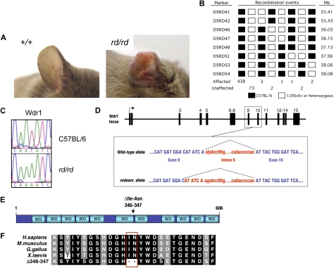
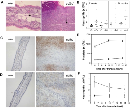
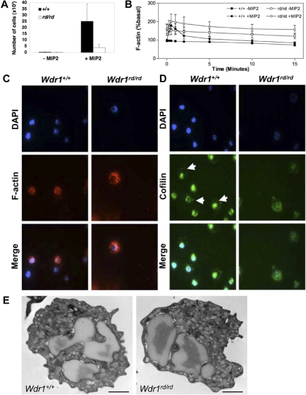
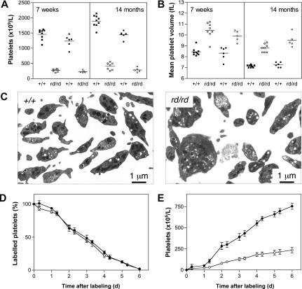
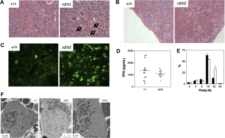
A pivotal mediator of actin dynamics is the protein cofilin, which promotes filament severing and depolymerization, facilitating the breakdown of existing filaments, and the enhancement of filament growth from newly created barbed ends. It does so in concert with actin interacting protein 1 (Aip1), which serves to accelerate cofilin's activity. While progress has been made in understanding its biochemical functions, the physiologic processes the cofilin/Aip1 complex regulates, particularly in higher organisms, are yet to be determined. We have generated an allelic series for WD40 repeat protein 1 (Wdr1), the mammalian homolog of Aip1, and report that reductions in Wdr1 function produce a dramatic phenotype gradient. While severe loss of function at the Wdr1 locus causes embryonic lethality, macrothrombocytopenia and autoinflammatory disease develop in mice carrying hypomorphic alleles. Macrothrombocytopenia is the result of megakaryocyte maturation defects, which lead to a failure of normal platelet shedding. Autoinflammatory disease, which is bone marrow-derived yet nonlymphoid in origin, is characterized by a massive infiltration of neutrophils into inflammatory lesions. Cytoskeletal responses are impaired in Wdr1 mutant neutrophils. These studies establish an essential requirement for Wdr1 in megakaryocytes and neutrophils, indicating that cofilin-mediated actin dynamics are critically important to the development and function of both cell types.


Figures

Figure 1
Figure 1
Identification of an N-ethyl-N-nitrosourea–induced mutation in Wdr1 causing spontaneous inflammation and thrombocytopenia. (A) Typical ear lesion observed in mice homozygous for the redears mutation at 6 months of age. A full histopathologic survey of major organs detected no other gross abnormalities. (B) Haplotype panel summarizing 521 meiotic events generated by outcrossing rd to 129S6/SvEv and collecting progeny from subsequent intercross and backcross matings. The final candidate interval for rd, defined by D5RD47 and D5RD51, was 1.81 Mb. (C) Electropherograms of DNA sequence from the Wdr1 exon 9/intron 9 splice boundary showing the presence of a T-to-A transversion in the second nucleotide of the splice donor in homozygous rd mice. (D) Schematic of the Wdr1 locus, with splicing of intron 9 indicated by dashed lines. The mutated nucleotide in intron 9 is underlined. Wdr1 intron 9 is a noncanonical AT–AC intron, a rare (0.036%) class of intron whose intron splice donor and acceptor dinucleotide sequences are AT and AC, respectively. AT–AC intron splice donors possess a highly conserved consensus sequence, ATATCCT, and the rd mutation affects the second nucleotide of this consensus. This causes activation of a cryptic AT–AC splice donor consensus, ATATCAA, present 6 bp upstream in exon 9, producing a transcript containing a 6-bp in-frame deletion. This transcript was identified in the bone marrow and spleen of Wdr1rd/rd mice. (E) Domain structure of Wdr1. The protein comprises 11 WD40 repeats, which, from studies of Aip1 in yeast and C. elegans, are predicted to form 2 beta-propellers. The location of the 2 residues deleted in the mutant rd Wdr1 protein is indicated. (F) Amino acid sequence of Wdr1/Aip1 at the carboxy terminus of the sixth WD40 repeat. The sequence of the mutant rd Wdr1 protein, which lacks the highly conserved isoleucine and asparagine residues, is shown.
Figure 2
Figure 2
Spontaneous inflammatory disease in Wdr1 mutant mice. (A) Hematoxylin and eosin–stained ear sections showing the epidermal hyperplasia and leukocytic infiltration characteristic of inflamed ears, with the cartilaginous scaffold of the ear indicated (black arrow). To illustrate the scale of the ongoing inflammatory process, wild-type and rd/rd images were taken at 200 × and 100 × magnification, respectively. (B) Peripheral blood neutrophil numbers are elevated in Wdr1rd/rd mice compared with wild-type littermates. Individual male (■+/+, □ rd/rd) and female (● +/+, ○ rd/rd) mice are shown. (C) Anti-Gr1 immunohistochemistry on frozen sections of ear demonstrating the presence of large numbers of neutrophils within the lesion. (D) Anti-F4/80 immunohistochemistry demonstrating that macrophages are also present within the inflammatory lesion. Isotype controls for both Gr1 and F4/80 exhibited no staining (data not shown). (E,F) Peripheral blood platelet (E) and neutrophil (F) numbers in lethally irradiated recipients of wild-type or Wdr1rd/rd bone marrow (■+/+, □ rd/rd). The CD45.1/CD45.2 leukocyte polymorphism system was used to distinguish donor from recipient hematopoiesis. Donor engraftment levels, as measured by contribution to peripheral blood leukocytes, were more than 90% in each recipient; n = 8 recipient mice per donor genotype. Data shown in (E,F) represent the mean plus or minus a standard deviation.
Figure 3
Figure 3
Wdr1 deficiency causes cytoskeletal defects in neutrophils. (A) Neutrophil chemotaxis toward MIP-2 in 3-micron transwells, demonstrating defective migration of Wdr1rd/rd neutrophils. (B) Basal and MIP-2–stimulated actin polymerization, showing elevated resting filamentous actin levels and impaired actin depolymerization rates in Wdr1rd/rd neutrophils. Data shown represent the means plus or minus a standard deviation; n equals 6 mice per genotype. (C) Fluorescence microscopy showing the cellular distribution of filamentous actin in neutrophils. In wild-type cells, filamentous actin is distributed throughout the cytosol and is more concentrated at the cell cortex. In Wdr1rd/rd neutrophils, the formation of filamentous actin is enhanced with no obvious cortical actin. (D) Cellular localization of cofilin in neutrophils. In wild-type cells, cofilin is localized to the nucleus and to the periphery (white arrows). In contrast, in Wdr1rd/rd neutrophils cofilin is delocalized from the cell cortex and becomes diffused throughout the cytosol. (E) Transmission electron microscopy of peripheral blood neutrophils. No gross morphologic changes were seen in Wdr1rd/rd neutrophils. Scale bar indicates 1 μm.
Figure 4
Figure 4
Defective platelet production in Wdr1rd/rd mice. (A) Thrombocytopenia in Wdr1rd/rd mice at 7 weeks and 14 months of age. Individual male (■+/+, □ rd/rd) and female (●+/+, ○ rd/rd) mice are shown. Platelet counts for males at 7 weeks of age were Wdr1+/+ 1460 (± 172) vs. Wdr1rd/rd 271 (± 45); at 14 months of age Wdr1+/+ 1892 (± 194) vs. Wdr1rd/rd 417 (± 113). Platelet counts for females at 7 weeks of age were Wdr1+/+ 1232 (± 202) vs. Wdr1rd/rd 230 (± 37); at 14 months of age Wdr1+/+ 1442 (± 123) vs. Wdr1rd/rd 283 (± 73). Horizontal bars indicate the mean. (B) Elevated mean platelet volume in Wdr1rd/rd mice. The platelet counts and MPV values shown in (A,B) were calculated in the same cohort of mice, aged specifically for the purpose. Data from unrelated C57BL/6 control animals bled on the same days as these mice indicate that machine drift over the intervening 12 months was primarily responsible for the decrease in baseline MPV observed. Horizontal bars indicate the mean. (C) Transmission electron microscopy of blood platelets. Platelets from Wdr1rd/rd mice exhibit dramatic morphologic abnormalities characterized by increased size, loss of discoid shape, and the irregular distribution of granules and microtubule coil. (D) Platelet clearance rates are unaffected in Wdr1rd/rd mice. N-hydroxysuccinimide-biotin was injected intravenously and the disappearance of labeled platelets and emergence of labeled platelets were measured twice daily. (E) Platelet production rates are markedly reduced in Wdr1rd/rd mice. Absolute number of unlabeled platelets was calculated by multiplying % unlabeled platelets by total circulating platelet count at each time point. Data shown in panels D and E represent mean (± standard deviation); n equals 8 female mice per genotype (● +/+, ○ rd/rd). Subsequent studies with male mice produced similar results (data not shown). The ordinate of panel E refers to the number of unlabeled platelets in the circulation. Because biotin labeling levels of between 72% and 96% were achieved, the data shown have been normalized to reflect the fact that a population of unlabeled platelets existed at time 0. Hence, the normalized mean platelet count shown at day 6 for wild-type mice is 754 (± 87) × 109/L. Actual mean platelet counts at day 6 were 953 (± 81) × 109/L, Wdr1+/+; 287 (± 97) × 109/L, Wdr1rd/rd.
Figure 5
Figure 5
Defective megakaryocytopoiesis in Wdr1rd/rd mice. (A,B) Hematoxylin and eosin stained spleen sections illustrating megakaryocytosis in the bone marrow (A) and spleen (B) of Wdr1rd/rd mice. Arrows indicate examples of the fragments of megakaryocyte cytoplasm observed. (C) Anti-CD41 immunofluorescence of frozen sections, demonstrating the presence of many fragments of megakaryocyte cytoplasm in the spleen of Wdr1rd/rd mice. (D) Serum thrombopoietin levels as measured by enzyme-linked immunosorbent assay in 12-week-old male mice. Horizontal bars indicate the mean. (E) Ploidy distribution of bone marrow megakaryocytes demonstrating a shift toward 32N in Wdr1rd/rd mice; n equals 12 male mice per genotype. Solid bars represent the mean; errors bars, standard deviation. (F) Transmission electron microscopy studies of bone marrow megakaryocytes. A typical mature wild-type megakaryocyte is shown at left. In contrast, Wdr1rd/rd megakaryocytes are smaller and exhibit gross abnormalities, particularly failure of the demarcation membrane system to develop, and large peripheral zones devoid of organelles and granules. The Wdr1rd/rd megakaryocyte shown in the right panel contains no nucleus; this class of cell constituted approximately 25% of the more than 60 individual Wdr1rd/rd megakaryocytes that were viewed. It may be representative of the abundant cytoplasmic fragments seen in the bone marrow and spleen of Wdr1rd/rd mice.

References

    1. Ono S, Baillie DL, Benian GM. UNC-60B, an ADF/cofilin family protein, is required for proper assembly of actin into myofibrils in Caenorhabditis elegans body wall muscle. J Cell Biol. 1999;145:491–502. - PMC - PubMed
    1. Dawe HR, Minamide LS, Bamburg JR, Cramer LP. ADF/cofilin controls cell polarity during fibroblast migration. Curr Biol. 2003;13:252–257. - PubMed
    1. Chen J, Godt D, Gunsalus K, Kiss I, Goldberg M, Laski FA. Cofilin/ADF is required for cell motility during Drosophila ovary development and oogenesis. Nat Cell Biol. 2001;3:204–209. - PubMed
    1. Ghosh M, Song X, Mouneimne G, Sidani M, Lawrence DS, Condeelis JS. Cofilin promotes actin polymerization and defines the direction of cell motility. Science. 2004;304:743–746. - PubMed
    1. Lappalainen P, Drubin DG. Cofilin promotes rapid actin filament turnover in vivo. Nature. 1997;388:78–82. - PubMed

Publication types

MeSH terms

Substances