. 2007 Dec;213(3):801-8.
doi: 10.1002/jcp.21153.

Patch-clamp 'mapping' of ion channel activity in human sperm reveals regionalisation and co-localisation into mixed clusters

Affiliations

Patch-clamp 'mapping' of ion channel activity in human sperm reveals regionalisation and co-localisation into mixed clusters

M C Jiménez-González et al. J Cell Physiol. 2007 Dec.

Abstract

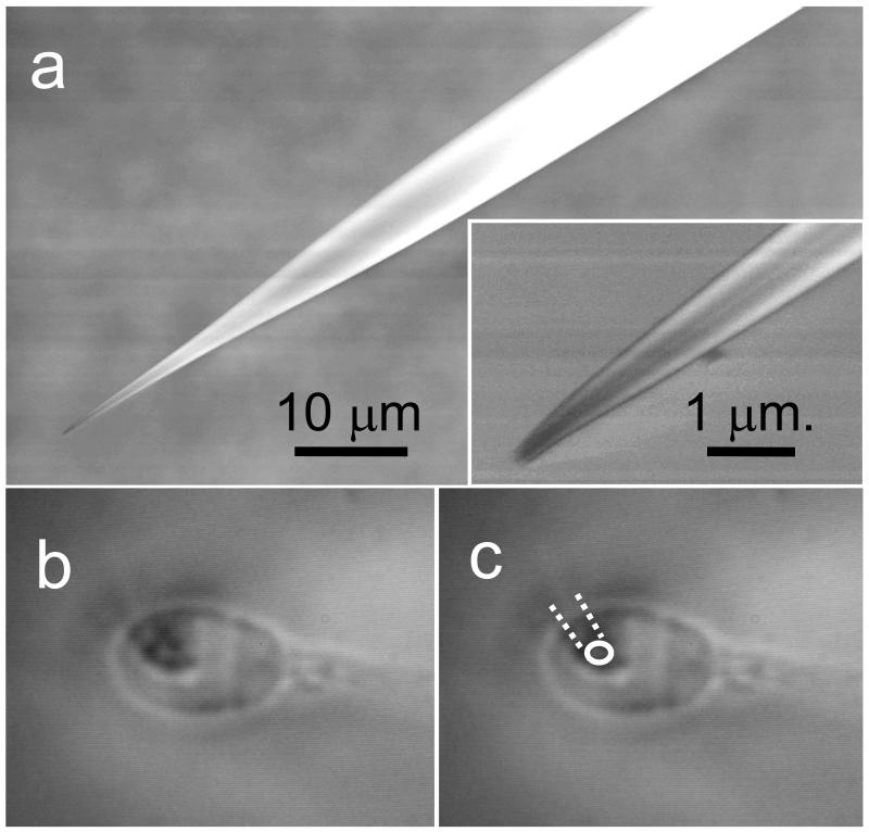
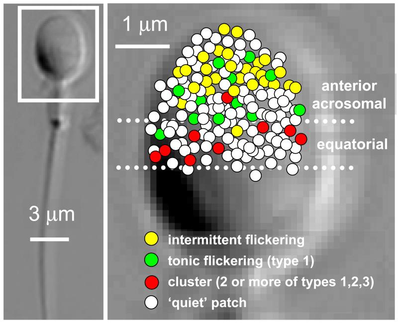
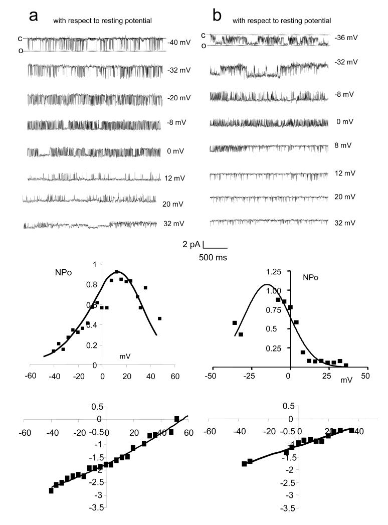
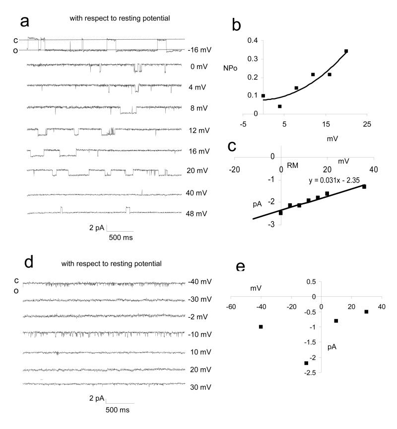
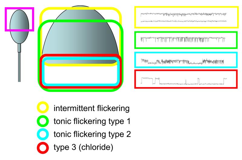
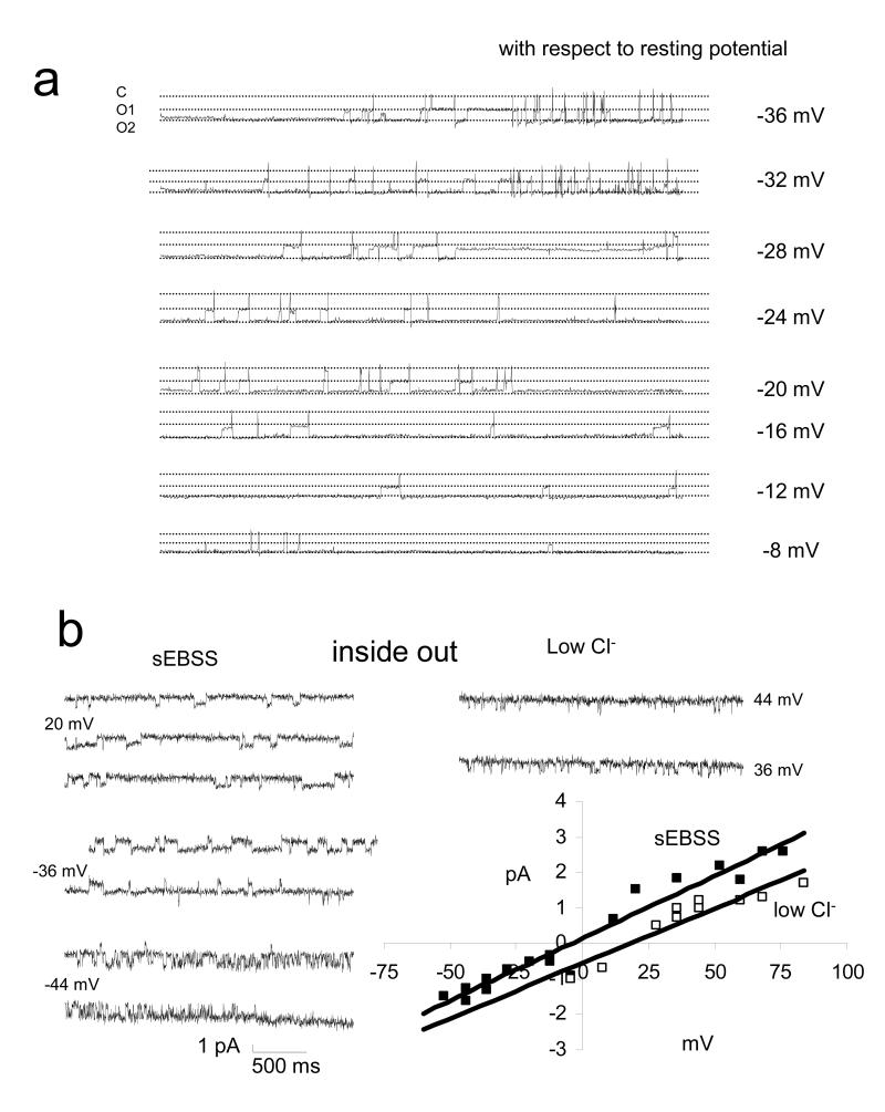
Ion channels are pivotal to many aspects of sperm physiology and function. We have used the patch clamp technique to investigate the distribution of ion channels in the plasma membrane of the head of human spermatozoa. We report that three types of activity are common in the equatorial and acrosomal regions of the sperm head. Two of these (a chloride-permeable anion channel showing long stable openings and a second channel which flickered between open and closed states and was dependent upon cytoplasmic factors for activity) were localised primarily to the equatorial segment. A third type, closely resembling the flickering activity but with different voltage sensitivity of P(open), was more widely distributed but was not detectable over the anterior acrosome. In the anterior acrosomal area channels were present but showed very low levels of spontaneous activity. A unique feature of channel activity in the sperm equatorial region was co-localisation into mixed clusters, most patches were devoid of activity but 'active' patches typically contained two or more types of activity (in a single 200-300 nM diameter patch). We conclude that ion channels in the sperm membrane show regionalisation of type and activity and that the channels are clustered into functional groups, possibly interacting through local effects on membrane potential.


Figures

Fig 1
Fig 1
Obtaining patches on the head of human spermatozoa. (a) Scanning electron micrograph of the fine patch pipettes used in this study. The final 20 μM of the pipette tapers gently to a tip of ≈300 nm (see inset), (b) Example of video image used during approach to the cell surface. Picture shows a sperm head with the tip of the pipette visible as a ring. The diameter is exaggerated due to light diffraction around the tip but the centre can be clearly located. In this instance the pipette is slightly off vertical and the shank is just visible (highlighted in ‘c’).
Fig. 2
Fig. 2
Mapping of ion channel distribution on the head of human sperm.. The position of each patch was recorded using the image as shown in fig 1c and combined to produce the composite image. All patches shown are in the equatorial and acrosomal areas. Colours show patches containing channel type 1 (green); mixed clusters of at least two of types 1, 2 and 3 (red) or intermittent flickering activity (yellow). White circles are ‘quiet’ patches. Dashed line show boundaries used to allocate patches to anterior-acrosomal and equatorial areas.
Fig. 3
Fig. 3
Characteristics of activity type 1 (a: patches shown green and in red in fig 2) and type 2 (b: most red patches in fig 2). (a) upper panel - representative traces from a type 1 channel at a series of patch potentials from −40 mV (with respect to resting potential) to 32 mV positive to resting potential. Downward transitions are opening (inward current). Channel shows constant flickering activity at all potentials. Centre panel panel - open probability (NPo) of type I activity is voltage dependent and is highest at 20-30 mV positive to resting potential. Lower panel - current-voltage plot for single type 1 channel currents. Gradient gives a conductance of ≈28 pS and the current reverses at ≈resting potential +55 mV. (b) Upper panel - representative traces from a type 2 channel at a series of patch potentials from potentials from −36 mV (with respect to membrane potential) to 32 mV positive to resting potential. Downward transitions are opening (inward current). Channel shows constant flickering activity at all potentials. Centre panel - open probability (NPo) of type 2 activity is voltage dependent and is highest at 10-20 mV negative to resting potential. Lower panel - current-voltage plot for single type 2 channel currents. Gradient gives a conductance of ≈23 pS and the current reverses at ≈resting potential +55 mV.
Figure 4
Figure 4
Characteristics of channel type 3 (a-c; patches shown red in fig 2) and intermittent/variable ‘flickering’ activity (d-e: yellow patches in fig 2).. (a) representative traces from a channels at a series of pipette potentials from potentials from −16 mV (with respect to membrane potential) to 48 mV positive to resting potential. Downward transitions are opening (inward current). Channel shows constant flickering activity at all potentials. (b) Open probability (NPo) is voltage dependent and increases at potentials positive to resting potential. (c) Current-voltage plot for single channel currents. Gradient gives a conductance of ≈31 pS and the current reverses at ≈resting potential +75 mV. (d) representative traces from one patch showing intermittent/variable ‘flickering’ activity. Records from −40 mV (with respect to membrane potential) to 30 mV positive to resting potential. Downward transitions are opening (inward current). Activity was very rare and the four traces in which activity was discernible were not all collected during a single voltage-stepping protocol. (e) Current-voltage scattergram for the single channel currents from the patch with intermittent/variable ‘flickering’ activity shown in ‘d’. The data cannot be fitted to a single conductance plot indicating that the patch contains more than one channel and/or channels have sub-conductance states.
Figure 5
Figure 5
Scematic to show distribution of the four type of activity reported here. Intermittent flickering, tonic flickering type 1, tonic flickering type 2 and type 3 activity are shown by the yellow, green, blue and red contours respectively. Examples of ‘typical’ activity are shown to the right, using the same colour code.
Figure 6
Figure 6
Complex records and characterisation of type 3 channel. (a) Coincident activity of two channels with conductances of ≈25 pS (probably one each of type 1 and type 2, both of which were observed in this patch). Coincident activity shows more stable open states and opening is usually synchronous. (b) Modulation of current through type 3 channel by substitution of Cl with gluconate. Left panel shows currents recorded at 3 different pipette potentials in an inside-out patch (symmetrical conditions). Upper right panel shows currents recorded after replacement of sEBSS ([Cl] =129 mM) in the bath (cytoplasmic face of patch) with modified saline in which NaCl was replaced with Na gluconate ([Cl] = 39 mM). Estimated conductance falls from 33 pS to 29 pS and reversal potential is shifted by +24 mV.

References

    1. Alavi SM, Cosson J. Sperm motility in fishes. I. Effects of temperature and pH: a review. Cell Biol Int. 2005;29:101–110. - PubMed
    1. Arnoult C, Kazam IG, Visconti PE, Kopf GS, Villaz M, Florman HM. Control of the low voltage-activated calcium channel of mouse sperm by egg ZP3 and by membrane hyperpolarization during capacitation. Proc Natl Acad Sci U S A. 1999;96:6757–6762. - PMC - PubMed
    1. Barfield JP, Yeung CH, Cooper TG. Characterization of potassium channels involved in volume regulation of human spermatozoa. Mol Hum Reprod. 2005;11:891–897. - PubMed
    1. Barratt CL, Publicover SJ. Interaction between sperm and zona pellucida in male fertility. Lancet. 2001;358:1660–1662. - PubMed
    1. Bedford JM, Moore HD, Franklin LE. Significance of the equatorial segment of the acrosome of the spermatozoon in eutherian mammals. Exp Cell Res. 1979;119:119–126. - PubMed

Publication types

LinkOut - more resources