

Age-dependent changes in Ca2+ homeostasis in peripheral neurones: implications for changes in function
- PMID: 17517039
- PMCID: PMC1974774
- DOI: 10.1111/j.1474-9726.2007.00298.x
Age-dependent changes in Ca2+ homeostasis in peripheral neurones: implications for changes in function
Abstract
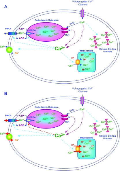
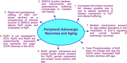
Calcium ions represent universal second messengers within neuronal cells integrating multiple cellular functions, such as release of neurotransmitters, gene expression, proliferation, excitability, and regulation of cell death or apoptotic pathways. The magnitude, duration and shape of stimulation-evoked intracellular calcium ([Ca2+]i) transients are determined by a complex interplay of mechanisms that modulate stimulation-evoked rises in [Ca2+]i that occur with normal neuronal function. Disruption of any of these mechanisms may have implications for the function and health of peripheral neurones during the aging process. This review focuses on the impact of advancing age on the overall function of peripheral adrenergic neurones and how these changes in function may be linked to age-related changes in modulation of [Ca2+]i regulation. The data in this review suggest that normal aging in peripheral autonomic neurones is a subtle process and does not always result in dramatic deterioration in their function. We present studies that support the idea that in order to maintain cell viability peripheral neurones are able to compensate for an age-related decline in the function of at least one of the neuronal calcium-buffering systems, smooth endoplasmic reticulum calcium ATPases, by increased function of other calcium-buffering systems, namely, the mitochondria and plasmalemma calcium extrusion. Increased mitochondrial calcium uptake may represent a 'weak point' in cellular compensation as this over time may contribute to cell death. In addition, we present more recent studies on [Ca2+]i regulation in the form of the modulation of release of calcium from smooth endoplasmic reticulum calcium stores. These studies suggest that the contribution of the release of calcium from smooth endoplasmic reticulum calcium stores is altered with age through a combination of altered ryanodine receptor levels and modulation of these receptors by neuronal nitric oxide containing neurones.
Figures
References
-
- Abbott RD, Curb JD, Rodriguez BL, Masaki K, Popper JS, Ross GW, Petrovich H. Age-related changes in risk factor effects on the incidence of thromboembolic and hemorrhagic stroke. J. Clin. Epidemiol. 2003;56:479–486. - PubMed
-
- Amenta F, Bronzetti E, Ferrante F, Ricci A. The noradrenergic innervation of spinal cord blood vessels in old rats. Neurobiol. Aging. 1990;11:47–50. - PubMed
-
- Balshaw DM, Yamaguchi N, Meissner G. Modulation of intracellular calcium-release channels by calmodulin. J. Membr. Biol. 2002;185:1–8. - PubMed
Publication types
MeSH terms
Substances
Grants and funding
LinkOut - more resources
Full Text Sources
Medical
Miscellaneous
