. 2007 May 21:7:53.
doi: 10.1186/1471-213X-7-53.

Cdkn1c (p57Kip2) is the major regulator of embryonic growth within its imprinted domain on mouse distal chromosome 7

Affiliations

Cdkn1c (p57Kip2) is the major regulator of embryonic growth within its imprinted domain on mouse distal chromosome 7

Stuart C Andrews et al. BMC Dev Biol. .

Abstract

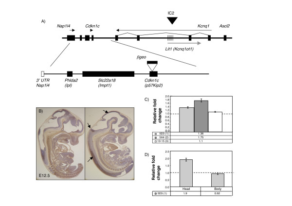
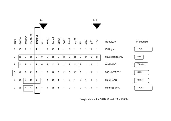
Background: Cdkn1c encodes an embryonic cyclin-dependant kinase inhibitor that acts to negatively regulate cell proliferation and, in some tissues, to actively direct differentiation. This gene, which is an imprinted gene expressed only from the maternal allele, lies within a complex region on mouse distal chromosome 7, called the IC2 domain, which contains several other imprinted genes. Studies on mouse embryos suggest a key role for genomic imprinting in regulating embryonic growth and this has led to the proposal that imprinting evolved as a consequence of the mismatched contribution of parental resources in mammals.

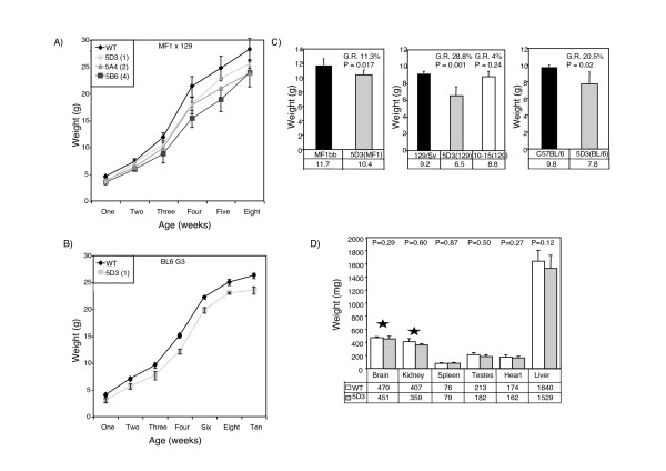
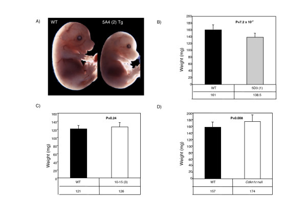
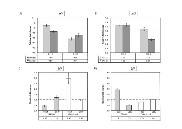
Results: In this study, we characterised the phenotype of mice carrying different copy number integrations of a bacterial artificial chromosome spanning Cdkn1c. Excess Cdkn1c resulted in embryonic growth retardation that was dosage-dependent and also responsive to the genetic background. Two-fold expression of Cdkn1c in a subset of tissues caused a 10-30% reduction in embryonic weight, embryonic lethality and was associated with a reduction in the expression of the potent, non-imprinted embryonic growth factor, Igf1. Conversely, loss of expression of Cdkn1c resulted in embryos that were 11% heavier with a two-fold increase in Igf1.

Conclusion: We have shown that embryonic growth in mice is exquisitely sensitive to the precise dosage of Cdkn1c. Cdkn1c is a maternally expressed gene and our findings support the prediction of the parental conflict hypothesis that that the paternal genome silences genes that have an inhibitory role in embryonic growth. Within the IC2 imprinted domain, Cdkn1c encodes the major regulator of embryonic growth and we propose that Cdkn1c was the focal point of the selective pressure for imprinting of this domain.


Figures

Figure 1
Figure 1
Physical map of the telomeric end of the imprinted domain in mouse distal chromosome 7 in relation to the position of the BAC transgene and expression analysis of transgenic lines. A) Top line shows a genomic map of the IC2 region on distal mouse chromosome 7. Hatched region marks the KvDMR1 region that is methylated only on the maternal allele. The black arrowhead marks the position of the IC2 control region for the domain. Arrows indicate direction of transcription. Below is the map of the 85 kb Cdkn1c transgene (BAC144D14). Filled boxes show the positions of the intact genes. In addition to Phlda2, Slc22a18 and Cdkn1c, the transgene includes the 3' UTR of Napl14 (white box) but not the 5' UTR. The modified BAC used to generate line 10–15 has a β-galactosidase-neomycin (β-geo) fusion gene inserted into Cdkn1c indicated by the white arrowhead and black filled box. B) Localisation of the Cdkn1c protein expressed from the BAC transgene. Sagittal section of E12.5 wild type and Cdkn1c-null/BAC transgene (KO/Tg) embryos stained with a Cdkn1c-specific antibody. Endogenous Cdkn1c is most strongly expressed in skeletal and cardiac muscle, cartilage and the developing pituitary with lower expression in neuronal tissues. The transgenic expression is predominantly only neural, indicated by the black arrows, and in a subset of cells in the pituitary, kidney, lung and adrenal gland. C) Quantitative RT-PCR data for Cdkn1c at E13.5 for 5D3 (single copy line) and 5A4 (two copy line) transgenic embryos and 10–15 (Cdkn1c-βgeo) at E12.5. Cdkn1c levels were normalised against three reference genes: Gapdh, Actin and 18S RNA. Cdkn1c was at 1.36-fold wild type levels in the single copy line and 1.75-fold endogenous in the two copy line. Cdkn1c levels were not significantly raised in the control line, 10–15 (Cdkn1c-βgeo). D) Quantitative RT-PCR data for Cdkn1c at E12.5 for 5D3 (single copy line) showing the difference in expression between head, where the transgene is predominantly expressed, and the body, where the transgene is only expressed in a small subset of tissues. This is consistent with copy number dependent expression of Cdkn1c from the transgene in a subset of tissues.
Figure 2
Figure 2
Postnatal growth retardation in mice carrying Cdkn1c transgene. A) Post natal growth curves of males from one to eight weeks for lines 5D3 (one copy, n = 2), 5A4 (two copy, n = 3) and 5B6 (four copies, n = 2) and their wild type littermates (n = 9). Mice carrying the transgene are significantly smaller in all lines (Statistical significance using Student's t-test P < 0.017 for all lines). All weights were obtained from F1 mice born from a cross between a chimaeric male founder (129/Sv) and a MF1 female. B) Post natal weight data from one to ten weeks of males for the single copy line, 5D3, bred onto the C57/BL6 background for three generation showing persistence of growth retardation after weaning (one copy, Tg n = 9, WT n = 8). C) Weights at 3 weeks of male mice from line 5D3 (one copy, Tg n = 4, WT n = 9) on a 50%:50%; 129/Sv × MF1 background labelled as 5D3/MF1) compared with a 75%:25% background labelled as 5D3/129 (Tg n = 6, WT n = 8). Interaction with a genetic factor within the 129/Sv background results in a more severe growth retardation, increasing from 11.3% to 28.8% (Statistically significant change using Student's t-test. P = 0.014). On a C57BL/6 background, the growth retardation is intermediate at 20.5% (Tg n = 9, WT n = 8). No significant growth retardation is observed in transgenic line 10–15 on a 129/Sv background (Tg n = 8, WT n = 4, P = 0.24). This line carries three copies of the modified BAC where transgenic Cdkn1c is not functional demonstrating that growth retardation is likely due to excess Cdkn1c. C) Comparison of organ weights in wild type and 5D3 transgenic adult males at 8–10 weeks. Growth retardation is not restricted to organs in which there was excess embryonic Cdkn1c WT n = 9 and Tg n = 6). *denotes tissues that were exposed to excess Cdkn1c during embryogenesis.
Figure 3
Figure 3
Embryonic growth is exquisitely sensitive to Cdkn1c levels. A) Image of a wild type and a transgenic embryo from line 5A4 (two copies) at E16.5 shows demonstrable growth retardation. The embryos appear normally proportioned. B) A comparison of wild type and transgenic embryo weights at E13.5 for line 5D3 (single copy unmodified BAC, Tg n = 22, WT n = 21). These embryos are from 129/Sv females mated with transgenic males maintained on an MF1 background. Transgenic embryos weigh significantly less than wild type embryos (86%, P = 7.2 × 10⁻⁷). C) A comparison of wild type and transgenic weight at E13.5 of embryos for line 10–15 (three copy BAC with an inactive transgenic Cdkn1c locus and no excess Cdkn1c, Tg n = 30, WT n = 22). The line was maintained on a 129/Sv background. Transgenic embryos are not significantly different in weight to wild type embryos (104%, P = 0.24). D) A comparison of wild type and transgenic weights at E13.5 of embryos inheriting a targeted Cdkn1c allele maternally (null for Cdkn1c, Tg n = 19, WT n = 16). The line was maintained on a 129/Sv background. Transgenic embryos weigh significantly more than wild type embryos (111%, P = 0.008).
Figure 4
Figure 4
Quantitative analysis of growth factor expression levels in response to excess embryonic Cdkn1c. A) Quantitative RT-PCR data for Igf1 on whole embryos from lines 5D3 and 5A4. At E11.5, there is a slight reduction in Igf1 in the two-copy line. At E13.5, there is a reduction in Igf1 levels in both lines. B) Quantitative RT-PCR data for Igf2 on whole embryos from lines 5D3 and 5A4. At E11.5, there is a slight increase in Igf2 in both lines while at E13.5, there is a significant decrease in Igf2 in the two-copy line. C) Comparison of expression of Igf1 at E12.5 in the single copy line, 5D3, head compared with body and whole Cdkn1c null embryos. In line 5D3, Igf1 levels are 60% less than wild type levels in the head where transgenic Cdkn1c is predominantly expressed but relatively unaffected in the body. In null embryos, Igf1 levels are raised by three-fold in the head. D) Comparison of expression of Igf2 at E12.5. In line 5D3, Igf2 levels are raised by two-fold in the head but 50% wild type levels in the body. In null embryos, Igf2 levels are similar to wild type.
Figure 5
Figure 5
Cdkn1c is the key regulator of embryonic growth within the IC2 imprinted domain on mouse distal chromosome 7. Summary of the reported genotypes and phenotypes associated with the imprinted genes on mouse distal chromosome 7. Black arrowheads indicate the positions of the known IC for the two domains. The number of expressed gene copies present in each genotype is shown with the top row being the normal, wild type state. The reported weight phenotypes are for maternal disomy distal chromosome 7 [22], paternal inheritance of the KvDMR1 deletion [25], maternal inheritance of an 800 kb YAC [42] and data from this paper. The strain backgrounds are C57BL/6 where indicated.

References

    1. Surani MA. Imprinting and the initiation of gene silencing in the germ line. Cell. 1998;93:309–312. doi: 10.1016/S0092-8674(00)81156-3. - DOI - PubMed
    1. DeChiara TM, Robertson EJ, Efstratiadis A. Parental imprinting of the mouse insulin-like growth factor II gene. Cell. 1991;64:849–859. doi: 10.1016/0092-8674(91)90513-X. - DOI - PubMed
    1. Efstratiadis A. Genetics of mouse growth. Int J Dev Biol. 1998;42:955–976. - PubMed
    1. Guillemot F, Caspary T, Tilghman SM, Copeland NG, Gilbert DJ, Jenkins NA, Anderson DJ, Joyner AL, Rossant J, Nagy A. Genomic imprinting of Mash2, a mouse gene required for trophoblast development. Nature Genetics. 1995;9:235–242. doi: 10.1038/ng0395-235. - DOI - PubMed
    1. Frank D, Fortino W, Clark L, Musalo R, Wang W, Saxena A, Li CM, Reik W, Ludwig T, Tycko B. Placental overgrowth in mice lacking the imprinted gene Ipl. Proc Natl Acad Sci U S A. 2002;99:7490–7495. doi: 10.1073/pnas.122039999. - DOI - PMC - PubMed

Publication types

Substances