. 2007 May 29;104(22):9266-71.
doi: 10.1073/pnas.0702427104. Epub 2007 May 21.

Nanoprobes with near-infrared persistent luminescence for in vivo imaging

Affiliations

Nanoprobes with near-infrared persistent luminescence for in vivo imaging

Quentin le Masne de Chermont et al. Proc Natl Acad Sci U S A. .

Abstract

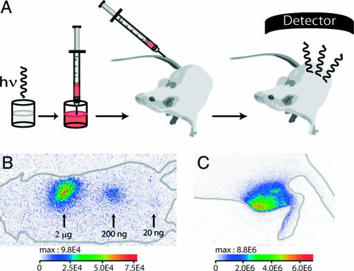
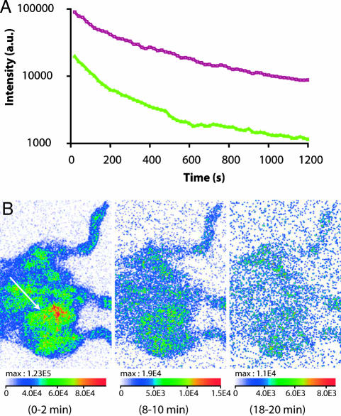
Fluorescence is increasingly used for in vivo imaging and has provided remarkable results. Yet this technique presents several limitations, especially due to tissue autofluorescence under external illumination and weak tissue penetration of low wavelength excitation light. We have developed an alternative optical imaging technique by using persistent luminescent nanoparticles suitable for small animal imaging. These nanoparticles can be excited before injection, and their in vivo distribution can be followed in real-time for more than 1 h without the need for any external illumination source. Chemical modification of the nanoparticles' surface led to lung or liver targeting or to long-lasting blood circulation. Tumor mass could also be identified on a mouse model.


Conflict of interest statement

The authors declare no conflict of interest.

Figures

Fig. 1.
Fig. 1.
Physical characteristics of long afterglow nanoparticles. (A) NPs showed a clinoenstatite-like structure. (B) Transmission electronic microscopy images of the synthesized NPs. (Scale bar: 200 nm.) (C) Excitation spectrum. (D) Long afterglow emission spectrum. (E) Time dependence of the luminescence intensity of the NPs. NPs (10 mg) were put in 96-well plates under direct exposure to a 6-W UV lamp for 5 min. The luminous intensity was quantified straightforward by using an intensified charge-coupled device (ICCD) camera (PhotonImager; Biospace). Data analysis was performed by signal integration for 5 s. The luminous decay data were fit by a power law function for time >100 s.
Fig. 2.
Fig. 2.
Principles of in vivo experiments and first in vivo images. (A) A suspension containing a proper amount of NPs is excited with a 6-W UV lamp and is directly injected to an anesthetized mouse. The signal is then acquired with an intensified charge-coupled device (CCD) camera. (B) Image of three s.c. injections of NPs (2 μg, 200 ng, 20 ng). The different localizations are labeled with arrows, and the corresponding NP amounts are indicated. The acquisition was performed during the 2 min after injection. (C) Image of an intramuscular injection (200 μg) corresponding to a 90-s acquisition. The luminous intensity is expressed in photons per s·cm2·steradians (sr).
Fig. 3.
Fig. 3.
NP surface modification and in vivo biodistristribution. (A) Schematic representation of NP surface modification. (i) Amino-NPs were synthesized by reaction with 3-aminopropyltriethoxysilane. (ii) Carboxyl-NPs resulted from a reaction of amino-NPs with diglycolic anhydride. (iii) PEG-NPs were achieved by a peptidic coupling of amino-NPs with PEG5000COOH (25). (B–F) Optical imaging of mouse with 1-mg tail vein injections of differently charged NPs. (B) Amino-NPs. (C) Carboxyl-NPs. (D) Preinjection of anionic liposomes (6 μmol, 100 μl) 5 min before carboxyl-NP injection. (E) PEG-NPs. (F) Preinjection of anionic liposomes (6 μmol, 100 μl) 5 min before PEG-NP injection. All images correspond to a 2-min signal acquisition performed between the times indicated above each image. The luminous intensity is expressed in photons per s·cm2·steradians (sr). Similar results were obtained in triplicate experiments.
Fig. 4.
Fig. 4.
Imaging of a tumor-bearing mouse. (A) Overall luminous intensity detected in a Swiss mouse (purple curve) and a body-shaved C57BL/6 mouse bearing a 3LL tumor (green curve). Tail vein i.v preinjection of anionic liposomes (6 μmol, 100 μl injected 5 min before NP injection) was followed by tail vein injection of 1 mg of PEG-NPs. A region of interest covering the whole mouse was manually selected and analyzed by a period of 20 s. (B) Visualization of the hypervascularization of a 3LL tumor (white arrow) with rapid clearance.

Comment in

References

    1. Cherry SR. Annu Rev Biomed Eng. 2006;8:35–62. - PubMed
    1. Michalet X, Pinaud FF, Bentolila LA, Tsay JM, Doose S, Li JJ, Sundaresan G, Wu AM, Gambhir SS, Weiss S. Science. 2005;307:538–544. - PMC - PubMed
    1. Dubertret B, Skourides P, Noriis DJ, Noireaux V, Brivanlou AH, Libchader A. Science. 2002;298:1759–1762. - PubMed
    1. So MK, Xu C, Loening AM, Gambhir SS, Rao J. Nat Biotech. 2006;24:339–343. - PubMed
    1. Ballou B, Lagerholm BC, Ernst LA, Bruchez MP, Waggoner AS. Bioconjugate Chem. 2004;15:79–86. - PubMed

Publication types

Substances

LinkOut - more resources